. 2018 Dec;22(12):6202-6212.
doi: 10.1111/jcmm.13907. Epub 2018 Sep 25.

Increase of PRPP enhances chemosensitivity of PRPS1 mutant acute lymphoblastic leukemia cells to 5-Fluorouracil

Affiliations

Increase of PRPP enhances chemosensitivity of PRPS1 mutant acute lymphoblastic leukemia cells to 5-Fluorouracil

Dan Wang et al. J Cell Mol Med. 2018 Dec.

Abstract

Relapse-specific mutations in phosphoribosyl pyrophosphate synthetase 1 (PRPS1), a rate-limiting purine biosynthesis enzyme, confer significant drug resistances to combination chemotherapy in acute lymphoblastic leukemia (ALL). It is of particular interest to identify drugs to overcome these resistances. In this study, we found that PRPS1 mutant ALL cells specifically showed more chemosensitivity to 5-Fluorouracil (5-FU) than control cells, attributed to increased apoptosis of PRPS1 mutant cells by 5-FU. Mechanistically, PRPS1 mutants increase the level of intracellular phosphoribosyl pyrophosphate (PRPP), which causes the apt conversion of 5-FU to FUMP and FUTP in Reh cells, to promote 5-FU-induced DNA damage and apoptosis. Our study not only provides mechanistic rationale for re-targeting drug resistant cells in ALL, but also implicates that ALL patients who harbor relapse-specific mutations of PRPS1 might benefit from 5-FU-based chemotherapy in clinical settings.

Keywords: 5-FU; PRPP; PRPS1; acute lymphoblastic leukemia; nucleotide metabolism.


Figures

Figure 1
Figure 1
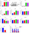
5‐FU sensitizes PRPS1 mutant ALL cells. (A and B) Stable expression of exogenous PRPS1 wild type or PRPS1 mutants (A190T and S103T) in Reh cells. The relative gray scale determination was analyzed using ImageJ software. (C–F) Screening of chemosensitivity of Reh cells (PRPS1 WT or mutants) to the chemotherapeutic drugs commonly used in ALL treatment by using cell viability assay measuring half maximal inhibitory concentration (IC50). **P < 0.01, and ***P < 0.001, two‐tailed Student's t tests. 6‐MP, 6‐mecaptopurine; 6‐TG, 6‐thioguanine; DXR, doxorubicin; VCR, vincristine. (G) Reh cells harboring PRPS1 mutants showed much more chemosensitivity to 5‐fluorouracil. *P < 0.05, **P < 0.01, two‐tailed Student's t tests. (H and I) Both of hydroxyurea (HU) and cisplatin showed little effect on the viability of Reh cells (IC50). (J) The primary ALL cells harboring PRPS1 mutant showed more chemosensitivity to 5‐FU. **P < 0.01, two‐tailed Student's t tests. (K) Percentage of GFP‐positive cells were dramatically reduced by 5‐FU in vivo. *P < 0.05, **P < 0.01, two‐tailed Student's t tests
Figure 2
Figure 2
5‐FU accelerates apoptosis of PRPS1 mutant Reh cells. (A) PRPS1 mutant cells proliferated more slowly than PRPS1 WT and control cells. Cell viability was measured at 0, 24, 48, and 72 hours after adding 1.5 μg/mL 5‐FU. ****P < 0.0001, two‐way ANOVA (simultaneous multiple comparison‐analysis of different treatment groups). (B) Apoptosis induced by 5‐FU was markedly increased in PRPS1 mutant cells. Percentage of apoptotic cells was detected at 72 hours after treatment with 1.5 μg/mL 5‐FU by annexin‐V/PI staining assay. ***P < 0.001, two‐tailed Student's t tests. (C‐E) 5‐FU caused enhancement of DNA damage response and apoptosis in PRPS1 mutant cells compared to that in PRPS1 WT and control cells. Cells were harvested at 48 hours after treatment with 1.5 μg/mL 5‐FU and the sample wasanalyzed by Western blot. Quantification of protein level was conducted by using ImageJ software. *P < 0.05, **P < 0.01, and ****P < 0.0001, two‐tailed Student's t tests. (F) DDR marker yH2AX was upregulated in PRPS1 mutant cells as detected byimmunofluorescence assay at 48 hours after treatment with 1.5 μg/mL 5‐FU
Figure 3
Figure 3
Apt conversion of 5‐FU to FUMP and FUTP in PRPS1 mutant ALL cells. (A) diagrammatic sketch of 5‐FU catabolism. OPRT, orotate phosphoribosyltransferase; TK, thymidylate kinase; TS, thymidylate synthase. (B–D) The outputs of FUMP and FUTP but not FdUMP in PRPS1 mutant cells were much higher than that in PRPS1 WT and control cells. Reh cells were harvested at 24 hours after treatment with 10 μg/mL 5‐FU. *P < 0.05, **P < 0.01, two‐tailed Student's t tests. (E) The ratio of 5‐FU metabolites in Reh, Nalm6, HCT116 and SW480. All cells were harvested at 24 hours after treatment with 10 μg/mL5‐FU
Figure 4
Figure 4
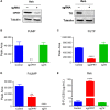
OPRT is required for the chemosensitivity of ALL cells to 5‐FU. (A) OPRT and TK in Ren cells were knocked down by using CRISPR‐Cas9 technology, respectively. (B–D) Conversion of 5‐FU to FUMP, FUTP and FdUMP in Reh cells when OPRT or TK was knocked down. **P < 0.01, ****P < 0.0001, two‐tailed Student's t tests. (E) Knocking down OPRT caused a dramatic resistance of Reh cells to 5‐FU analyzed by measuring IC50. ****P < 0.0001, two‐tailed Student's t tests
Figure 5
Figure 5
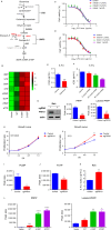
Accumulated PRPP enhances chemosensitivity of PRPS1 mutant ALL cells to 5‐FU. (A) Schematic depicting pyrimidine biosynthesis. (B) Pyrimidine nucleotides were increased in PRPS1 mutant cells compared to PRPS1 WT or control cells as showed by Heatmap. (C and D) Extra orotate and uracil could not sensitize Reh ALL cells to 5‐FU. (E and F) Extra R‐5‐P or PRPP enhanced Reh cell sensitivity to 5‐FU. **P < 0.01, ***P < 0.001, two‐tailed Student's t tests. (G) Knocking down PRPS1 by using CRISPR‐Cas9 technology reduced the levels of intracellular PRPP in Reh cells. PRPP was detected by LCMS, **P < 0.01, ***P < 0.001, two‐tailed Student's t tests. (H) PRPS1 knockdown cells proliferated much faster than control cells with the treatment of 5‐FU. Cell proliferation was measured at 0, 24, 48 and 72 hours after adding 1.5 μg/mL 5‐FU. **P < 0.01, two‐way ANOVA (multiple comparison‐analysis of different treatment groups at the same time). (I and J) Knocking down PRPS1 resulted in impaired conversion of 5‐FU to FUMP and FUTP in Reh cells. *P < 0.05, two‐tailed Student's t tests. (K) Knocking down PRPS1 caused a significant resistance of Reh cells to 5‐FU. **P < 0.01, two‐tailed Student's t tests. (L) Intracellular PRPP was significantly accumulated in Reh cells harboring PRPS1 mutants compared to control cells. *P < 0.05, **P < 0.01, ***P < 0.001, ****P < 0.0001, two‐tailed Student's t tests

References

    1. Siegel DA, King J, Tai E, Buchanan N, Ajani UA, Li J. Cancer incidence rates and trends among children and adolescents in the United States, 2001‐2009. Pediatrics. 2014;134:e945‐e955. - PMC - PubMed
    1. Pui CH, Carroll WL, Meshinchi S, Arceci RJ. Biology, risk stratification, and therapy of pediatric acute leukemias: an update. J Clin Oncol. 2011;29:551‐565. - PMC - PubMed
    1. Karran P, Attard N. Thiopurines in current medical practice: molecular mechanisms and contributions to therapy‐related cancer. Nat Rev Cancer. 2008;8:24‐36. - PubMed
    1. Raetz EA, Bhatla T. Where do we stand in the treatment of relapsed acute lymphoblastic leukemia?. Hematology Am Soc Hematol Educ Program. 2012;2012:129‐136. - PubMed
    1. Li B, Li H, Bai Y, et al. Negative feedback‐defective PRPS1 mutants drive thiopurine resistance in relapsed childhood ALL. Nat Med. 2015;21:563‐571. - PMC - PubMed

Publication types

MeSH terms

LinkOut - more resources