GABA levels and TSPO expression in people at clinical high risk for psychosis and healthy volunteers: a PET-MRS study
- PMID: 30255837
- PMCID: PMC6397035
- DOI: 10.1503/jpn.170201
GABA levels and TSPO expression in people at clinical high risk for psychosis and healthy volunteers: a PET-MRS study
Abstract
Background: γ-Aminobutyric acidergic (GABAergic) dysfunction and immune activation have been implicated in the pathophysiology of schizophrenia. Preclinical evidence suggests that inflammation-related abnormalities may contribute to GABAergic alterations in the brain, but this has never been investigated in vivo in humans. In this multimodal imaging study, we quantified cerebral GABA plus macromolecule (GABA+) levels in antipsychotic-naive people at clinical high risk for psychosis and in healthy volunteers. We investigated for the first time the association between GABA+ levels and expression of translocator protein 18 kDa (TSPO; a marker of microglial activation) using positron emission tomography (PET).
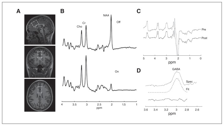
Methods: Thirty-five people at clinical high risk for psychosis and 18 healthy volunteers underwent 3 T proton magnetic resonance spectroscopy to obtain GABA+ levels in the medial prefrontal cortex (mPFC). A subset (29 people at clinical high risk for psychosis and 15 healthy volunteers) also underwent a high-resolution [18F]FEPPA PET scan to quantify TSPO expression. Each participant was genotyped for the TSPO rs6971 polymorphism.
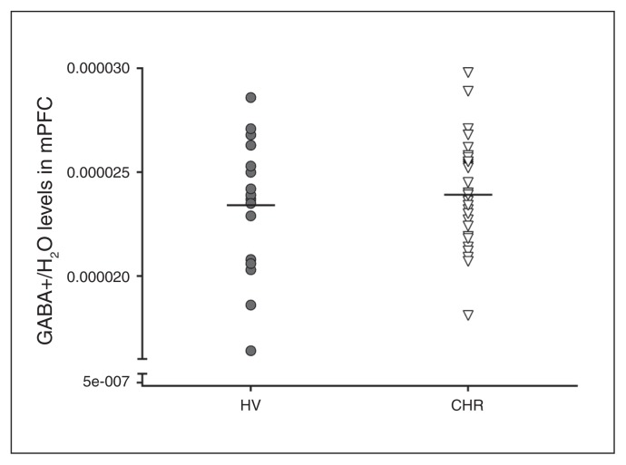
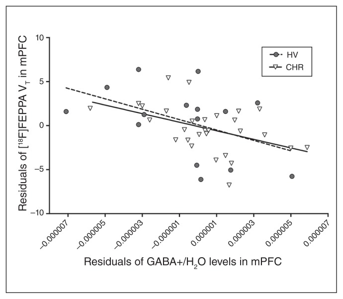
Results: We found that GABA+ levels were significantly associated with TSPO expression in the mPFC (F1,40 = 10.45, p = 0.002). We found no significant differences in GABA+ levels in the mPFC (F1,51 = 0.00, p > 0.99) between people at clinical high risk for psychosis and healthy volunteers. We found no significant correlations between GABA+ levels or residuals of the association with TSPO expression and the severity of prodromal symptoms or cognition.
Limitations: Given the cross-sectional nature of this study, we could determine no cause-and-effect relationships for GABA alterations and TSPO expression.
Conclusion: Our findings suggest that TSPO expression is negatively associated with GABA+ levels in the prefrontal cortex, independent of disease status.
© 2019 Joule Inc. or its licensors
Conflict of interest statement
R. Mizrahi has received speaker fees from Otsuka Lundbeck Canada. No other competing interests declared.
Figures
References
-
- Wassef A, Baker J, Kochan LD. GABA and schizophrenia: a review of basic science and clinical studies. J Clin Psychopharmacol. 2003;23:601–40. - PubMed
-
- Akbarian S, Kim JJ, Potkin SG, et al. Gene expression for glutamic acid decarboxylase is reduced without loss of neurons in prefrontal cortex of schizophrenics. Arch Gen Psychiatry. 1995;52:258–66. - PubMed
-
- Lewis DA, Hashimoto T, Volk DW. Cortical inhibitory neurons and schizophrenia. Nat Rev Neurosci. 2005;6:312–24. - PubMed
Publication types
MeSH terms
Substances
Grants and funding
LinkOut - more resources
Full Text Sources
Other Literature Sources
Medical