

Expanding the Spectrum of Genetic Alterations in Pseudomyogenic Hemangioendothelioma With Recurrent Novel ACTB-FOSB Gene Fusions
- PMID: 30256258
- PMCID: PMC6608746
- DOI: 10.1097/PAS.0000000000001147
Expanding the Spectrum of Genetic Alterations in Pseudomyogenic Hemangioendothelioma With Recurrent Novel ACTB-FOSB Gene Fusions
Abstract
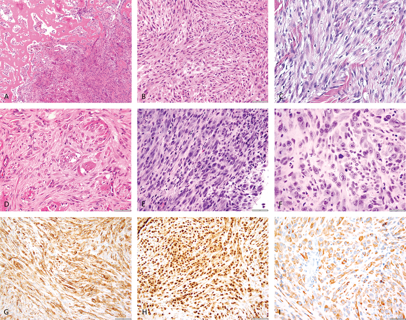
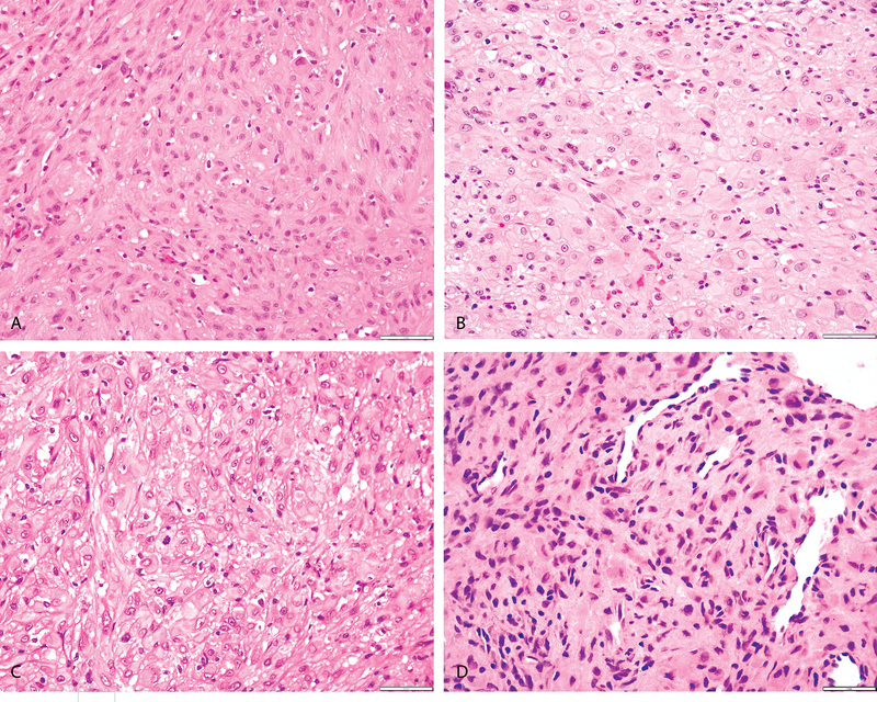
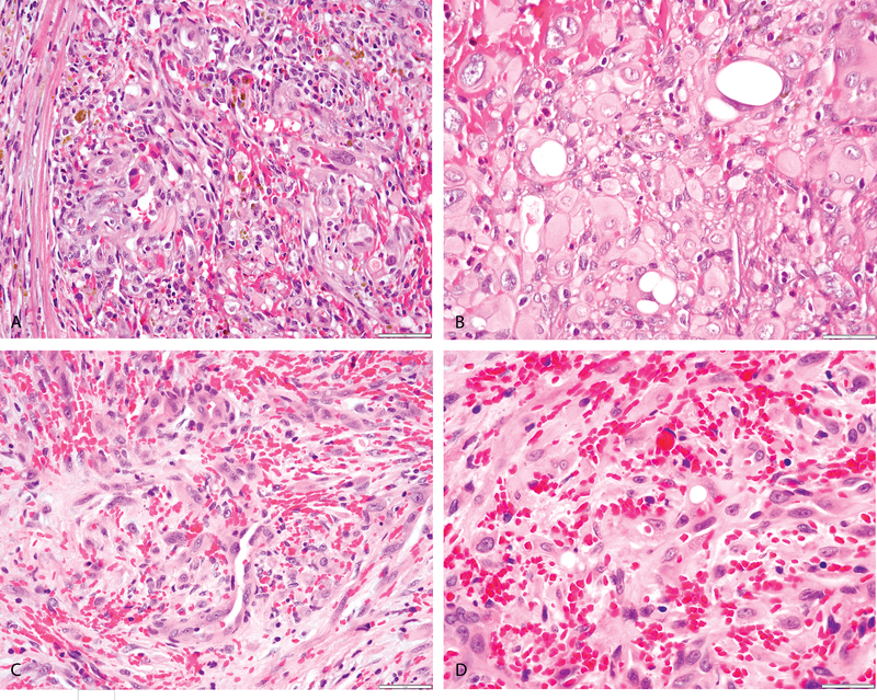
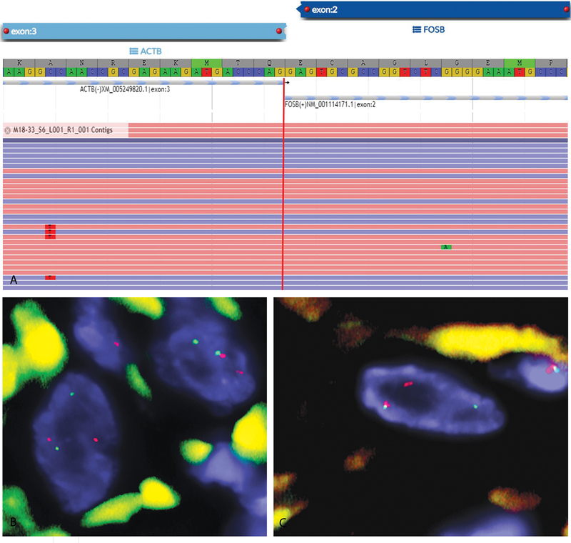
Pseudomyogenic hemangioendothelioma (PHE) is an uncommon, rarely metastasizing vascular neoplasm with predilection to affect young adults. The tumors often present as multiple nodules involving various tissue planes, including superficial and deep soft tissues as well as bone. Recurrent SERPINE1-FOSB gene fusions have been reported as the hallmark genetic abnormality in PHE, however, in our experience, a number of cases with typical histology lack this genetic abnormality. In this study, we identify a novel ACTB-FOSB gene fusion, which is as prevalent as the initial translocation reported. We selected 15 consecutive cases of PHE with typical morphologic features which had material for molecular testing. The cohort included 10 males and 5 females, ranging in age from 17 to 58 years (median age: 33 y; mean age: 35.3 y). Eight (53%) cases were located in the lower extremities (foot, calf, tibia, thigh), 5 (33%) were located in the trunk, abdomen or pelvis (abdominal wall-2, shoulder, back, ischium) and 2 (13%) were located in the upper extremity (humerus and hand). Ten (67%) cases had multifocal presentation and 5 (33%) presented as solitary lesions. Three (20%) cases were located only in the superficial dermis and subcutaneous tissues, 4 (27%) involved the superficial and deep soft tissue and 8 (53%) cases involved only the deep soft tissue and bone. Using fluorescence in situ hybridization and ARCHER fusionplex analysis we identified a novel ACTB-FOSB gene fusion in 7 cases, while the remaining 8 had the previously described SERPINE1-FOSB fusion. The clinicopathologic features and behavior of PHE associated with the ACTB-FOSB gene fusion were similar to those harboring the SERPINE1-FOSB; except that tumors with the ACTB variant were more often associated with solitary presentation. In conclusion, our results expand the spectrum of genetic alterations in PHE with a novel gene fusion identified in half of the cases. We speculate that some of the novel targeted therapies that have shown promise in SERPINE1-FOSB-positive PHE might also be beneficial in this molecular subset.
Figures
References
-
- Amary MF, O’Donnell P, Berisha F, Tirabosco R, Briggs T, Pollock R, et al. Pseudomyogenic (epithelioid sarcoma-like) hemangioendothelioma: characterization of five cases. Skeletal radiology. 2013;42(7):947–57. - PubMed
-
- Gabor KM, Sapi Z, Tiszlavicz LG, Fige A, Bereczki C, Bartyik K. Sirolimus therapy in the treatment of pseudomyogenic hemangioendothelioma. Pediatric blood & cancer. 2018;65(2). - PubMed
-
- Hornick JL, Fletcher CD. Pseudomyogenic hemangioendothelioma: a distinctive, often multicentric tumor with indolent behavior. Am J Surg Pathol. 2011;35(2):190–201. - PubMed
-
- Ide YH, Tsukamoto Y, Ito T, Watanabe T, Nakagawa N, Haneda T, et al. Penile pseudomyogenic hemangioendothelioma/epithelioid sarcoma-like hemangioendothelioma with a novel pattern of SERPINE1-FOSB fusion detected by RT-PCR--report of a case. Pathol Res Pract. 2015;211(5):415–20. - PubMed
-
- Inyang A, Mertens F, Puls F, Sumathi V, Inwards C, Folpe A, et al. Primary Pseudomyogenic Hemangioendothelioma of Bone. Am J Surg Pathol. 2016;40(5):587–98. - PubMed
Publication types
MeSH terms
Substances
Grants and funding
LinkOut - more resources
Full Text Sources
Other Literature Sources
Miscellaneous
