. 2018 Aug;99(4):158-171.
doi: 10.1111/iep.12283.

Histopathological changes in androgenized ovaries are recovered by melatonin treatment

Affiliations

Histopathological changes in androgenized ovaries are recovered by melatonin treatment

Bianca R de Souza et al. Int J Exp Pathol. 2018 Aug.

Abstract

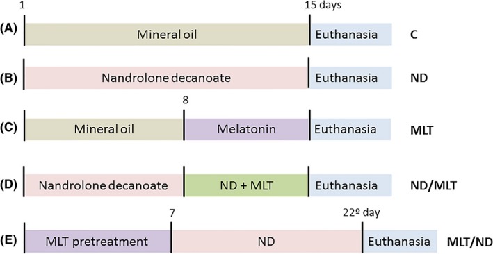
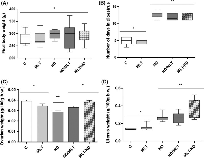
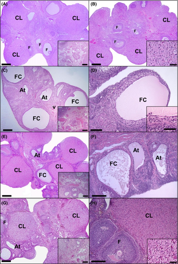
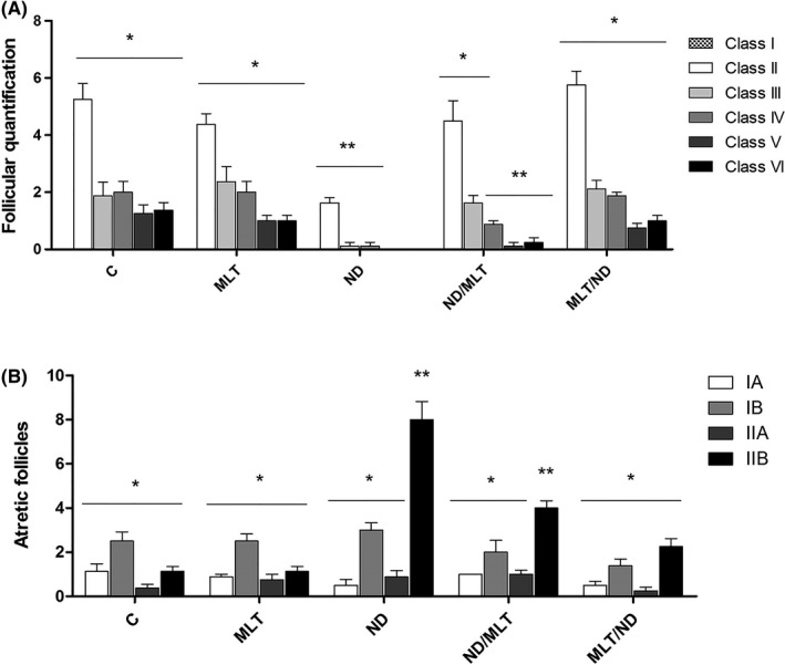
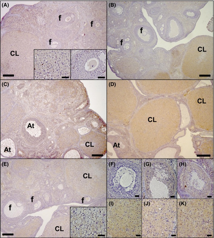
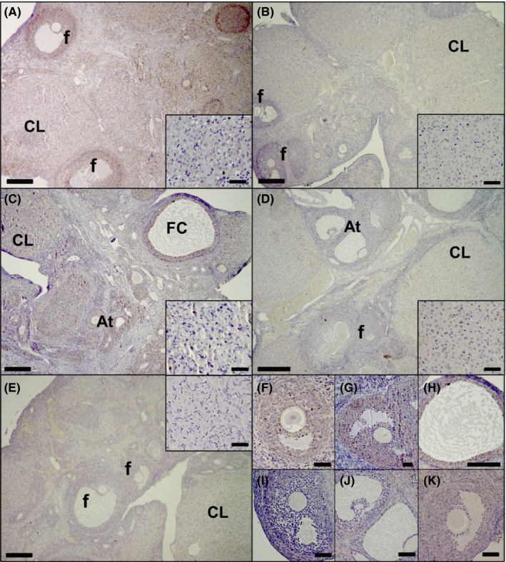
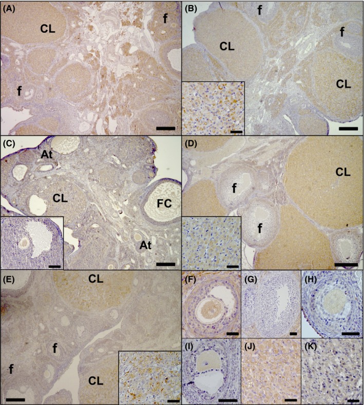
Nandrolone decanoate (ND) is a synthetic steroid, which promotes adverse effects on the ovarian tissue, and melatonin (MLT) exhibits a number of beneficial properties in the reproductive system. This study evaluated the general features of the ovarian tissue and the immunoexpression of sex steroid receptors in ND-treated rats that were submitted to short-term melatonin treatment. Adult rats received mineral oil (control group) and ND at doses of 7.5 mg/kg for 15 days (ND-treated group). The treatment with MLT (10mg/kg for 7 days) was given alone, before or in combination with ND. All ND-treated animals showed persistent dioestrus. In the androgenized groups that received MLT, ovarian morphology and size, and the number/area of corpora lutea were recovered. The number of healthy and atretic follicles was recovered when MLT was administered prior to ND; this was similar to the ovaries of control and MLT groups. There was a decrease in estrogen receptors immunostaining in the follicles of androgenized rats that were treated with MLT, and pretreatment with MLT reduced the expression of androgen receptor in atretic follicles and corpora lutea, when compared with ND-treated group. We conclude that MLT treatment recovered the histopathological aspects of the androgenized ovaries, and MLT pretreatment was the most effective.

Keywords: melatonin; morphometry; nandrolone decanoate; ovarian histology; sex steroid receptors.


Figures

Figure 1
Figure 1
Experimental protocol used for the treatment of animals. (A) Control (C), treated with mineral oil during 15 days; (B) treated with nandrolone decanoate during 15 days (ND); (C) treated with mineral oil for eight days and with melatonin during the last seven days of the experimental treatment (MLT); (D) treated with nandrolone decanoate for 15 days and with melatonin in the last seven days of steroid treatment (ND/MLT); (E) pretreatment with melatonin for seven days followed by the 15‐day steroid treatment (MLT/ND) [Colour figure can be viewed at http://wileyonlinelibrary.com]
Figure 2
Figure 2
Measurement of the final body weight, oestrous cycle and reproductive organs weight. (A) Final body weight (g) in animals of the different experimental groups. Values are expressed as the mean ± standard deviation; ANOVA. (B) Number of days in dioestrus phase. Values are expressed as the median ± interquartile deviation; Kruskal‐Wallis, Student‐Newman‐Keuls test. (C) Relative weight of the ovaries in animals of the different experimental groups. Values are expressed as the mean ± standard deviation; ANOVA, Tukey's test. (D) Relative weight of the uterus in animals of the different experimental groups. Values are expressed as the median ± interquartile deviation; Kruskal‐Wallis, Student‐Newman‐Keuls test. Different symbols indicate significant difference among the groups (P < 0.05). Groups: C (Control), MLT (treated with melatonin), ND (treated with nandrolone decanoate), ND/MLT (treated with ND and melatonin) and MLT/ND (pretreatment with melatonin and subsequent treatment with ND)
Figure 3
Figure 3
Photomicrographs of the ovaries in the different experimental groups: Control (A); treated with melatonin (B); treated with nandrolone decanoate (C, D); treated with nandrolone decanoate and melatonin (E, F); and pretreatment with melatonin followed by treatment with nandrolone decanoate (G, H). Images A and B show the same morphological aspect of the ovaries with many corpora lutea (CL) and follicles (F) in different stages of maturation. The medullary vascularization (A) and the morphology of the lutein cells (B) are shown in detail. Image C shows gonadal atrophy, intense follicular atresia (At), lack of corpora lutea, and medullary vasodilation (v; in detail). Image (D) shows a follicular cyst (FC) with a thin layer of flat cells surrounding the follicular wall (in detail). In the ND/MLT group (E, F), corpora lutea (CL), atretic follicles (At), follicular cysts (FC) and the medullary vasodilation (in detail) are shown. In the MLT/ND group (G, H), the healthy aspect of the CL, and the presence of follicles in maturation (F) and degeneration (At) are evidenced. Non‐dilated blood vessels in the medulla and lutein cells with normal morphology are shown in detail (G, H respectively). Haematoxylin‐eosin. Bars = 600 μm (A‐C, E, G); 100 μm (D, F, H); 50 μm (details in B, D, H); 200 μm (details in A, C, E, G) [Colour figure can be viewed at http://wileyonlinelibrary.com]
Figure 4
Figure 4
Evaluation of follicular kinetics. (A) Ovarian follicular quantitation in the different maturation classes. (B) Quantification of the atretic follicles in different degenerative stages. Values are expressed as the median ± interquartile deviation. Kruskal‐Wallis and Student‐Newman‐Keuls test (n = 4 sections/ovary/female/group). Different symbols indicate significant difference among the groups (P < 0.05). Groups: C (Control), MLT (treated with melatonin), ND (treated with nandrolone decanoate), ND/MLT (treated with ND and melatonin) and MLT/ND (pretreatment with melatonin and subsequent treatment with ND)
Figure 5
Figure 5
Immunostaining for AR in the ovaries of the different experimental groups. Control group (A) showed moderate staining intensity in healthy follicles (f; in detail) and corpora lutea (CL; in detail). In the MLT group (B), the follicles (f) and corpora lutea (CL) presented weak and strong immunostaining respectively. In C is shown the strong staining intensity in atretic follicles (At) and corpora lutea (CL) of the ND group. In the ND/MLT group (D), the reaction for AR in corpora lutea (CL) and atretic follicles (At) was strong while in MLT/ND group (E), it was moderate in follicles (f) and corpora lutea (CL; in detail). In (F), atretic follicles exhibited low marking in the MLT group, while in the ND (G), and ND/MLT (H) groups, the atretic follicles were strongly marked. In (I, J and K), the lutein cells of the MLT, ND and ND/MLT groups, presented strong immunostaining for AR. Bars = 200 μm (A‐E) and 50 μm (F‐K and details in A, E) [Colour figure can be viewed at http://wileyonlinelibrary.com]
Figure 6
Figure 6
Immunostaining for ERα in the ovaries of the different experimental groups. In the control group (A), the healthy follicles (f) and corpora lutea (CL) showed moderate staining intensity. In the MLT group (B), the corpora lutea (CL) presented moderate reaction, whereas a low reaction appeared in the follicles. In the ND group (C), atretic follicles (At), corpora lutea (CL) and follicular cysts (FC) exhibited moderate staining. In the ND/MLT (D) and MLT/ND (E) groups, the follicles (f) showed weak immunostaining and corpora lutea (CL) exhibited moderate staining. In detail (A‐E) is shown the immunostaining for ERα in lutein cells of each group. In F (control group) and G (ND group), antral follicles showed a moderate immunoreaction to ERα. In the MLT (I), ND/MLT (J) and MLT/ND (K) groups, the reaction was weak in the antral follicles. In H (ND group) is shown a follicular cyst with moderate ERα immunolabelling. Bars = 200 μm (A‐E, H) and 50 μm (F, G, I‐K and details in A‐E) [Colour figure can be viewed at http://wileyonlinelibrary.com]
Figure 7
Figure 7
Immunostaining for ERβ in the ovaries of the different experimental groups. In (A), the moderate immunostaining in healthy follicles (f) and strong staining in corpora lutea (CL) in the control group is observed. In the MLT group (B), the follicles (f) exhibited weak immunoreaction and the corpora lutea (CL) presented strong staining intensity (lutein cells in detail). In (C) (ND group), the moderate intensity in the corpora lutea (CL), atretic follicles (At; in detail) and follicular cysts is observed (FC). In the ND/MLT (D) and MLT/ND (E) groups, the corpora lutea (CL) maintained the strong staining intensity (lutein cells in detail), but the follicles (f) were weakly marked. In F (control group) is shown the moderate reaction in the antral follicle, compared to antral follicles of the MLT (G), ND/MLT (H) and MLT/ND (I) groups, which exhibit low staining intensity. In the lutein cells of the control group (J), there was strong ERβ immunolabelling, while in the ND group (K), this immunolabelling was moderate. Bars = 200 μm (A‐E) and 50 μm (F‐K and details in B‐E) [Colour figure can be viewed at http://wileyonlinelibrary.com]

Similar articles

Cited by

References

    1. Arlt W. Androgen therapy in women. Eur J Endocrinol. 2006;154:1‐11. - PubMed
    1. Kicman AT. Pharmacology of anabolic steroids. Br J Pharmacol. 2008;154:502‐521. - PMC - PubMed
    1. Amsterdam J, Opperhuizen A, Hartgens F. Adverse health effects of anabolic–androgenic steroids. Regul Toxicol Pharmacol. 2010;57:117‐123. - PubMed
    1. Cannavò S, Curtò L, Trimarchi F. Exercise‐related female reproductive dysfunction. J Endocrinol Invest. 2001;24:823‐832. - PubMed
    1. Camargo ICC, Leite GAA, Pinto T, Ribeiro‐Paes JT. Histopathologycal findings in the ovaries and uterus of albino female rats promoted by co‐administration of synthetic steroids and nicotine. Exp Toxicol Pathol. 2014;66:195‐202. - PubMed

LinkOut - more resources