

STK33 alleviates gentamicin-induced ototoxicity in cochlear hair cells and House Ear Institute-Organ of Corti 1 cells
- PMID: 30256516
- PMCID: PMC6201369
- DOI: 10.1111/jcmm.13792
STK33 alleviates gentamicin-induced ototoxicity in cochlear hair cells and House Ear Institute-Organ of Corti 1 cells
Abstract
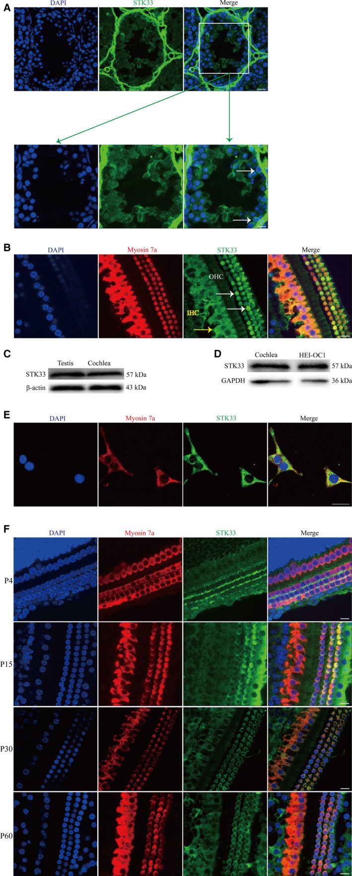
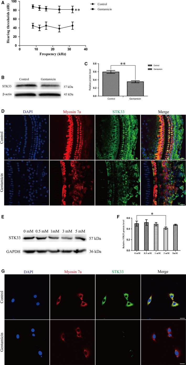
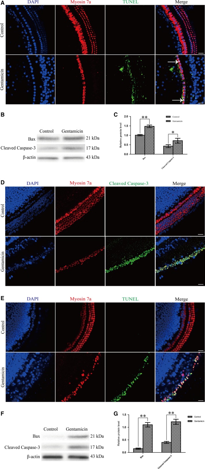
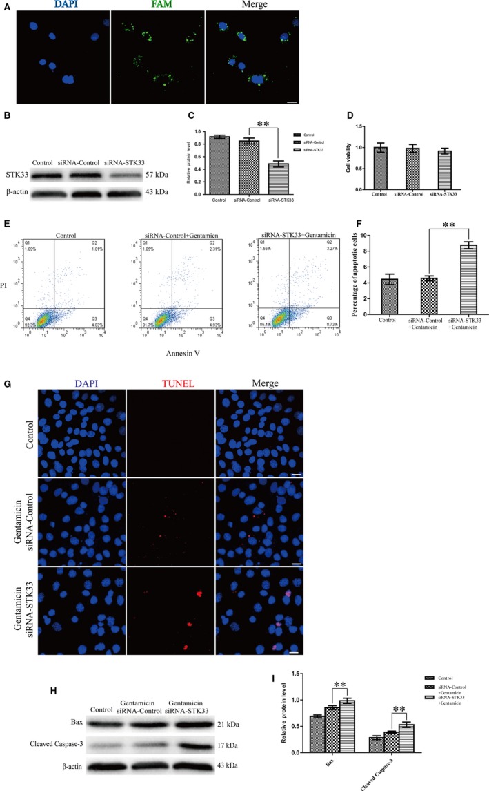
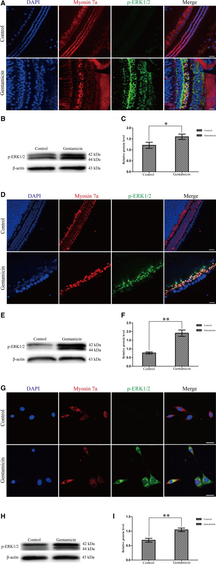
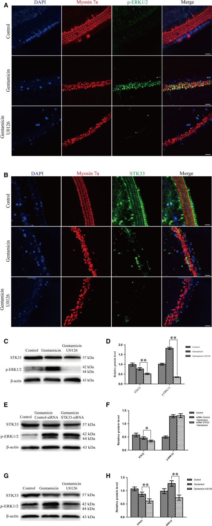
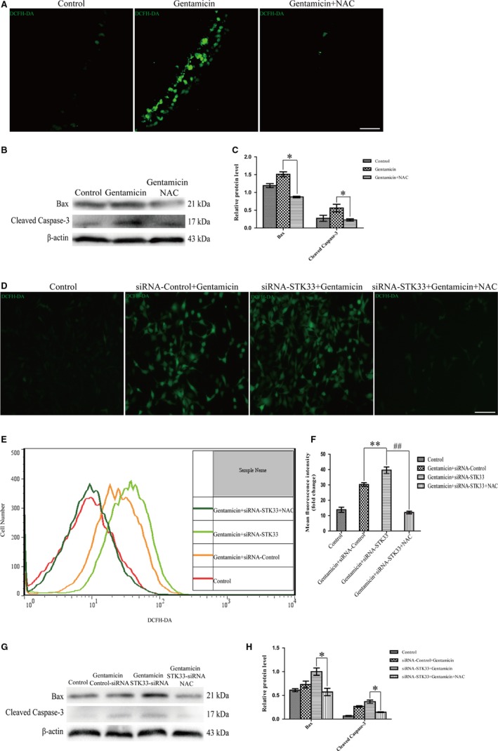
Serine/threonine kinase 33 (STK33), a member of the calcium/calmodulin-dependent kinase (CAMK), plays vital roles in a wide spectrum of cell processes. The present study was designed to investigate whether STK33 expressed in the mammalian cochlea and, if so, what effect STK33 exerted on aminoglycoside-induced ototoxicity in House Ear Institute-Organ of Corti 1 (HEI-OC1) cells. Immunofluorescence staining and western blotting were performed to investigate STK33 expression in cochlear hair cells (HCs) and HEI-OC1 cells with or without gentamicin treatment. CCK8, flow cytometry, immunofluorescence staining and western blotting were employed to detect the effects of STK33 knockdown, and/or U0126, and/or N-acetyl-L-cysteine (NAC) on the sensitivity to gentamicin-induced ototoxicity in HEI-OC1 cells. We found that STK33 was expressed in both mice cochlear HCs and HEI-OC1 cells, and the expression of STK33 was significantly decreased in cochlear HCs and HEI-OC1 cells after gentamicin exposure. STK33 knockdown resulted in an increase in the cleaved caspase-3 and Bax expressions as well as cell apoptosis after gentamicin damage in HEI-OC1 cells. Mechanistic studies revealed that knockdown of STK33 led to activated mitochondrial apoptosis pathway as well as augmented reactive oxygen species (ROS) accumulation after gentamicin damage. Moreover, STK33 was involved in extracellular signal-regulated kinase 1/2 pathway in primary culture of HCs and HEI-OC1 cells in response to gentamicin insult. The findings from this work indicate that STK33 decreases the sensitivity to the apoptosis dependent on mitochondrial apoptotic pathway by regulating ROS generation after gentamicin treatment, which provides a new potential target for protection from the aminoglycoside-induced ototoxicity.
Keywords: apoptosis; extracellular signal-regulated kinase 1/2; gentamicin; reactive oxygen species; serine/threonine kinase 33.
© 2018 The Authors. Journal of Cellular and Molecular Medicine published by John Wiley & Sons Ltd and Foundation for Cellular and Molecular Medicine.
Figures
Similar articles
-
NaHS Protects Cochlear Hair Cells from Gentamicin-Induced Ototoxicity by Inhibiting the Mitochondrial Apoptosis Pathway.PLoS One. 2015 Aug 21;10(8):e0136051. doi: 10.1371/journal.pone.0136051. eCollection 2015. PLoS One. 2015. PMID: 26295804 Free PMC article.
-
Induction of mitophagy in the HEI-OC1 auditory cell line and activation of the Atg12/LC3 pathway in the organ of Corti.Hear Res. 2018 Apr;361:52-65. doi: 10.1016/j.heares.2018.01.003. Epub 2018 Jan 11. Hear Res. 2018. PMID: 29352609
-
Evaluation of apoptotic markers in HEI-OC1 cells treated with gentamicin with and without the mitochondria-targeted antioxidant mitoquinone.Otol Neurotol. 2015 Mar;36(3):526-30. doi: 10.1097/MAO.0000000000000517. Otol Neurotol. 2015. PMID: 25076226
-
Hair Cell Protection from Ototoxic Drugs.Neural Plast. 2021 Jul 11;2021:4909237. doi: 10.1155/2021/4909237. eCollection 2021. Neural Plast. 2021. PMID: 34335732 Free PMC article. Review.
-
Signaling pathways regulating the immune function of cochlear supporting cells and their involvement in cochlear pathophysiology.Glia. 2024 Apr;72(4):665-676. doi: 10.1002/glia.24476. Epub 2023 Nov 7. Glia. 2024. PMID: 37933494 Review.
Cited by
-
Liproxstatin-1 Protects Hair Cell-Like HEI-OC1 Cells and Cochlear Hair Cells against Neomycin Ototoxicity.Oxid Med Cell Longev. 2020 Dec 1;2020:1782659. doi: 10.1155/2020/1782659. eCollection 2020. Oxid Med Cell Longev. 2020. PMID: 33343803 Free PMC article.
-
Ivacaftor attenuates gentamicin-induced ototoxicity through the CFTR-Nrf2-HO1/NQO1 pathway.Redox Rep. 2024 Dec;29(1):2332038. doi: 10.1080/13510002.2024.2332038. Epub 2024 Apr 2. Redox Rep. 2024. PMID: 38563333 Free PMC article.
-
Inhibition of ferroptosis protects House Ear Institute-Organ of Corti 1 cells and cochlear hair cells from cisplatin-induced ototoxicity.J Cell Mol Med. 2020 Oct;24(20):12065-12081. doi: 10.1111/jcmm.15839. Epub 2020 Sep 14. J Cell Mol Med. 2020. PMID: 32929878 Free PMC article.
-
Characterization of quinoxaline derivatives for protection against iatrogenically induced hearing loss.JCI Insight. 2021 Mar 8;6(5):e141561. doi: 10.1172/jci.insight.141561. JCI Insight. 2021. PMID: 33476306 Free PMC article.
-
Prediction of the Molecular Mechanisms Underlying Erlong Zuoci Treatment of Age-Related Hearing Loss via Network Pharmacology-Based Analyses Combined with Experimental Validation.Front Pharmacol. 2021 Nov 23;12:719267. doi: 10.3389/fphar.2021.719267. eCollection 2021. Front Pharmacol. 2021. PMID: 34887749 Free PMC article.
References
-
- Mujica AO, Brauksiepe B, Saaler‐Reinhardt S, Reuss S, Schmidt ER. Differential expression pattern of the novel serine/threonine kinase, STK33, in mice and men. Febs J. 2005;272:4884‐4898. - PubMed
-
- Wang P, Cheng H, Wu J, Yan A, Zhang L. STK33 plays an important positive role in the development of human large cell lung cancers with variable metastatic potential. Acta Biochim Biophys Sin (Shanghai). 2015;47:214‐223. - PubMed
-
- Martins LR, Bung RK, Koch S, et al. Stk33 is required for spermatid differentiation and male fertility in mice. Dev Biol. 2018;433:84‐93. - PubMed
Publication types
MeSH terms
Substances
LinkOut - more resources
Full Text Sources
Other Literature Sources
Molecular Biology Databases
Research Materials
