. 2018 Nov 15;29(23):2887-2897.
doi: 10.1091/mbc.E18-07-0433. Epub 2018 Sep 26.

Rab17 regulates apical delivery of hepatic transcytotic vesicles

Affiliations

Rab17 regulates apical delivery of hepatic transcytotic vesicles

Anneliese C Striz et al. Mol Biol Cell. .

Abstract

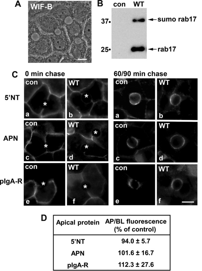
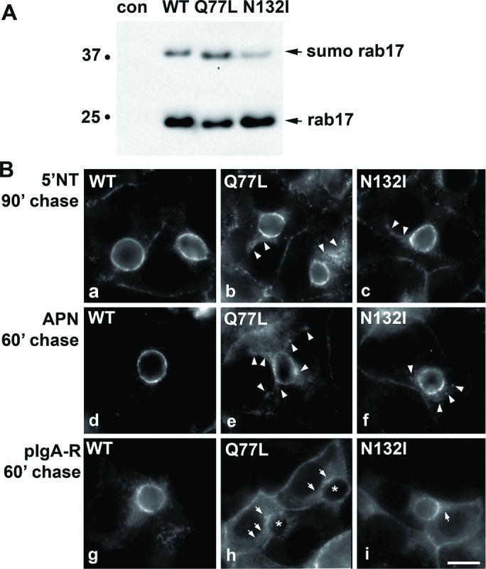
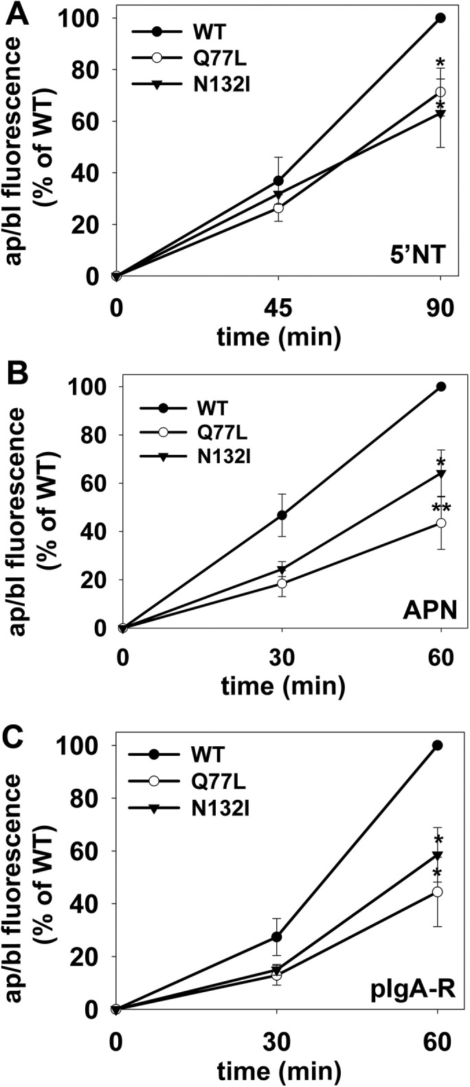
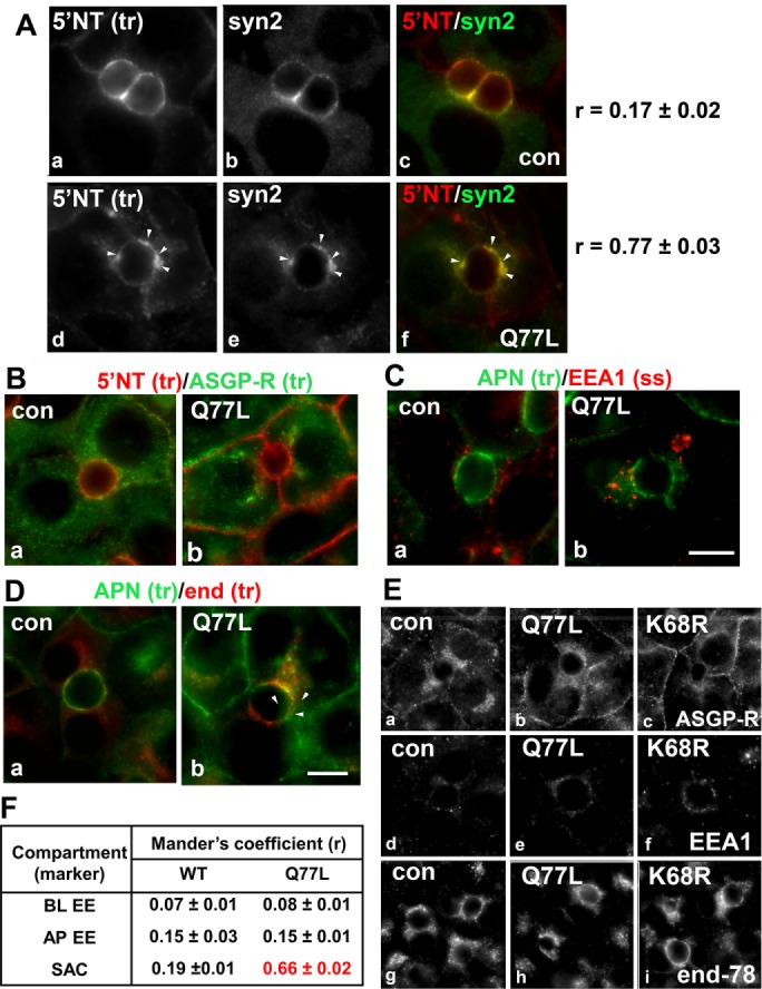
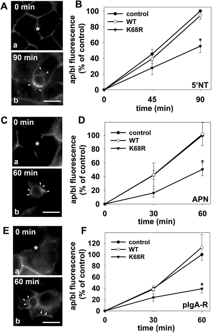
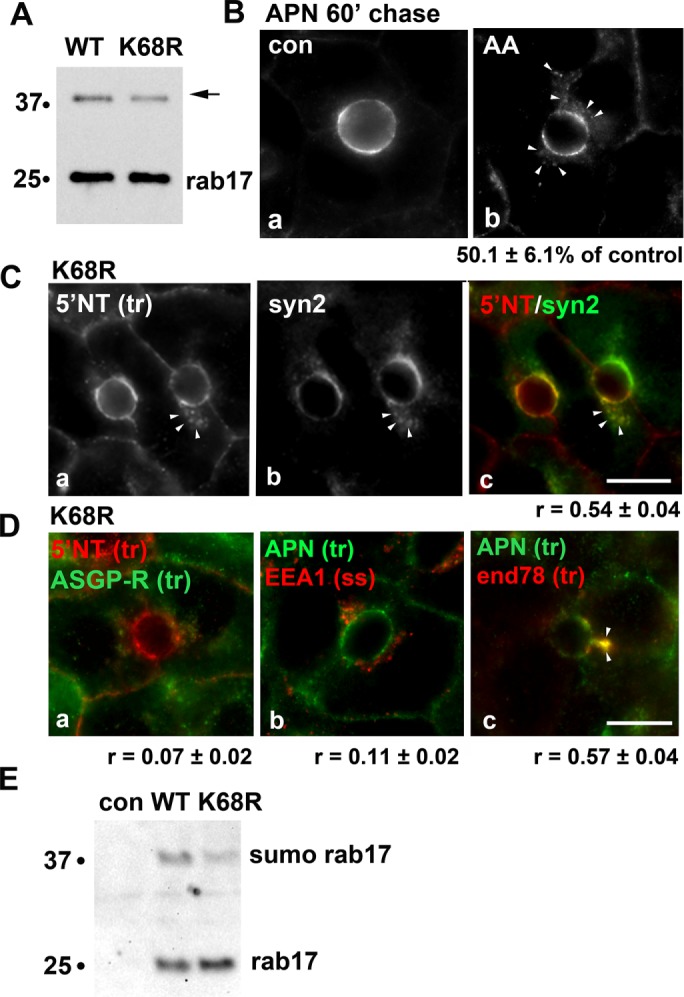
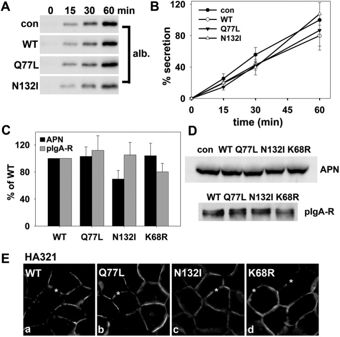
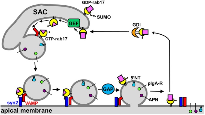
A major focus for our laboratory is identifying the molecules and mechanisms that regulate basolateral-to-apical transcytosis in polarized hepatocytes. Our most recent studies have focused on characterizing the biochemical and functional properties of the small rab17 GTPase. We determined that rab17 is a monosumoylated protein and that this modification likely mediates selective interactions with the apically located syntaxin 2. Using polarized hepatic WIF-B cells exogenously expressing wild-type, dominant active/guanosine triphosphate (GTP)-bound, dominant negative/guanosine diphosphate (GDP)-bound, or sumoylation-deficient/K68R rab17 proteins, we confirmed that rab17 regulates basolateral-to-apical transcytotic vesicle docking and fusion with the apical surface. We further confirmed that transcytosis is impaired from the subapical compartment to the apical surface and that GTP-bound and sumoylated rab17 are likely required for apical vesicle docking. Because expression of the GTP-bound rab17 led to impaired transcytosis, whereas wild type had no effect, we further propose that rab17 GTP hydrolysis is required for vesicle delivery. We also determined that transcytosis of three classes of newly synthesized apical residents showed similar responses to rab17 mutant expression, indicating that rab17 is a general component of the transcytotic machinery required for apically destined vesicle docking and fusion.


Figures

FIGURE 1:
FIGURE 1:
Basolateral-to-apical transcytosis is not altered by overexpression of wild-type rab17. (A) The characteristic, polarized hepatic morphology of WIF-B cells is shown in a phase image. (B) Total cell lysates were prepared from uninfected (control) WIF-B cells or cells expressing FLAG-tagged WT rab17 and immunoblotted with anti-FLAG antibodies. The monosumoylated form of rab 17 is indicated (sumo rab17). Molecular weight markers are indicated on the left of each immunoblot in kDa. (C) Uninfected cells or cells expressing wild-type rab 17 were basolaterally labeled with antibodies specific for the extracellular epitopes of the indicated apical proteins at 4°C. Cells were additionally infected with recombinant adenoviruses expressing pIgA-R in panels e and f (C). After excess antibodies were washed away, antibody–antigen complexes were chased for 0, 90, or 60 min as indicated at 37°C. Cells were fixed, permeabilized, and labeled with secondary antibodies to detect the transcytosed proteins. Asterisks mark the unlabeled bile canaliculi. Images are representative of at least three experiments. Bar = 10 μm. (D) Control (uninfected) WIF-B cells or cells expressing wild-type rab17 were basolaterally labeled for the indicated apical proteins and chased as described in C. Random fields were visualized by indirect immunofluorescence. From micrographs, the average pixel intensity of each marker at selected regions of interest placed at the apical or basolateral membrane of the same WIF-B cell was measured. The averaged background pixel intensity was subtracted from each value and the ratio of apical- (ap) to-basolateral (bl) fluorescence intensity was determined. Wild-type values were normalized to control values that were set to 100%. Values are expressed as the mean ± SEM. Measurements were performed on at least three independent experiments.
FIGURE 2:
FIGURE 2:
Transcytosis is impaired in cells expressing GTP-bound/Q77L or GDP-bound/N132I rab17. (A) Total cell lysates were prepared from WIF-B cells expressing FLAG-tagged WT, GTP-bound/Q77L, or GDP-bound/N132I rab17 and immunoblotted with anti-FLAG antibodies. The monosumoylated form of rab 17 is indicated (sumo rab17). Molecular weight markers are indicated on the left of each immunoblot in kDa. (B) Cells expressing wild-type, GTP-bound/Q77L, or GDP-bound/N132I rab17 were basolaterally labeled with antibodies specific for the extracellular epitopes of the indicated apical proteins at 4°C. Cells were additionally infected with recombinant adenoviruses expressing pIgA-R in panels g–i. After excess antibodies were washed away, antibody–antigen complexes were chased for 90 min (a–c) or 60 min (d–i) at 37°C. Cells were fixed, permeabilized, and labeled with secondary antibodies to detect the transcytosed proteins. Arrows indicate subapically accumulated transcytosing proteins in cells expressing mutant rab17. Images are representative of at least three experiments. Bar = 10 μm.
FIGURE 3:
FIGURE 3:
Transcytosis is impaired to a similar extent for different classes of apical proteins in cells expressing GTP-bound/Q77L or GDP-bound/N132I rab17. WIF-B cells expressing wild-type, GDP-bound/Q77L, or GDP-bound/N132I rab17 were basolaterally labeled for the indicated apical proteins as described in Figure 2 and chased for 0, 45, or 90 min (A) or 0, 30, or 60 min (B, C) at 37°C. Cells were additionally infected with recombinant adenoviruses expressing pIgA-R in C. Cells were fixed, permeabilized, and labeled with secondary antibodies to detect the transcytosed proteins. Random fields were visualized by indirect immunofluorescence and digitized. From micrographs, the average pixel intensity of each marker at selected regions of interest placed at the apical or basolateral membrane of the same WIF-B cell was measured. The averaged background pixel intensity was subtracted from each value and the ratio of apical (ap) to basolateral (bl) fluorescence intensity was determined. Values are expressed as the mean ± SEM for 5′NT (A), APN (B), and pIgA-R (C) and are from at least three independent experiments. *p ≤ 0.05, **p ≤ 0.005.
FIGURE 4:
FIGURE 4:
Transcytosing proteins accumulate in syntaxin 2–positive SAC structures in cells expressing GTP-bound/Q77L rab17. (A) Control (uninfected) WIF-B cells or cells expressing GTP-bound/Q77L rab17 were basolaterally labeled with antibodies against 5′NT and antigen-antibody complexes were chased for 60 min. Cells were fixed and double labeled for steady-state syntaxin 2 distributions. Merged images are shown in panels c and f Arrows indicate subapically accumulated transcytosing proteins in cells expressing mutant rab17. Bar = 10 μm. Mander’s coefficients of colocalization are indicated on the right. Values are expressed as the mean ± SEM from at least three independent experiments. Control (uninfected) WIF-B cells or cells expressing GTP-bound/Q77L rab17 were basolaterally labeled for 5′NT and ASGP-R (B) or APN (C) or APN and endolyn-78 (D) and allowed to continuously chase for 60 min. Cells were fixed and stained for the corresponding trafficked antibody–antigen complexes. In C, cells were labeled for steady-state distributions of EEA1. Merged images are shown for each. Arrows indicate subapically accumulated transcytosing proteins in cells expressing mutant rab17. Bar = 10 µm. In E, control (uninfected) WIF-B cells or cells expressing GTP-bound/Q77L or sumo-deficient/K68R rab17 were labeled for the steady-state distributions of ASGP-R, EEA1, and endolyn-78 as indicated. No changes in distributions were observed for any of the proteins confirming the validity of their use as compartment markers. Bar = 10 µm. In F, Mander’s coefficients of colocalization for the experiments shown in B, C, and D are shown. Values are expressed as the mean ± SEM from at least three independent experiments. BL EE, basolateral early endosome; AP EE, apical early endosome; SAC, subapical compartment.
FIGURE 5:
FIGURE 5:
Transcytosis is impaired to a similar extent for different classes of apical proteins in cells expressing sumo-deficient/K68R rab17. (A–F) Control (uninfected) cells and cells expressing wild-type or sumo-deficient/K68R rab17 were basolaterally labeled with antibodies specific for the extracellular epitopes of the indicated apical proteins at 4°C. Cells were additionally infected with recombinant adenoviruses expressing pIgA-R in E and F. After excess antibodies were washed away, antibody–antigen complexes were chased for 0, 60 min, or 90 min as indicated (A, C, and E) at 37°C. Cells were fixed, permeabilized and labeled with secondary antibodies to detect transcytosed APN (A), 5′NT (C) or pIgA-R (E). Asterisks are marking unlabeled canaliculi. Images are representative of at least three experiments. Bar = 10 μm. (B, D, F) Control (uninfected) WIF-B cells or cells expressing wild-type or sumo-deficient/K68R rab17 were basolaterally labeled for the indicated apical proteins as described in Figure 1 and chased for 0, 45, or 90 min (B) or 0, 30 or 60 min (D and F) at 37°C. Cells were fixed, permeabilized, and labeled with secondary antibodies to detect the transcytosed proteins. Random fields were visualized by indirect immunofluorescence. From micrographs, the average pixel intensity of each marker at selected regions of interest placed at the apical or basolateral membrane of the same WIF-B cell was measured. The averaged background pixel intensity was subtracted from each value, and the ratio of apical (ap) to basolateral (bl) fluorescence intensity was determined. Values are expressed as the mean ± SEM for 5′NT (B), APN (D), and pIgA-R (F). Measurements were performed on at least three independent experiments. *p ≤ 0.05.
FIGURE 6:
FIGURE 6:
Transcytosing proteins accumulate in syntaxin 2–positive SAC structures in cells expressing sumo-deficient rab17. (A) Total cell lysates were prepared from WIF-B cells expressing FLAG-tagged WT or sumo-deficient/K68R rab17 and immunoblotted with anti-FLAG antibodies. The mono-sumoylated form of rab17 is indicated with an arrow. Molecular-weight markers are indicated on the left of each immunoblot in kDa. (B) Uninfected WIF-B cells were pretreated with 5 μM anacardic acid (AA) for 60 min at 37°C before APN antibody labeling at 4°C for 20 min. APN-antibody complexes were chased for 60 min in the continued presence of the drug. The amount of impaired transcytosis is indicated below the panel as the percent of control. Values are expressed as the mean ± SEM from at least three independent experiments. (C) WIF-B cells expressing sumo-deficient/K68R rab17 were basolaterally labeled with antibodies against 5′NT, and antigen-antibody complexes were chased for 60 min. Cells were fixed and double labeled for steady-state syntaxin 2 distributions. A merged image is shown in panel c. Arrows indicate subapically accumulated transcytosing proteins in cells expressing mutant rab17. Bar = 10 μm. The Mander’s coefficient of colocalization is indicated below the merged image. Values are expressed as the mean ± SEM from at least three independent experiments. (D) WIF-B cells expressing sumo-deficient/K68R rab17 were basolaterally labeled for 5′NT and ASGP-R (a) or APN (b) or APN and endolyn-78 (c) and allowed to continuously chase for 60 min. Cells were fixed and stained for the corresponding trafficked antibody–antigen complexes. In D (panel b), cells were labeled for steady-state distributions of EEA1. Merged images are shown. Arrows indicate subapically accumulated transcytosing proteins in cells expressing mutant rab17. Mander’s coefficients of colocalization are indicated below. Values are expressed as the mean ± SEM from at least three independent experiments. Bar = 10 µm. (E) Uninfected WIF-B cells (con) or cells expressing WT of K68R rab16 were lysed in ice-cold GTP binding buffer. Cleared lysates were mixed with GTP-agarose for 2 h at 4°C. Bound fractions were recovered, washed, and immunoblotted for rab17 using anti-FLAG epitope antibodies. The monosumoylated form of rab17 is indicated with an arrow. Molecular-weight markers are indicated on the left of each immunoblot in kDa. A representative immunoblot from three independent experiments is shown.
FIGURE 7:
FIGURE 7:
Rab17 selectively regulates transcytosis. (A, B) Control (uninfected) cells or cells expressing wild-type, GTP-bound/Q77L, or GDP-bound/N132I rab17 were washed in PBS and reincubated in serum-free medium. At 0, 15, 30, and 60 min after reincubation, aliquots of media were collected and immunoblotted for albumin (A). Densitometric analysis of the immunoreactive species was performed and albumin secretion plotted as a percentage of control at 60 min (B). Values are expressed as the average ± SEM from at least three independent experiments. (C) Cells expressing wild-type, GTP-bound/Q77L, GDP-bound/N132I, or sumo-deficient/K68R rab17 were surface labeled for pIgA-R or APN for 30 min at 4°C. Cells were additionally infected with recombinant adenoviruses expressing pIgA-R. Lysates were immunoblotted with primary antibodies to detect the entire population of pIgA-R or APN. On a parallel immunoblot, lysates were probed directly with secondary antibodies to detect only the surface-bound primary antibodies. The amount of surface-bound antibodies was normalized to the total antigen amount. In all cases, control ratios were set to 100%. Values are expressed as the mean ± SEM from at least three independent experiments. (D) Total cell lysates were prepared from uninfected (con) WIF-B cells or cells expressing WT, GTP-bound/Q77L, GDP-bound/N132I rab17, or sumo-deficient K68R rab17 and immunoblotted for APN or pIgA-R as indicated. In E, cells expressing wild-type, GTP-bound/Q77L, GDP-bound/N132I, or sumo-deficient K68R rab17 were labeled for HA321. Asterisks mark selected bile canaliculi. Bar = 10 μm.
FIGURE 8:
FIGURE 8:
Model for the role of rab17 in regulation of apical vesicle docking and fusion. The prenylated, monosumoylated, and GDP-bound rab17 is activated by its specific GEF at the SAC. The GTP-bound rab17 associates with a budding vesicle via associations with a transcytotic vesicle-specific VAMP or a yet-to-be-identified coat protein (not shown). The budded vesicle is delivered to the apical membrane where the GTP-bound, sumoylated rab17 interacts with syntaxin 2 to initiate vesicle docking. A rab17-specific GAP at the apical surface activates GTP hydrolysis required for vesicle fusion. The GDP-bound, sumoylated rab17 is extracted from the apical membrane by a GDI and recycled to the SAC. GAP, GTPase-activating protein; GEF, guanine exchange factor; syn2, syntaxin 2; VAMP.

Similar articles

Cited by

References

    1. Alvarez C, Fujita H, Hubbard A, Sztul E. (1999). ER to Golgi transport: Requirement for p115 at a pre-Golgi VTC stage. J Cell Biol , 1205–1222. - PMC - PubMed
    1. Aquea G, Bresky G, Lancellotti D, Madariaga JA, Zaffiri V, Urzua U, Haberle S, Bernal G. (2014). Increased expression of P2RY2, CD248 and EphB1 in gastric cancers from Chilean patients. Asian Pac J Cancer Prev , 1931–1936. - PubMed
    1. Barr VA, Hubbard AL. (1993). Newly synthesized hepatocyte plasma membrane proteins are transported in transcytotic vesicles in the bile duct-ligated rat. Gastroenterology , 554–571. - PubMed
    1. Bartles JR, Braiterman LT, Hubbard AL. (1985a). Biochemical characterization of domain-specific glycoproteins of the rat hepatocyte plasma membrane. J Biol Chem , 12792–12802. - PubMed
    1. Bartles JR, Braiterman LT, Hubbard AL. (1985b). Endogenous and exogenous domain markers of the rat hepatocyte plasma membrane. J Cell Biol , 1126–1138. - PMC - PubMed

Publication types

Substances

LinkOut - more resources