

Molecular characterisation and expression analysis of SEREX-defined antigen NUCB2 in gastric epithelium, gastritis and gastric cancer
- PMID: 30256860
- PMCID: PMC3167273
- DOI: 10.4081/ejh.2009.e2
Molecular characterisation and expression analysis of SEREX-defined antigen NUCB2 in gastric epithelium, gastritis and gastric cancer
Abstract
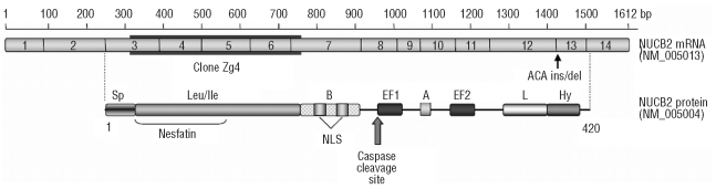
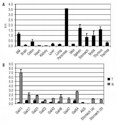
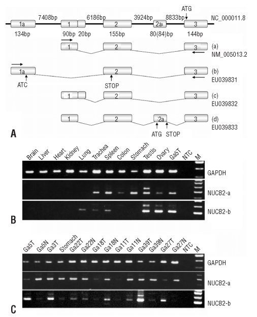
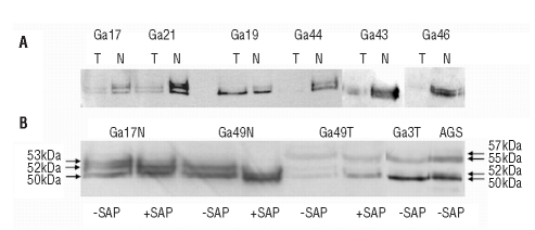
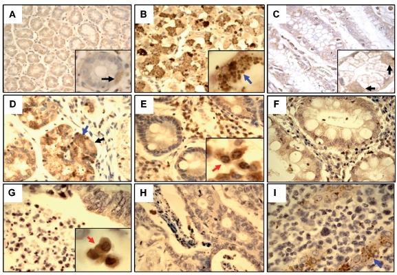
NUCB2 is an EF-hand Ca2+ binding protein that has been implicated in various physiological processes like calcium homeostasis, hypothalamic regulation of feeding and TNF receptor shedding. In our previous study we identified NUCB2 as a potential tumour antigen eliciting autoantibody responses in 5.4% of gastric cancer patients but not in the healthy individuals. The current study aimed to elucidate the molecular mechanism underlying NUCB2 immunogenicity and to gain an insight into the physiological functions of NUCB2 in the stomach. mRNA expression analysis demonstrated that NUCB2 is ubiquitously expressed in normal tissues, including lymphoid tissues, and downregulated in gastric tumours when compared with the adjacent relatively normal stomach tissues. The search for molecular alterations resulted in the identification of novel mRNA variants transcribed from an alternative promoter and expressed predominantly in gastric cancers. Western blot analysis demonstrated that the protein levels correspond to mRNA levels and revealed that NUCB2 is phosphorylated in gastric mucosa. Furthermore, a 55 kDa isoform, generated presumably by yet an unidentified post-translational modification was detected in gastric tumours and AGS gastric cancer cells but was absent in the relatively normal gastric mucosa and thereby might have served as a trigger for the immune response against NUCB2. Staining of stomach tissue microarray with anti-NUCB2 antibody revealed that it is expressed in the secretory granules of chief cells and in the cytoplasm of parietal cells in the functioning gastric glands which are lost in atrophic glands and tumour cells. Hence we propose that NUCB2 may be implicated in gastric secretion by establishing an agonist-releasable Ca2+ store in ER or Golgi apparatus, signalling via heterotrimeric Gα proteins and/or mediating the exocytosis of the secretory granules.
Keywords: NUCB2; SEREX; chief cells; gastric cancer; parietal cells; pepsinogen secretion.; tumour-associated antigens.
Figures
Similar articles
-
Molecular characterisation and expression analysis of SEREX-defined antigen NUCB2 in gastric epithelium, gastritis and gastric cancer.Eur J Histochem. 2009 Jan-Mar;53(1):7-18. doi: 10.4081/ejh.2009.7. Eur J Histochem. 2009. PMID: 19351608
-
Identification and characterization of nesfatin-1 immunoreactivity in endocrine cell types of the rat gastric oxyntic mucosa.Endocrinology. 2009 Jan;150(1):232-8. doi: 10.1210/en.2008-0747. Epub 2008 Sep 25. Endocrinology. 2009. PMID: 18818289 Free PMC article.
-
Pharmacological inhibition of cannabinoid receptor 1 stimulates gastric release of nesfatin-1 via the mTOR pathway.World J Gastroenterol. 2017 Sep 21;23(35):6403-6411. doi: 10.3748/wjg.v23.i35.6403. World J Gastroenterol. 2017. PMID: 29085189 Free PMC article.
-
Regulation of NUCB2/nesfatin-1 production in rat's stomach and adipose tissue is dependent on age, testosterone levels and lactating status.Mol Cell Endocrinol. 2015 Aug 15;411:105-12. doi: 10.1016/j.mce.2015.04.016. Epub 2015 Apr 23. Mol Cell Endocrinol. 2015. PMID: 25916958
-
Pepsinogens: physiology, pharmacology pathophysiology and exercise.Pharmacol Res. 2000 Mar;41(3):265-81. doi: 10.1006/phrs.1999.0586. Pharmacol Res. 2000. PMID: 10675278 Review.
Cited by
-
Nucleobindin 2 inhibits senescence in gastric carcinoma.Sci Rep. 2024 May 17;14(1):11261. doi: 10.1038/s41598-024-61111-5. Sci Rep. 2024. PMID: 38760405 Free PMC article.
-
NUCB2: roles in physiology and pathology.J Physiol Biochem. 2022 Aug;78(3):603-617. doi: 10.1007/s13105-022-00895-4. Epub 2022 Jun 9. J Physiol Biochem. 2022. PMID: 35678998 Review.
References
-
- Barnikol-Watanabe S, Gross NA, Gotz H, Henkel T, Karabinos A, Kratzin H, et al. Human protein NEFA, a novel DNA binding/EF-hand/leucine zipper protein. Molecular cloning and sequence analysis of the cDNA, isolation and characterization of the protein. Biol Chem Hoppe Seyler. 1994;375:497–512. - PubMed
-
- Bos R, van DS, van HT, Kaaijk P, Taubert R, Kyewski B, et al. Expression of a natural tumor antigen by thymic epithelial cells impairs the tumor-protective CD4+ T-cell repertoire. Cancer Res. 2005;65:6443–9. - PubMed
-
- Brevini TA, Cillo F, Antonini S, Tosetti V, Gandolfi F. Temporal and spatial control of gene expression in early embryos of farm animals. Reprod Fertil Dev. 2007;19:35–42. - PubMed
Publication types
LinkOut - more resources
Full Text Sources
Miscellaneous
