. 2018 Dec 14;46(22):12022-12039.
doi: 10.1093/nar/gky862.

The structural basis of CstF-77 modulation of cleavage and polyadenylation through stimulation of CstF-64 activity

Affiliations

The structural basis of CstF-77 modulation of cleavage and polyadenylation through stimulation of CstF-64 activity

Petar N Grozdanov et al. Nucleic Acids Res. .

Abstract

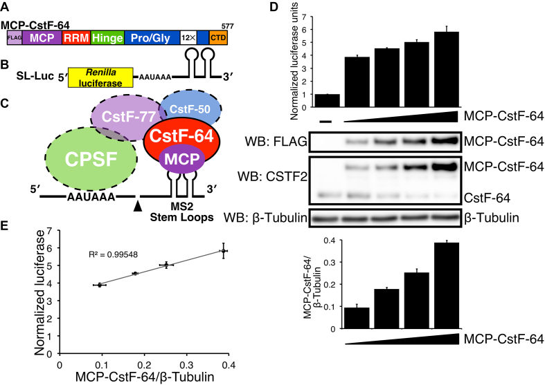
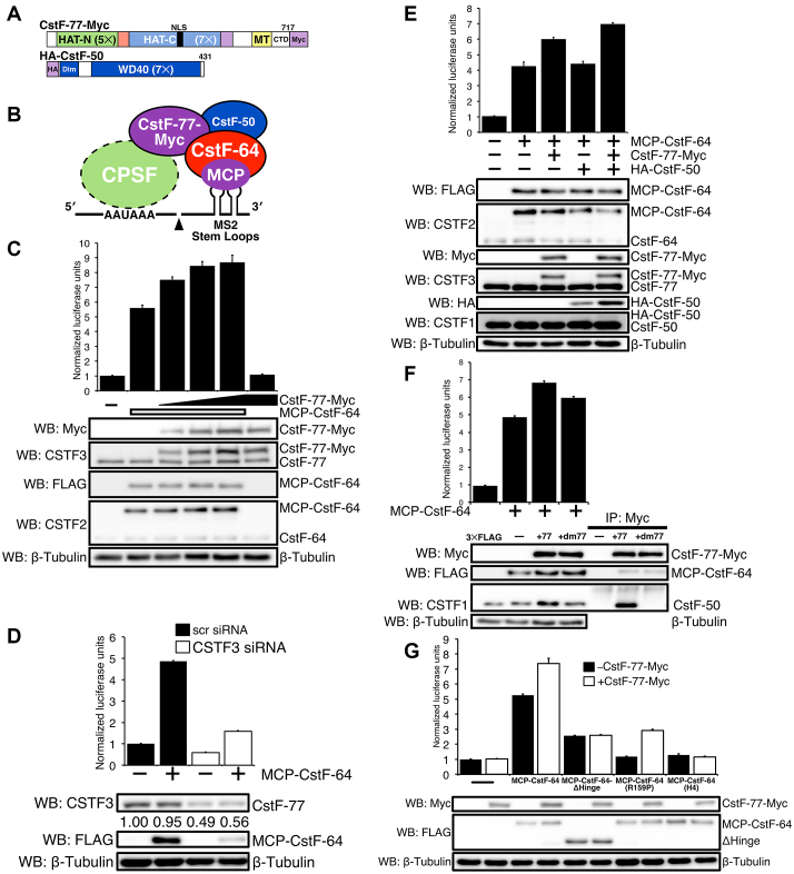
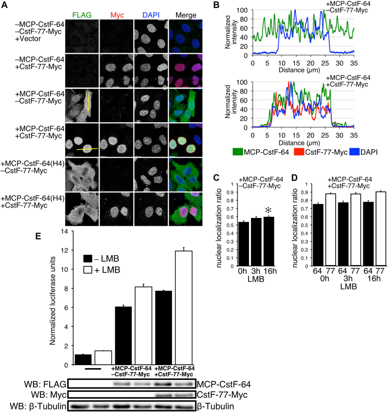
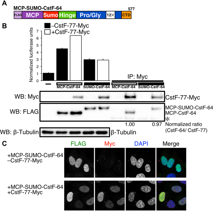
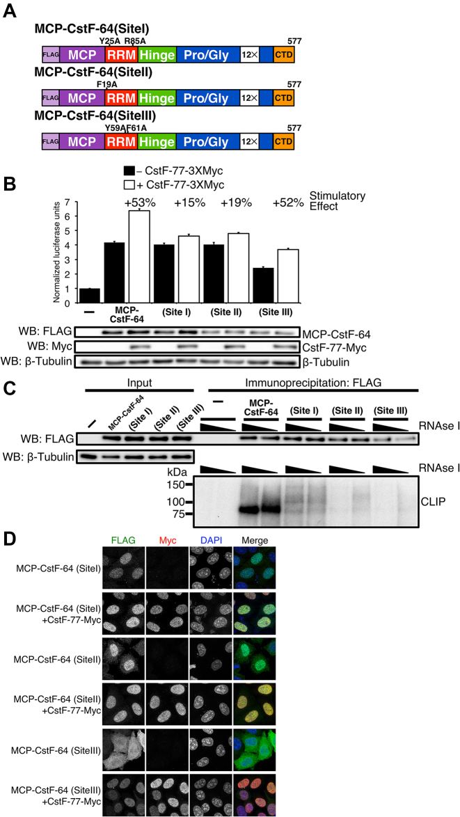
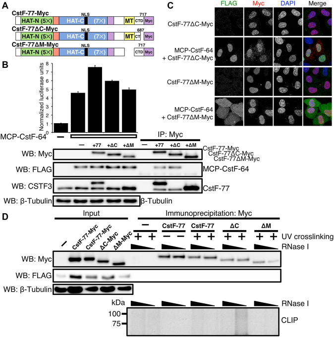
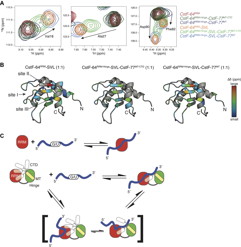
Cleavage and polyadenylation (C/P) of mRNA is an important cellular process that promotes increased diversity of mRNA isoforms and could change their stability in different cell types. The cleavage stimulation factor (CstF) complex, part of the C/P machinery, binds to U- and GU-rich sequences located downstream from the cleavage site through its RNA-binding subunit, CstF-64. Less is known about the function of the other two subunits of CstF, CstF-77 and CstF-50. Here, we show that the carboxy-terminus of CstF-77 plays a previously unrecognized role in enhancing C/P by altering how the RNA recognition motif (RRM) of CstF-64 binds RNA. In support of this finding, we also show that CstF-64 relies on CstF-77 to be transported to the nucleus; excess CstF-64 localizes to the cytoplasm, possibly via interaction with cytoplasmic RNAs. Reverse genetics and nuclear magnetic resonance studies of recombinant CstF-64 (RRM-Hinge) and CstF-77 (monkeytail-carboxy-terminal domain) indicate that the last 30 amino acids of CstF-77 increases the stability of the RRM, thus altering the affinity of the complex for RNA. These results provide new insights into the mechanism by which CstF regulates the location of the RNA cleavage site during C/P.


Figures

Figure 1.
Figure 1.
CstF-64 stimulates cleavage and polyadenylation in correlation to the amount of CstF-64 protein produced in SLAP. (A) Schematic representation of the coat binding protein from bacteriophage MS2 (MCP) fused to human CstF-64 (MCP-CstF-64). Functional domains in CstF-64 and three FLAG tags are indicated. (B) Schematic representation of the reporter construct (SL-Luc) containing the Renilla luciferase and two MS2 stem-loop (SL) sequences downstream of the cleavage and polyadenylation site. (C) The interaction between MCP-CstF-64 and the SL-Luc reporter construct is mediated through the interaction between MCP and MS2 stem-loops. (D) SLAP results showing NLUs in HeLa cells without MCP-CstF-64 (–), and in cells transfected with increasing amounts of MCP-CstF-64. Western blots with the designated antibodies to show the expression of the FLAG-tagged version of MCP-CstF-64 (WB: FLAG) and endogenous CstF-64 (WB: CSTF2). β-tubulin was used as a loading control. A bar graph of the quantified expression of MCP-CstF-64 normalized to β-tubulin (bottom). (E) A linear regression between the amount of MCP-CstF-64 protein normalized to β-tubulin (x-axis) to the NLU from SLAP (y-axis). The correlation value (R2 = 0.99548) is shown.
Figure 2.
Figure 2.
CstF-77 and CstF-50 enhance MCP-CstF-64-dependent cleavage and polyadenylation. (A) Schematic representation of CstF-77-Myc (CstF-77 with three Myc tags at its carboxy-terminal end) and HA-CstF-50 (CstF-50 with three hemagglutinin tags at its amino-terminal end). The position of the nuclear localization sequence (NLS), position and the number of the carboxy- and amino-terminal HATs, MT and the CTD of CstF-77 are shown. Similarly, the seven WD40 (WD40) repeats and the dimerization (Dim) domains are shown for CstF-50. (B) Cartoon of the interactions between MCP-CstF-64, CstF-77, CstF-50 and SL-Luc reporter construct. (C) Increased amounts of CstF-77-Myc increase C/P as measured by SLAP. Western blots with an antibody against Myc (WB: Myc), against CstF-77 (WB: CSTF3), against MCP-CstF-64 (WB: FLAG) or CstF-64 (WB: CSTF2) are shown. Antibodies against β-tubulin were used as a loading control. (D) Decreased CstF-77 reduces C/P as measured by SLAP. Hela cells were transfected with either a non-specific (scr) or CSTF3-specific siRNAs. Twelve hours after siRNA transfection, cells were transfected for SLAP. Western blots show the reduced expression of the endogenous CstF-77, MCP-CstF-64 (WB: FLAG). (E) HA-CstF-50 co-expressed with MCP-CstF-64 and CstF-77-Myc increases C/P as measured by SLAP. Western blots with the respective antibodies to verify expression of the exogenous proteins through their tags (WB: FLAG, Myc, HA) and antibodies to show relative expression to the endogenous proteins (WB: CSTF1, CSTF2, CSTF3). (F) Mutations in CstF-77-Myc that disrupt the interaction of CstF-77 with CstF-50 reduce C/P as measured by SLAP. Two-point mutations were made in CstF-77 (P584A, M589A, collectively called +dm77) that disrupt the interaction of CstF-77 with CstF-50. Immunoprecipitation with an antibody against Myc tag: no CstF-77, CstF-77-Myc (+77) and CstF-77-Myc-dm77 (+dm77). Western blots for Myc tag (WB: Myc), and endogenous CstF-50 (WB: CSTF1) and β-tubulin were presented. (G) Mutations in MCP-CstF-64 that disrupt the interaction of CstF-64 with CstF-77 decrease both SLAP and the stimulatory effect of CstF-77. MCP-CstF-64-ΔHinge lacks amino acids 96–216 from CstF-64. MCP-CstF-64 (R159P) is a point mutation in the Hinge domain that interrupts the interaction with CstF-77, MCP-CstF-64 (H4) is a series of point mutations in the fourth α-helix of the Hinge domain that interrupt interaction with CstF-77.
Figure 3.
Figure 3.
MCP-CstF-64 distributes equally between the cytoplasm and nucleus and does not shuttle between these cellular compartments. (A) Immunofluorescent confocal images of the described constructs stained with antibodies against the FLAG tag for MCP-CstF-64 (green) and Myc tag for CstF-77 (red). Cells were counterstained with DAPI to delineate the nucleus. Yellow lines through the cells indicate the position of the intensity profile of these cells shown in B. (B) Intensity signal profile of the cells shown in A. Left —a cell transfected only with MCP-CstF-64, right cell co-transfected with MCP-CstF-64 and CstF-77-Myc. (C) Quantification of the nuclear to cytoplasmic ratio of cells that were not treated with LMB (0 h), after 3 and 16 h of treatment (3 and 16 h, respectively). (D) Effect on addition of CstF-77-Myc on the nuclear to cytoplasm ratio of MCP-CstF-64 (black bars) with or without treatment with LMB at the indicated time points. Open bars—nuclear to cytoplasmic ratio for CstF-77-Myc. (E) LMB increases SLAP. A bar graph of cells with no additional MCP-CstF-64, with MCP-CstF-64 and additional CstF-77-Myc, with treatment for 16 h or no treatment. Representative western blots are shown to demonstrate the amount of exogenous proteins present in the assay.
Figure 4.
Figure 4.
The RRM of CstF-64 contributes to SLAP. (A) Illustration of the MCP-SUMO-CstF-64 construct. The first 107 amino acids of the CstF-64 (RRM) is replaced by the SUMO domain (103 amino acids). (B) SLAP with the MCP-CstF-64 and MCP-SUMO-CstF-64 alone (black bars) and co-transfected with CstF-77-Myc (open bars). Western blots with antibody recognizing FLAG and Myc tags. On right, immunoprecipitation with an antibody against the Myc tag in the order of inputs: MCP-CstF-64 alone, MCP-CstF-64 co-transfected with CstF-77-Myc, MCP-SUMO-CstF-64 alone co-transfected with CstF-77-Myc. Numbers beneath the immunoprecipitations show the normalized ratio between MCP-CstF-64, MCP-SUMO-CstF-64 and CstF-77. (C) Immunohistochemistry on HeLa cells transfected with the MCP-SUMO-CstF-64 (green) alone and co-transfected with CstF-77-Myc (red). DNA in the nucleus was counterstained with DAPI.
Figure 5.
Figure 5.
RNA binding of RRM is needed for the enhancement of SLAP provided by CstF-77. (A) Schematic illustration of point mutants in sites I, II and III of the RRM of MCP-CstF-64. (B) SLAP of the wild- type and I, II and III mutants of the RRM of CstF-64 alone and co-expressed with CstF-77-Myc. Western blots were performed to show expression of MCP-CstF-64 constructs and CstF-77-Myc. β-tubulin was used as a loading control. (C) CLIP experiment verifying that site I, II and III MCP-CstF-64 mutants do not bind or bind minimally to RNA (bottom). Western blot of the immunoprecipitated samples is also shown (top). (D) Immunohistochemistry staining of site I, II and III MCP-CstF-64 mutants. Site I and II localize to the nucleus when expressed alone (green). Site III demonstrates a cytoplasmic localization when expressed alone (green). All MCP-CstF-64 mutants locate to the nucleus when co-expressed with CstF-77-Myc (red).
Figure 6.
Figure 6.
The CTD of CstF-77 is involved in modulating the binding affinity of the RRM of CstF-64. (A) Illustration of CstF-77-Myc deletion mutants: the last 30 amino acids of the carboxy terminal domain (amino acids deleted Δ688–717, CstF-77ΔC-Myc), and MT Δ607–664, CstF-77ΔM-Myc). (B) SLAP of the wild-type and CstF-77-Myc deletion mutants. Western blots to verify the expression of the proteins: antibodies against Myc tag to detect CstF-77-Myc construct, FLAG to detect MCP-CstF-64 and against CSTF3 to detect endogenous and exogenous CstF-77 protein. Note that the antibody against CSTF3 is raised against a peptide within the last 30 amino acids of the CstF-77 and therefore does not recognize the CstF-77ΔC-Myc construct. (C) Immunohistochemistry staining of the CstF-77-Myc deletion mutants (red) alone or co-transfected with MCP-CstF-64 (green). DNA in the nucleus is counterstained with DAPI. (D) CLIP (bottom) of the CstF-77-Myc deletion mutants. UV-crosslinking or the lack of it is indicated. The increasing amount of RNase I is also indicated. Amount of the proteins in the input is shown for MCP-CstF-64 (WB: FLAG), wild-type CstF-77-Myc and deletion mutants (WB: Myc). β-tubulin was used as a loading control. Note that in the conditions of CLIP using RIPA buffer, MCP-CstF-64 does not co-immunoprecipitated with CstF-77-Myc.
Figure 7.
Figure 7.
The CTD of CstF-77 perturbs CstF-64RRM-RNA binding. (A) Overlay of 2D 15N-1H HSQC spectra for four different residues experiencing CSPs upon the addition of SVL RNA. Red, dark green and dark blue contours represent the apo form of CstF-64RRM, CstF-64RRM-Hinge-CstF-77MT-CTD and CstF-64RRM-Hinge-CstF-77MT, respectively, whereas the orange, light green and light blue contours represent the 1:1 protein—SVL RNA complexes for CstF-64RRM, CstF-64RRM-Hinge-CstF-77MT-CTD and CstF-64RRM-Hinge-CstF-77MT, respectively. (B) Ribbon diagrams of the CstF-64RRM structure depicting the changes in 15N-1H chemical shift upon titration of SVL RNA into CstF-64RRM (left), CstF-64RRM-Hinge-CstF-77MT-CTD (middle) and CstF-64RRM-Hinge-CstF-77MT (right), respectively. The dark blue-to-red gradient represents backbone amide groups that experience a small-to-large weighted, averaged CSP (0.037–0.24 ppm) (55). Arrows point to the three RNA binding sites (site I, II, III) described by Taylor and co-workers (18). (C) Model of illustrating a possible effect on the CTD of CstF-77 on RNA binding. The top scheme shows the simple two-state model employed by CstF-64RRM and CstF-64RRM-Hinge-CstF-77MT complex. The bottom scheme includes a dynamic complex where the CTD of CstF-77 can adopt multiple conformations, which can occlude binding to sites within the RRM of CstF-64.

Similar articles

Cited by

References

    1. Fu Y., Sun Y., Li Y., Li J., Rao X., Chen C., Xu A.. Differential genome-wide profiling of tandem 3′ UTRs among human breast cancer and normal cells by high-throughput sequencing. Genome Res. 2011; 21:741–747. - PMC - PubMed
    1. Singh P., Alley T.L., Wright S.M., Kamdar S., Schott W., Wilpan R.Y., Mills K.D., Graber J.H.. Global changes in processing of mRNA 3′ untranslated regions characterize clinically distinct cancer subtypes. Cancer Res. 2009; 69:9422–9430. - PMC - PubMed
    1. Mayr C., Bartel D.P.. Widespread shortening of 3′ UTRs by alternative cleavage and polyadenylation activates oncogenes in cancer cells. Cell. 2009; 138:673–684. - PMC - PubMed
    1. Reyes A., Huber W.. Alternative start and termination sites of transcription drive most transcript isoform differences across human tissues. Nucleic Acids Res. 2018; 46:582–592. - PMC - PubMed
    1. Tian B., Hu J., Zhang H., Lutz C.S.. A large-scale analysis of mRNA polyadenylation of human and mouse genes. Nucleic Acids Res. 2005; 33:201–212. - PMC - PubMed

Publication types

MeSH terms