. 2018 Sep 25;19(10):2900.
doi: 10.3390/ijms19102900.

Optimized Expression and Characterization of a Novel Fully Human Bispecific Single-Chain Diabody Targeting Vascular Endothelial Growth Factor165 and Programmed Death-1 in Pichia pastoris and Evaluation of Antitumor Activity In Vivo

Affiliations

Optimized Expression and Characterization of a Novel Fully Human Bispecific Single-Chain Diabody Targeting Vascular Endothelial Growth Factor165 and Programmed Death-1 in Pichia pastoris and Evaluation of Antitumor Activity In Vivo

Chenghao Xiong et al. Int J Mol Sci. .

Abstract

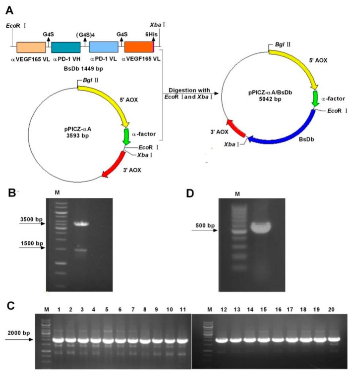
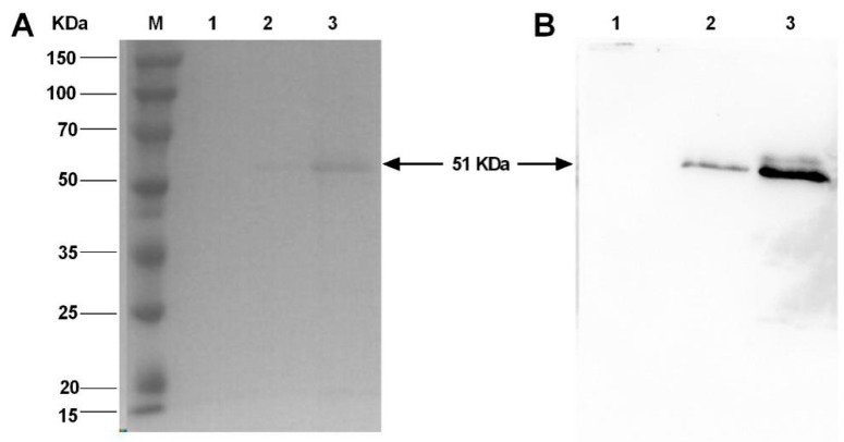
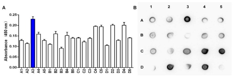
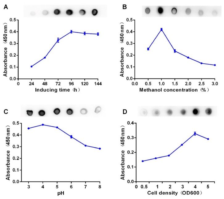
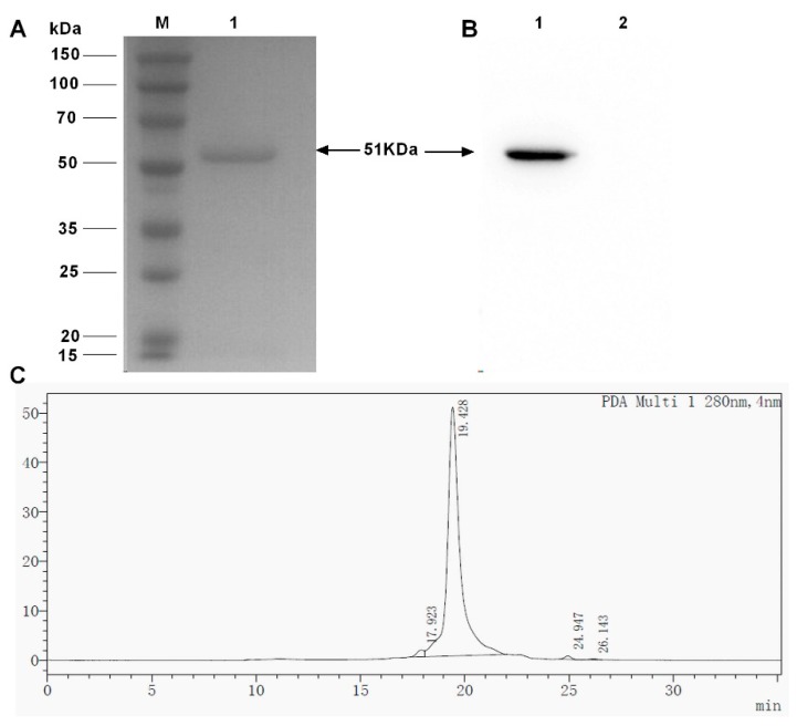
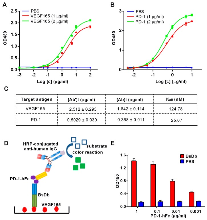
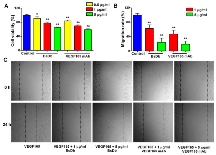
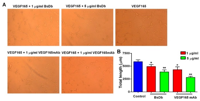
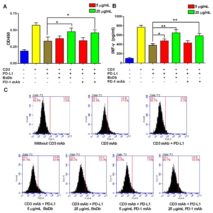
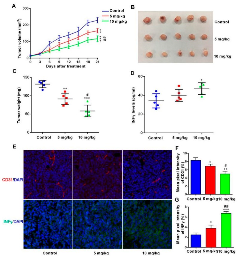
Bispecific antibodies, which can bind to two different epitopes on the same or different antigens simultaneously, have recently emerged as attractive candidates for study in various diseases. Our present study successfully constructs and expresses a fully human, bispecific, single-chain diabody (BsDb) that can bind to vascular endothelial growth factor 165 (VEGF165) and programmed death-1 (PD-1) in Pichia pastoris. Under the optimal expression conditions (methanol concentration, 1%; pH, 4.0; inoculum density, OD600 = 4, and the induction time, 96 h), the maximum production level of this BsDb is achieved at approximately 20 mg/L. The recombinant BsDb is purified in one step using nickel-nitrilotriacetic acid (Ni-NTA) column chromatography with a purity of more than 95%. Indirect enzyme-linked immune sorbent assay (ELISA) and sandwich ELISA analyses show that purified BsDb can bind specifically to VEGF165 and PD-1 simultaneously with affinities of 124.78 nM and 25.07 nM, respectively. Additionally, the BsDb not only effectively inhibits VEGF165-stimulated proliferation, migration, and tube formation in primary human umbilical vein endothelial cells (HUVECs), but also significantly improves proliferation and INF-γ production of activated T cells by blocking PD-1/PD-L1 co-stimulation. Furthermore, the BsDb displays potent antitumor activity in mice bearing HT29 xenograft tumors by inhibiting tumor angiogenesis and activating immune responses in the tumor microenvironment. Based on these results, we have prepared a potential bispecific antibody drug that can co-target both VEGF165 and PD-1 for the first time. This work provides a stable foundation for the development of new strategies by the combination of an angiogenesis inhibition and immune checkpoint blockade for cancer therapy.

Keywords: Pichia pastoris; anti-angiogenesis; bispecific single-chain diabody; immunotherapy; programmed death-1; vascular endothelial growth factor165.


Conflict of interest statement

The authors declare no conflict of interest.

Figures

Figure 1
Figure 1
Construction and transformation of the pPICZ-αA/BsDb expression plasmid. (A) Schematic diagram of the pPICZ-αA/BsDb expression plasmid generation. The gene fragment of BsDb containing a 6His-tag at the C-terminal end was cloned into pPICZαA by using EcoRI/XbaI digestion; (B) Restriction enzyme digestion of recombinant pPICZαA-BsDb expression vector. Lane M, 1 k bp marker (Thermo, Waltham, MA, USA); (C) Colony PCR analysis of 20 positive recombinants obtained from YPD plates containing zeocin. Lane M, 1 k bp marker (Thermo, Waltham, MA, USA); (D) Colony PCR analysis of a negative clone transformed pPICZαA empty vector. Lane M, 100 bp marker (Thermo, Waltham, MA, USA).
Figure 2
Figure 2
Analysis of recombinant BsDb expression in Pichia pastoris. (A) SDS-PAGE analysis: culture supernatant from a negative clone induced by 1% methanol for 96 h (lane 1); culture supernatant from the positive transformant grown under identical condition (lane 2); culture supernatant concentrated 5-fold from the positive transformant (lane 3); and protein molecular weight marker (Sangon, Shanghai, China) (Lane M); (B) Western blot analysis: supernatant from a negative clone (lane 1); culture supernatant from a positive transformant (lane 2); and culture supernatant concentrated 5-fold from a positive transformant (lane 3).
Figure 3
Figure 3
Analysis of relative expression levels of 20 positive clones. (A) Evaluation of the relative expression levels of various positive clones using ELISA. Recombinant BSDb relative expression levels were measured using the absorbance of 450 nm, clone A3 (blue column) possessed the highest production of the recombinant BsDb compared to the others; (B) Relative expression levels of BsDb in supernatants from twenty PCR positive clones were evaluated using spot Western blotting. The experiments were performed in triplicate, and the mean values ± standard deviation (SD) are presented.
Figure 4
Figure 4
Optimization of recombinant BsDb expression in Pichia pastoris. Supernatants collected at each evaluated condition were analyzed using ELISA and spot Western blotting analysis. (A) Optimization of the methanol inducing time; (B) optimization of the methanol concentration; (C) optimization of the pH value; and (D) optimization of the cell density. The experiment was performed in triplicate, and the mean values ± SD were presented.
Figure 5
Figure 5
Purification of recombinant BsDb using Ni-NTA affinity chromatography. (A) SDS-PAGE of purified recombinant BsDb; (B) Western blot analysis of purified recombinant BsDb (lane 1) and 5 µg BSA was used as a negative control for Western blot analysis (lane 2), protein molecular weight marker (Sangon, Shanghai, China) (Lane M); and (C) HPLC analysis of purified recombinant BsDb.
Figure 6
Figure 6
Measurement of affinities of recombinant BsDb to PD-1 and VEGF165. Binding curves of recombinant BsDb to VEGF165 (A) and PD-1 (B) tested using indirect ELISA; (C) The affinity constant was determined using the Beatty formula. [Ab]t and [Ab’]t represent the EC50 of high and low concentrations of antigen, respectively. PBS was used as a negative control; (D) Schematic diagram of sandwich ELISA; (E) Simultaneous binding of recombinant BsDb to both PD-1 and VEGF165 was evaluated using sandwich ELISA. PBS was used as a negative control. The experiments were performed in triplicate, and the mean values ± SD were presented.
Figure 7
Figure 7
The recombinant BsDb inhibited VEGF-induced proliferation and migration of primary HUVECs. (A) BsDb inhibited the primary HUVECs proliferation. Primary HUVECs were cultured in 96-well plates and stimulated with 100 ng/mL VEGF165 and various concentrations of recombinant BsDb or VEGF165 mAb. Cell growth was then determined using an MTT assay; (B,C) BsDb inhibited the primary HUVECs migration. The HUVECs monolayer was scratched and placed in fresh Endothelial Cell Medium (ECM) containing 1% FBS with 100 ng/mL VEGF165 and various concentrations of recombinant BSDb or VEGF165 mAb. The HUVECs migration was photographed using a microscope after 24 h (40×) and the migration rate was calculated. The experiment was done in triplicate and the mean values ± SD were presented. * p < 0.05 and ** p < 0.01 using a two-tailed students t-test versus the control group.
Figure 8
Figure 8
The recombinant BsDb inhibited tube formation of primary HUVECs. (A) HUVECs were seeded into 96-well plates that were coated with matrigel, then 100 ng/mL VEGF165 and various concentrations of BsDb or VEGF165 mAb were added to the 96-well plate, and the wells were photographed after incubation for 5 h at 37 °C (200×); (B) Quantification of the tube length by ImageJ software. The experiment was performed in triplicate and the mean values ± SD were shown. * p < 0.05 and ** p < 0.01 using a two-tailed students t-test versus the control group.
Figure 9
Figure 9
BsDb improved T cell proliferation and rescued T cell activation. (A) BsDb improved T cell proliferation. T cells were seeded into the 96-well plates precoated with an anti-CD3 antibody and human PD-L1. Subsequently, various concentrations of BsDb or PD-1 mAb were added into each well after incubation at 37 °C for 72 h. T cell proliferation was analyzed using the CCK-8 assay; (B) BsDb rescued T cell activation and increased INFγ secretion. T cells were seeded into the 96-well plates precoated with anti-CD3 antibody and human PD-L1. Subsequently, various concentrations of BsDb or PD-1 mAb were added into each well and, after incubation at 37 °C for 72 h, IFN-γ in-culture supernatants were evaluated using an ELISA kit; (C) BsDb improved intracellular IFN-γ of T cells using FCM analysis. The experiment was performed in triplicate and the mean values ± SD were presented. * p < 0.05 and ** p < 0.01 using a two-tailed students t-test versus the control group.
Figure 10
Figure 10
BsDb suppressed tumor growth in vivo. (A) The tumor volume of xenograft mice during the treatment. BsDb exerted robust antitumor activity in vivo with a dose-dependent manner; (B,C) mice were sacrificed after treatment for 21 days and tumors were collected and weighed; (D) Serum levels of INF-γ in different groups of mice were detected using ELISA after treatment for 21 days. Data were expressed as the mean ± SD (n = 5); (E) Immunofluorescence staining of CD31 (red fluorescence) and INF-γ (green fluorescence) in sections from tumors of different groups of mice. The cell nuclei were stained with 4′,6-diamidino-2-phenylindole (DAPI) in blue and the sections were photographed using a fluorescence microscope (200×); (F,G) quantitative analysis of CD31 and INF-γ fluorescence staining using ImageJ software. Data were expressed as the mean ± SD (n = 3). Statistical analysis was tested using one-way analysis variance (ANOVA) and post hoc Tukey honestly significant difference (HSD). * p < 0.05, ** p < 0.01, and *** p < 0.001 versus the control group; # p < 0.05 and ## p < 0.01 versus the 5 mg/kg group.

Similar articles

Cited by

References

    1. Strohl W.R. Current progress in innovative engineered antibodies. Protein Cell. 2018;9:86–120. doi: 10.1007/s13238-017-0457-8. - DOI - PMC - PubMed
    1. Przepiorka D., Ko C.W., Deisseroth A., Yancey C.L., Candau-Chacon R., Chiu H.J., Gehrke B.J., Gomez-Broughton C., Kane R.C., Kirshner S., et al. FDA Approval: Blinatumomab. Clin. Cancer Res. 2015;21:4035–4039. doi: 10.1158/1078-0432.CCR-15-0612. - DOI - PubMed
    1. Godar M., de Haard H., Blanchetot C., Rasser J. Therapeutic bispecific antibody formats: A patent applications review (1994–2017) Expert Opin. Ther. Pat. 2018;28:251–276. doi: 10.1080/13543776.2018.1428307. - DOI - PubMed
    1. Yancopoulos G.D., Davis S., Gale N.W., Rudge J.S., Wiegand S.J., Holash J. Vascular-specific growth factors and blood vessel formation. Nature. 2000;407:242–248. doi: 10.1038/35025215. - DOI - PubMed
    1. Ferrara N., Adamis A.P. Ten years of anti-vascular endothelial growth factor therapy. Nat. Rev. Drug Discov. 2016;15:385–403. doi: 10.1038/nrd.2015.17. - DOI - PubMed

MeSH terms

Substances