Review
. 2018 Sep 25;10(4):167.
doi: 10.3390/pharmaceutics10040167.

Porous Inorganic Carriers Based on Silica, Calcium Carbonate and Calcium Phosphate for Controlled/Modulated Drug Delivery: Fresh Outlook and Future Perspectives

Affiliations
Review

Porous Inorganic Carriers Based on Silica, Calcium Carbonate and Calcium Phosphate for Controlled/Modulated Drug Delivery: Fresh Outlook and Future Perspectives

Alexey D Trofimov et al. Pharmaceutics. .

Abstract

Porous inorganic nanostructured materials are widely used nowadays as drug delivery carriers due to their adventurous features: suitable architecture, large surface area and stability in the biological fluids. Among the different types of inorganic porous materials, silica, calcium carbonate, and calcium phosphate have received significant attention in the last decade. The use of porous inorganic materials as drug carriers for cancer therapy, gene delivery etc. has the potential to improve the life expectancy of the patients affected by the disease. The main goal of this review is to provide general information on the current state of the art of synthesis of the inorganic porous particles based on silica, calcium carbonate and calcium phosphate. Special focus is dedicated to the loading capacity, controllable release of drugs under internal biological stimuli (e.g., pH, redox, enzymes) and external noninvasive stimuli (e.g., light, magnetic field, and ultrasound). Moreover, the diverse compounds to deliver with silica, calcium carbonate and calcium phosphate particles, ranging from the commercial drugs to genetic materials are also discussed.

Keywords: calcium carbonate; calcium phosphate; drug delivery systems; drug loading; in vitro and in vivo delivery; silica-based particles.


Conflict of interest statement

The authors declare no conflict of interest.

Figures

Figure 1
Figure 1
Schematic overview of some advantages of porous carriers for the drug delivery applications.
Figure 2
Figure 2
TEM images of silica-based carriers with tunable diameters and pore size. (AE) Reproduced with permission from [31]. Royal Society of Chemistry, 2013; (FJ) Reproduced with permission from [33]. American Chemical Society, 2007; (KO) Reproduced with permission from [45]. American Chemical Society, 2017.
Figure 3
Figure 3
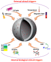
Schematic presentation of a variety of external and internal triggering mechanisms for delivery of bioactive compounds using porous silica-based carriers.
Figure 4
Figure 4
(A) Schematic illustration of CaCO3 crystallization process; (B) Schematic representation of ACC-vaterite-calcite crystallization pathway for the full crystallization reaction in the pure ACC system (the green triangles and full black squares represent the ACC and vaterite from this study, and the open squares and red triangles represent the vaterite and calcite). Reproduced with permission from [92]. American Chemical Society, 2012; (C) initial polymorphic composition of CaCO3 in dependence of the temperature. Reproduced with permission from [94]. Elsevier, 2014; (D) the typical shapes of calcium carbonate (CaCO3) particles. (A: Average Diameter; B: Crystalline Phase; C: Crystalline System). Reproduced with permission from [97]. MedCrave, 2017.
Figure 5
Figure 5
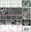
(A) Schematic presentation of the three types of synthesis with varied parameters as stirring time, the presence/absence of ethylene glycol, the salt ratio S, and the pH of the solutions and scanning electron microscopy (SEM) images for the obtained vaterite particles with size diameters of 3.15, 0.65, and 1.35 µm vaterite. Reproduced with permission from [122]. Frontiers Media S.A., 2018; (B) SEM images of the CaCO3 particles synthesized with varying stirring time and presence/absence of ethylene glycol and at pH values of 5, 7, 9 and plots depicted the dependence of particle size from the stirring time. Reproduced with permission from [96]. Springer, 2015; (C) SEM images of cross-section of CaCO3 microparticle. Reproduced with permission [113]. Royal Society of Chemistry, 2004; (D) SEM images of CaCO3 particle grown at different temperature. The pore sizes were found to be 19 ± 5, 28 ± 9, and 44 ± 13 nm for crystals prepared at 7.5, 22, and 45 °C, respectively. Reproduced with permission from [109]. American Chemical Society, 2016; (E) CaCO3 particle size distribution as a function of preparation conditions (salt concentration (c), speed (v) and time (t) of salt stirring). Parameter variations are on the top of the curves. Orange—salt concentration varied (v = 650 rpm, t = 30 s); violet—stirring speed is varied (c = 0.33 M, t = 30 s); green—stirring time is varied (c = 0.33 M, v = 650 rpm). Reproduced with permission from [123]. Wiley Publishing groups, 2012.
Figure 6
Figure 6
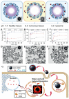
Schematic description of the pH-responsive drug release of ACC-DOX@silica nanoparticles. Reproduced with permission from [145]. Wiley Publishing Groups, 2012. Cumulative releases of doxorubicin (DOX) from ACC-DOX@silica suspensions in various aqueous buffers at (A) pH 7.4; (B) pH 6.5 and (C) pH 5.5 with different temperatures of 25, 37 and 50 °C. Representative TEM images of the suspensions under 37 °C at (D) pH 7.4; (E) pH 6.5 and (F) pH 5.5. Reproduced with permission from [145]. Wiley Publishing Groups, 2012. The scale bars are 500 nm. (G) Schematic illustration of assembly process and disassemble mechanism of PEG/OA-ACC-DOX within cancer cells. Reproduced with permission from [136]. Royal Society of Chemistry, 2017.
Figure 7
Figure 7
(A) SEM image of amorphous calcium phosphate particles synthesized by using CaCl2∙2H2O as the calcium source and ATP as both the phosphorous source and stabilizer by microwave-assisted hydrothermal method at 120 °C for 10 min. Reproduced with permission from [168]. Wiley Publishing Groups, 2013; (B) TEM images of calcium phosphate precipitates. Reproduced with permission from [177]. Bulletin of the Chemical Society of Japan, 2008; (C) TEM image of hollow calcium phosphate particles prepared using soybean lecithin, Na2ATP and CaCl2 by the microware-assisted hydrothermal method at 120 °C. Reproduced with permission from [171]. Royal Society of Chemistry, 2015; (D) TEM image of hollow calcium phosphate nanospheres. Reproduced with permission from [172]. Royal Society of Chemistry, 2012; (E) Transmission electron microscopy (TEM) image of calcium phosphate nanoshells synthesized with templates of DOPA. Reproduced with permission from [173]. Elsevier, 2012; (F) TEM image of calcium phosphate spheres. Reproduced with permission from [176]. IOPscience, 2009.
Figure 8
Figure 8
Schematic illustration of calcium phosphate particle degradation inside endosomal/lysosomal compartment (endosomal/lysosomal compartments are in orange color, enzymes/proteins are in green color, spherical calcium phosphate particle is in grey color, delivered cargoes are orange rhombs and green triangles).
Figure 9
Figure 9
In vivo distribution of calcium phosphate particles loaded with siRNA observed by the in vivo imaging system. The nude mice bearing the A549 tumor was given intravenous injection via tail vein. Reproduced with permission from [189]. Royal Society of Chemistry, 2016.

Similar articles

Cited by

References

    1. Croissant J.G., Fatieiev Y., Khashab N.M. Degradability and clearance of silicon, organosilica, silsesquioxane, silica mixed oxide, and mesoporous silica nanoparticles. Adv. Mater. 2017;29:1604634. doi: 10.1002/adma.201604634. - DOI - PubMed
    1. Wu M., Meng Q., Chen Y., Du Y., Zhang L., Li Y., Zhang L., Shi J. Large-Pore ultrasmall mesoporous organosilica nanoparticles: Micelle/precursor co-templating assembly and nuclear-targeted gene delivery. Adv. Mater. 2015;27:215–222. doi: 10.1002/adma.201404256. - DOI - PubMed
    1. Timin A.S., Khashirova S.Y., Zhansitov A., Rumyantsev E.V. Synthesis and application of silica hybrids grafted with new guanidine-containing polymers as highly effective adsorbents for bilirubin removal. Colloid Polym. Sci. 2015;293:1667–1674. doi: 10.1007/s00396-015-3555-2. - DOI
    1. Timin A.S., Rumyantsev E.V., Solomonov A.V., Musabirov I.I., Sergeev S.N., Ivanov S.P., Berlier G., Balantseva E. Preparation and characterization of organo-functionalized silicas for bilirubin removal. Colloids Surf. Physicochem. Eng. Asp. 2015;464:65–77. doi: 10.1016/j.colsurfa.2014.10.012. - DOI
    1. Gao X., Zhai M., Guan W., Liu J., Liu Z., Damirin A. Controllable synthesis of a smart multifunctional nanoscale metal-organic framework for magnetic resonance/optical imaging and targeted drug delivery. ACS Appl. Mater. Interfaces. 2017;9:3455–3462. doi: 10.1021/acsami.6b14795. - DOI - PubMed

LinkOut - more resources