

Interaction of systemic oxidative stress and mesial temporal network degeneration in Parkinson's disease with and without cognitive impairment
- PMID: 30257698
- PMCID: PMC6158841
- DOI: 10.1186/s12974-018-1317-z
Interaction of systemic oxidative stress and mesial temporal network degeneration in Parkinson's disease with and without cognitive impairment
Abstract
Background: To identify the vulnerable areas associated with systemic oxidative stress and further disruption of these vulnerable areas by measuring the associated morphology and functional network alterations in Parkinson's disease (PD) patients with and without cognitive impairment.
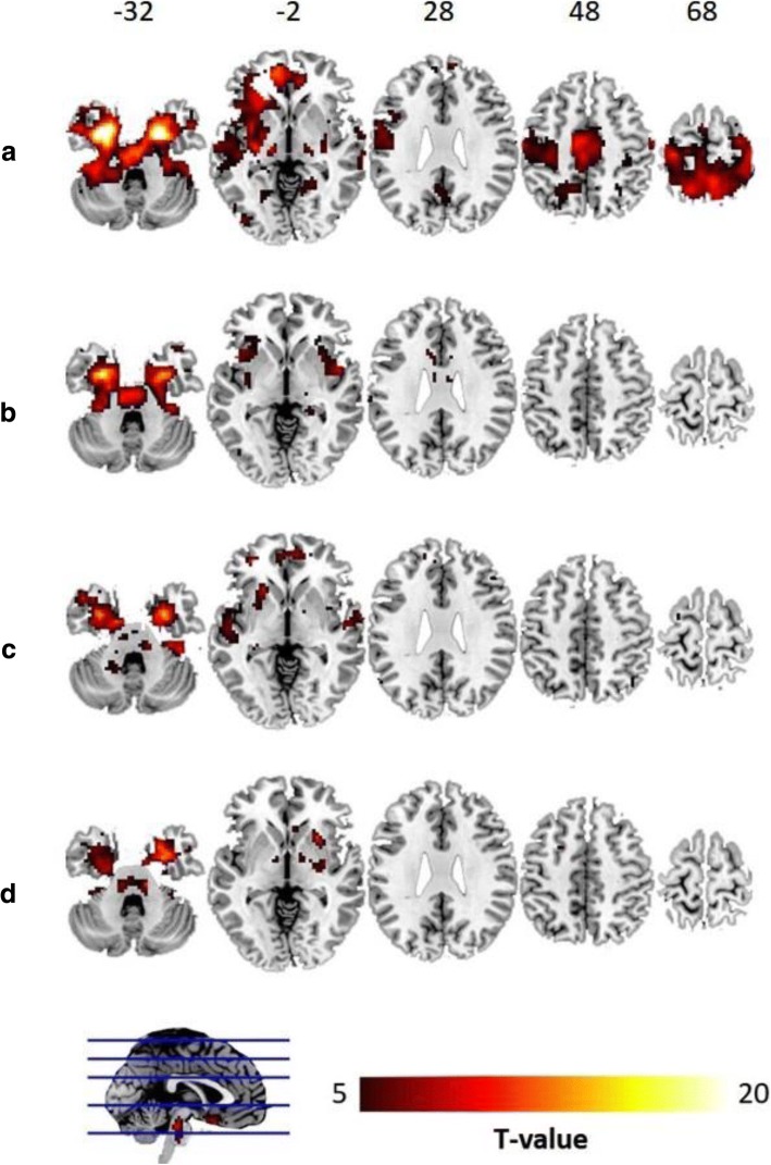
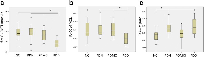
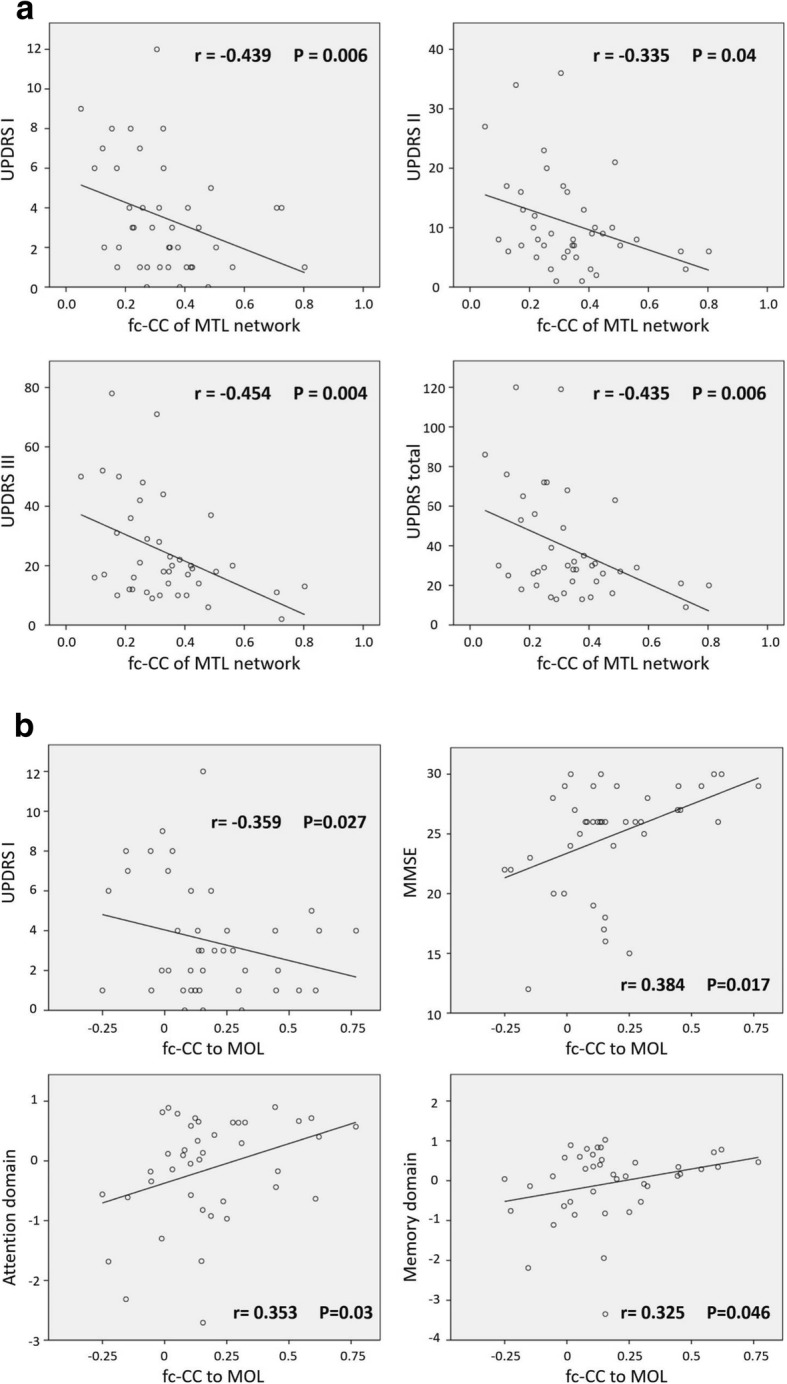
Methods: This prospective study was approved by the institutional review board of KCGMH, and written informed consent was obtained. Between December 2010 and May 2015, 41 PD patients with different levels of cognitive functions and 29 healthy volunteers underwent peripheral blood sampling to quantify systemic oxidative stress, as well as T1W volumetric and resting state functional MRI (rs-fMRI) scans. Rs-fMRI was used to derive the healthy intrinsic connectivity patterns seeded by the vulnerable areas associated with any of the significant oxidative stress markers. The two groups were compared in terms of the functional connectivity correlation coefficient (fc-CC) and gray matter volume (GMV) of the network seeded by the vulnerable areas.
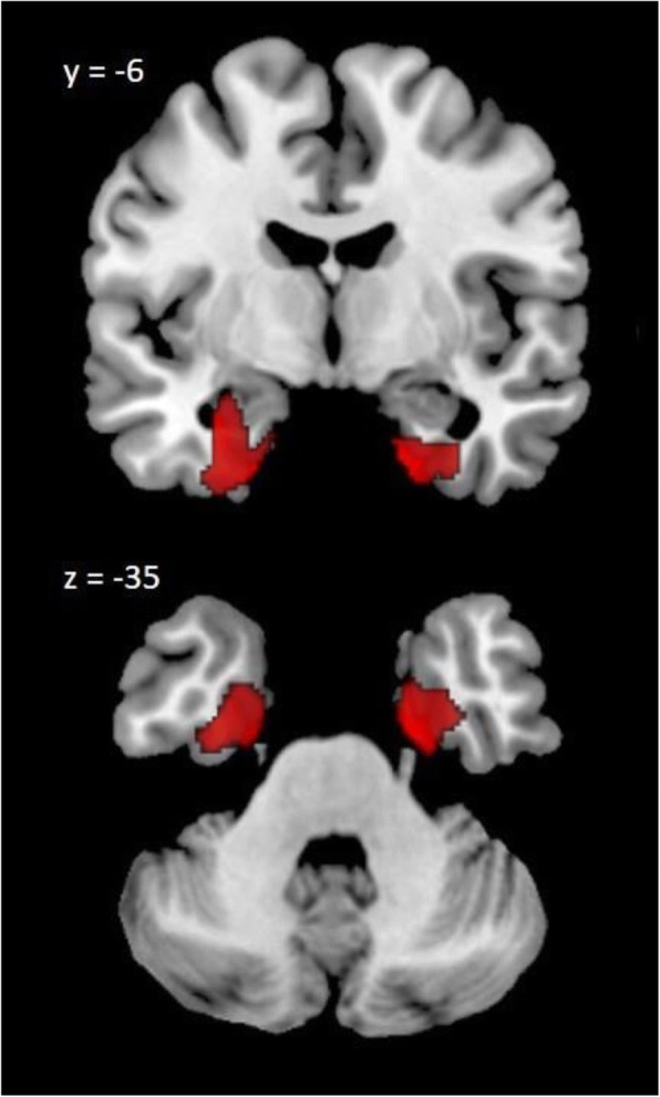
Results: The levels of oxidative stress markers, including leukocyte apoptosis and adhesion molecules, were significantly higher in the PD group. Using whole-brain VBM-based correlation analysis, the bilateral mesial temporal lobes (MTLs) were identified as the most vulnerable areas associated with lymphocyte apoptosis (P < 0.005). We found that the MTL network of healthy subjects resembled the PD-associated atrophy pattern. Furthermore, reduced fc-CC and GMV were further associated with the aggravated cognitive impairment.
Conclusion: The MTLs are the vulnerable areas associated with peripheral lymphocyte infiltration, and disruptions of the MTL functional network in both architecture and functional connectivity might result in cognitive impairments in Parkinson's disease.
Keywords: Cognitive impairment; Functional connectivity; Gray matter volume; Lymphocyte apoptosis; Mesial temporal network; Parkinson’s disease; Systemic oxidative stress.
Conflict of interest statement
Ethics approval and consent to participate
The Chang Gung Memorial Hospital Ethics Committee approved the study, and all of the participants provided written informed consent.
Consent for publication
All authors consent to the publication of the manuscript in Journal of Neuroinflammation.
Competing interests
The authors declare that they have no competing interests.
Publisher’s Note
Springer Nature remains neutral with regard to jurisdictional claims in published maps and institutional affiliations.
Figures
References
-
- Litvan I, Goldman JG, Troster AI, Schmand BA, Weintraub D, Petersen RC, Mollenhauer B, Adler CH, Marder K, Williams-Gray CH, et al. Diagnostic criteria for mild cognitive impairment in Parkinson's disease: Movement Disorder Society task force guidelines. Mov Disord. 2012;27(3):349–356. doi: 10.1002/mds.24893. - DOI - PMC - PubMed
MeSH terms
Substances
Grants and funding
LinkOut - more resources
Full Text Sources
Other Literature Sources
Medical
