. 2018 Sep 26;9(1):251.
doi: 10.1186/s13287-018-0981-3.

The effect of acute respiratory distress syndrome on bone marrow-derived mesenchymal stem cells

Affiliations

The effect of acute respiratory distress syndrome on bone marrow-derived mesenchymal stem cells

Ben Antebi et al. Stem Cell Res Ther. .

Abstract

Background: It is known that, following a physiological insult, bone marrow-derived mesenchymal stem cells (MSCs) mobilize and home to the site of injury. However, the effect of injury on the function of endogenous MSCs is unknown. In this study, MSCs harvested from the bone marrow of swine with or without acute respiratory distress syndrome (ARDS) were assessed for their characteristics and therapeutic function.

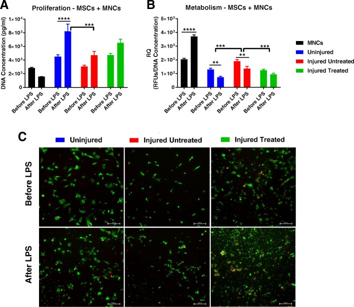
Methods: MSCs were harvested from three groups of anesthetized and mechanically ventilated swine (n = 3 in each group): 1) no ARDS ('Uninjured' group); 2) ARDS induced via smoke inhalation and 40% burn and treated with inhaled epinephrine ('Injured Treated' group); and 3) ARDS without treatment ('Injured Untreated' group). Cellular evaluation of the three groups included: flow cytometry for MSC markers; colony forming unit-fibroblast (CFU-F) assay; proliferative and metabolic capacity; gene expression using quantitative real-time polymerase chain reaction (qRT-PCR); and a lipopolysaccharide (LPS) challenge, with or without coculture with mononuclear cells (MNCs), for evaluation of their protein secretion profile using Multiplex. Statistical analysis was performed using one- or two-way analysis of variance (ANOVA) with a Tukey's post-test; a p-value less than 0.05 was considered statistically significant.

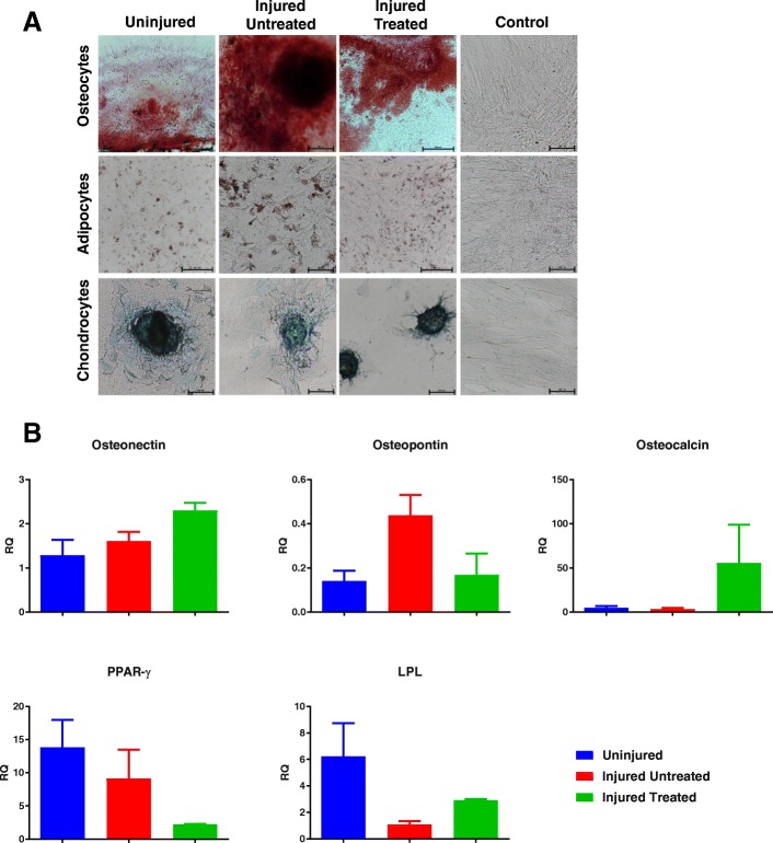
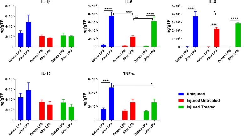
Results: Cells from all groups exhibited nearly 100% expression of MSC surface markers and retained their multidifferentiation capacity. However, the MSCs from the 'Injured Untreated' group generated a significantly higher number of colonies compared with the other two groups (p < 0.0001), indicative of increased clonogenic capacity following ARDS. Following an LPS challenge, the MSCs from the 'Injured Untreated' group exhibited a significant reduction in their proliferative capacity (p = 0.0002), significant downregulation in the expression of high-mobility group box 1 (HMGB1; p < 0.001), Toll-like receptor (TLR)-4 (p < 0.01), and vascular endothelial growth factor (VEGF; p < 0.05) genes, and significantly diminished secretory capacity for the inflammatory mediators interleukin (IL)-6 (p < 0.0001), IL-8 (p < 0.05), and tumor necrosis factor (TNF)-α (p < 0.05) compared with the 'Uninjured' group.

Conclusions: The results suggest that, following ARDS, there is an increase in the clonogenic capacity of MSCs to increase the available stem cell pool in vivo. However, MSCs harvested from subjects with ARDS seem to exhibit a diminished capacity to proliferate, express regenerative signals, and secrete pro/anti-inflammatory mediators.

Keywords: Acute respiratory distress syndrome (ARDS); Autologous therapy; Bone marrow; Immunomodulation; Injury; Mesenchymal stem cells (MSCs).


Conflict of interest statement

Ethics approval and consent to participate

Research was conducted in compliance with the Animal Welfare Act, the implementing Animal Welfare Regulations, and the principles of the Guide for the Care and Use of Laboratory Animals, National Research Council. The facility’s Institutional Animal Care and Use Committee approved all research conducted in this study. The facility where this research was conducted is fully accredited by AAALAC International.

Consent for publication

Not applicable.

Competing interests

The authors declare that they have no competing interests.

Publisher’s Note

Springer Nature remains neutral with regard to jurisdictional claims in published maps and institutional affiliations.

Figures

Fig. 1
Fig. 1
Surface marker expression of MSCs from different groups of swine: ‘Uninjured’; ‘Injured Untreated’; and ‘Injured Treated’. Cells expressed nearly 100% of common MSC surface markers, including CD29 (99.56 ± 0.23% (mean ± SEM) for ‘Uninjured’; 99 ± 0.64% for ‘Injured Untreated’; 99.47 ± 0.43% for ‘Injured Treated’), CD90 (99.96 ± 0.02% for ‘Uninjured; 100 ± 0.0% for ‘Injured Untreated’; 99.8 ± 0.13% for ‘Injured Treated’), CD105 (96.44 ± 0.92% for ‘Uninjured; 93.56 ± 2.03% for ‘Injured Untreated’; 95.57 ± 2.14% for ‘Injured Treated’), and a lack of expression of CD45 (1.36 ± 0.48% for ‘Uninjured; 0.74 ± 0.09% for ‘Injured Untreated’; 1.2 ± 0.18% for ‘Injured Treated’)
Fig. 2
Fig. 2
Multidifferentiation capacity of MSCs from the three groups of swine. a Histological images demonstrate that MSCs from all groups possess multidifferentiation capacity, exemplified by the ability to differentiate to osteocytes, adipocytes, and chondrocytes in vitro; scale bars = 250 μm. b Gene expression analysis revealed no significant differences in the ability of MSCs to differentiate down the osteogenic (osteonectin, osteopontin, and osteocalcin genes) and adipogenic (peroxisome proliferator-activated receptor (PPAR)-γ and lipoprotein lipase (LPL) genes) lineages. RQ relative quotient
Fig. 3
Fig. 3
Functional characteristics of MSCs from the different groups. a Clonogenic capacity of MSCs from the ‘Injured Untreated’ group as measured by the colony-forming unit fibroblast (CFU-F) assay was significantly higher than the other two groups. b Proliferation of MSCs from the ‘Uninjured’ and ‘Injured Untreated’ groups was significantly higher than MSCs from the ‘Injured Treated’ group (p < 0.01 and p < 0.001, respectively). c No differences were observed in the metabolic activity of the MSCs between the different groups. d Representative images of proliferation of the three groups of MSCs is shown throughout the 10-day study period through fluorescent staining of the cytoplasm of viable cells; scale bars = 100 μm. **p < 0.01, ***p < 0.001, ****p < 0.0001. RFUs - relative fluorescent units , RQ - relative quotient
Fig. 4
Fig. 4
Secretion profile of MSCs from the different groups before and after LPS treatment. MSCs from the ‘Injured’ groups demonstrated diminished capacity to secrete interleukin (IL)-6, IL-8, and tumor necrosis factor (TNF)-α compared with ‘Uninjured’ MSCs. Within each group, lipopolysaccharide (LPS) exposure induced significant upregulation of the inflammatory markers, which was most pronounced in the ‘Uninjured’ MSCs. *p < 0.05, **p < 0.01, ***p < 0.001, **** p < 0.0001. TP total protein
Fig. 5
Fig. 5
Gene expression of MSCs from the different groups after LPS treatment. Significant downregulation of high-mobility group box 1 (HMGB1) and Toll-like receptor (TLR)-4 genes in ‘Injured’ MSCs compared with ‘Uninjured’ MSCs (p < 0.01). Additionally, downregulation of angiopoietin 1 (Ang-1) and vascular endothelial growth factor (VEGF) genes in ‘Injured Untreated’ MSCs compared with ‘Injured Treated’ and ‘Uninjured’ MSCs, respectively (p < 0.05). Within each group, lipopolysaccharide (LPS) exposure induced significant downregulation of HMGB1 (p < 0.0001) and TLR-4 (p < 0.01) genes in ‘Injured’ MSC compared with upregulation in ‘Uninjured’ MSCs. For angiogenic genes, VEGF was upregulated in both ‘Uninjured’ and ‘Injured Treated’ MSCs (p < 0.0001) while Ang-1 was upregulated in ‘Injured Treated’ MSCs only (p < 0.05). *p < 0.05, **p < 0.01, ***p < 0.001, ****p < 0.0001. RQ relative quotient, SOX-2 sex determining region Y-box 2
Fig. 6
Fig. 6
Proliferation and metabolic activity of MSCs cocultured with MNCs before and after LPS treatment. a After lipopolysaccharide (LPS) exposure, there was an increase in proliferation in the cocultures with mesenchymal stem cells (MSCs) and cell death in the mononuclear cell (MNC) alone group; the ‘Uninjured’ group generated significantly more cells than the ‘Injured Untreated’ group (p < 0.001). Additionally, in the ‘Uninjured’ group, significantly more cells were generated after LPS exposure, whereas no differences were detected among the ‘Injured’ MSCs. b The metabolic activity was significantly higher (before and after LPS exposure) in ‘Injured Untreated’ MSCs (p < 0.001). Within each group, a significant increase in the metabolic activity of MNCs was observed after LPS exposure (p < 0.0001), while the metabolic activity was significantly decreased in ‘Uninjured’ and ‘Injured Untreated’ cocultures. c Overlaid fluorescent live/dead images showing viable cytoplasm in green, while the nuclei of dead cells are stained red. Increased cell death is seen following LPS exposures in all groups; scale bars = 500 μm. *p < 0.05, **p < 0.01, ***p < 0.001, ****p < 0.0001. Relative quotient
Fig. 7
Fig. 7
Secretion profile of MSCs cocultured with MNCs in response to LPS treatment. ‘Injured’ MSCs exhibit diminished capacity to secrete interleukin (IL)-1α and IL-1ra compared with mononuclear cells (MNCs) (p < 0.01) as well as reduced capacity to secrete IL-10 and IL-12 compared with ‘Uninjured’ MSCs (p < 0.05); all MSCs were able to significantly both secrete high levels of IL-6 and suppress tumor necrosis factor (TNF)-α production compared with MNCs alone (p < 0.0001). *p < 0.05, **p < 0.01, ***p < 0.001, ****p < 0.0001. TP total protein

References

    1. Antebi Ben, Mohammadipoor Arezoo, Batchinsky Andriy I., Cancio Leopoldo C. The promise of mesenchymal stem cell therapy for acute respiratory distress syndrome. Journal of Trauma and Acute Care Surgery. 2018;84(1):183–191. doi: 10.1097/TA.0000000000001713. - DOI - PubMed
    1. Trounson A, McDonald C. Stem cell therapies in clinical trials: progress and challenges. Cell Stem Cell. 2015;17(1):11–22. doi: 10.1016/j.stem.2015.06.007. - DOI - PubMed
    1. Rennert RC, Sorkin M, Garg RK, Gurtner GC. Stem cell recruitment after injury: lessons for regenerative medicine. Regen Med. 2012;7(6):833–850. doi: 10.2217/rme.12.82. - DOI - PMC - PubMed
    1. Belenkiy S, Ivey KM, Batchinsky AI, Langer T, Necsoiu C, Baker W, Salinas J, Cancio LC. Noninvasive carbon dioxide monitoring in a porcine model of acute lung injury due to smoke inhalation and burns. Shock. 2013;39(6):495–500. doi: 10.1097/SHK.0b013e318292c331. - DOI - PubMed
    1. McDaniel JS, Antebi B, Pilia M, Hurtgen BJ, Belenkiy S, Necsoiu C, Cancio LC, Rathbone CR, Batchinsky AI. Quantitative assessment of optimal bone marrow site for the isolation of porcine mesenchymal stem cells. Stem Cells Int. 2017;2017:1836960. doi: 10.1155/2017/1836960. - DOI - PMC - PubMed

Publication types

MeSH terms

LinkOut - more resources