Review
. 2019 Jan 7;218(1):39-54.
doi: 10.1083/jcb.201807179. Epub 2018 Sep 26.

Histone supply: Multitiered regulation ensures chromatin dynamics throughout the cell cycle

Affiliations
Review

Histone supply: Multitiered regulation ensures chromatin dynamics throughout the cell cycle

Shweta Mendiratta et al. J Cell Biol. .

Abstract

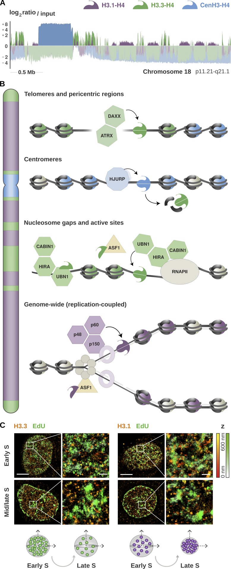
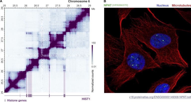
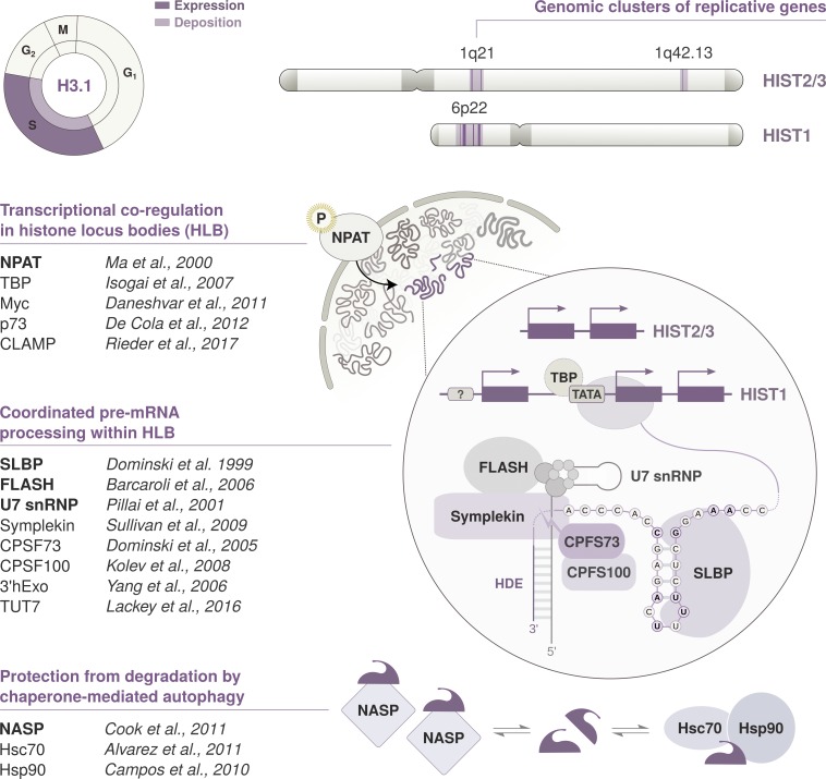
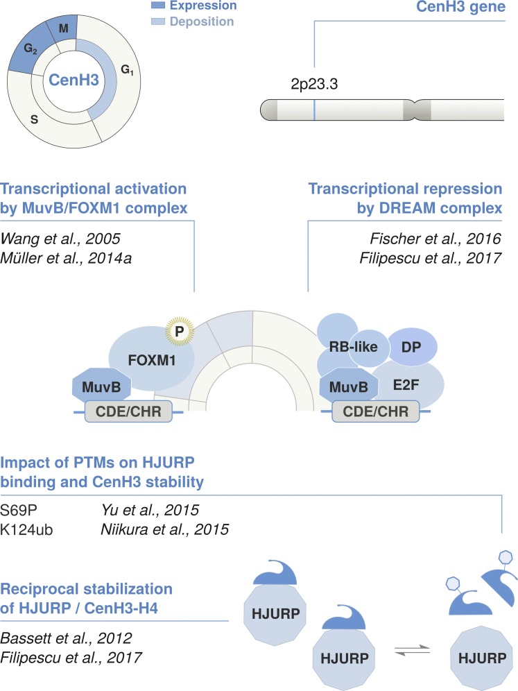
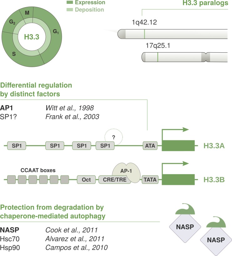
As the building blocks of chromatin, histones are central to establish and maintain particular chromatin states associated with given cell fates. Importantly, histones exist as distinct variants whose expression and incorporation into chromatin are tightly regulated during the cell cycle. During S phase, specialized replicative histone variants ensure the bulk of the chromatinization of the duplicating genome. Other non-replicative histone variants deposited throughout the cell cycle at specific loci use pathways uncoupled from DNA synthesis. Here, we review the particular dynamics of expression, cellular transit, assembly, and disassembly of replicative and non-replicative forms of the histone H3. Beyond the role of histone variants in chromatin dynamics, we review our current knowledge concerning their distinct regulation to control their expression at different levels including transcription, posttranscriptional processing, and protein stability. In light of this unique regulation, we highlight situations where perturbations in histone balance may lead to cellular dysfunction and pathologies.


Figures

Figure 1.
Figure 1.
Enrichment of histone H3 variants and their deposition by dedicated chaperones. (A) Genomic distribution of H3.1, H3.3, and CenH3 from published ChIP-Seq data in HeLa cells (Lacoste et al., 2014; Clément et al., 2018). The plot shows the enrichment relative to input for all variants at a representative region spanning the centromere and the proximal short and long arms of chromosome 18 (p11.21-q21.1). The enrichment is computed as the log2 ratio between the mean per-base number of reads from H3.1 (purple), H3.3 (green), and CenH3 (blue) and their respective input, at consecutive 10-kb bins (smoothed over five nonzero bins). Enriched regions are highlighted in darker colors, illustrating the partitioning of the genome into chromatin domains associated with specific variants. (B) Schematic representation of the histone chaperones involved in the deposition of H3 variants at distinct chromosomal locations. The DAXX/ATRX complex promotes the accumulation of the non-replicative variant H3.3 at telomeres and pericentric heterochromatin. H3.3 is also deposited at actively transcribed regions and regulatory sites by HIRA, a complex consisting of three subunits: CABIN1, HIRA, and UBN1. The non-replicative variant CenH3 is deposited specifically at centromeres by its dedicated chaperone HJURP, marking the site of kinetochore assembly. Both H3.1 and H3.3 are incorporated at centromeres during S phase, and H3.3 acts as a placeholder for CenH3 loading in late mitosis and early G1. H3.1 is deposited genome-wide by the CAF-1 complex during S phase, or at DNA repair sites throughout the cell cycle. The CAF-1 complex consists of three subunits: p48, p50, and p160. The p150 subunit interacts with PCNA and promotes CAF-1 recruitment at replication forks. This couples H3.1 deposition to DNA synthesis and ensures proper chromatin assembly during replication. ASF1 is a general chaperone that can handle both H3.1 and H3.3 and hands them over to their dedicated chaperones. (C) Illustration of high-resolution visualization by STORM of H3.1 and H3.3 along S phase (adapted from Clément et al., 2018). The STORM images show the nuclear distribution of H3.1 and H3.3 (HA staining, in red) at sites of DNA synthesis (EdU staining, in green) in early and mid/late S. Scale bars represent 5 µm. Insets represent enlarged images of selected areas where scale bars correspond to 600 nm. H3.3 clusters show stable volume, but there is a decrease in H3.3 density as S phase progresses; the late domains likely show a dilution of H3.3 during DNA replication. In contrast, H3.1 clusters change in both volume and density during S phase, with larger H3.1 clusters and low densities in early S and clusters with smaller volumes and higher density in mid/late S phase. See Clément et al. (2018) for further details.
Figure 2.
Figure 2.
Higher-order organization of replicative histone genes and compartmentalization of nuclear factors in HLBs. Replicative histones are redundantly encoded by multiple intronless genes that exhibit a conserved cluster organization across several lineages. (A) Location and spatial interactions among histone genes within the human histone cluster 1 (HIST1). The contact matrix was generated using iteratively corrected Hi-C data in GM12878 cells at 10-kb resolution (Rao et al., 2014) and plot with gcMapExplorer (Kumar et al., 2017). Genome bins where histone genes are located are marked in purple, illustrating the presence of three separate subsets within the HIST1 cluster. Neighboring genes in each subset interact within separate TADs, but all three subsets further engage in long-range interactions that bring distant genes together over an ∼1.5-Mb distance. This spatial organization might reflect the compartmentalization of replicative histone genes in the nucleus. Their pre-mRNAs are indeed transcribed and processed in dedicated nuclear bodies, called HLBs. (B) Nuclear distribution of NPAT, an essential factor driving HLB assembly and transcription initiation. The confocal image was retrieved from the Human Protein Atlas (v.18) and shows NPAT staining in U2-OS cells (in green). The nucleus and microtubules are stained in blue and red, respectively. NPAT concentrates at distinct subnuclear locations and marks the HLBs.
Figure 3.
Figure 3.
Cell cycle timing and regulation of replicative histone genes. The replicative variant H3.1 is deposited by CAF-1 in a DNA synthesis–coupled manner, mostly during replication in S phase. Expression peaks at the G1/S transition, and its transcription is regulated in concert with other replicative histone genes located within the histone clusters. Their pre-mRNAs are processed through a distinct pathway that involves the recognition of two unique cis-regulatory elements: a 3′ stem-loop structure and an HDE. Transcription and pre-mRNA processing are compartmentalized in the nucleus and coordinated in HLBs. HLB assembly and transcriptional activation is initiated by NPAT. NPAT is phosphorylated by the Cyclin E/CDK2 complex at the G1/S transition. The maturation of histone pre-mRNAs requires the endonucleolytic cleavage of its 3′ tail and is mediated by several factors recruited to HLBs. The U7 snRNP binds the HDE via hybridization of the U7 snRNA. It interacts with SLBP, a protein that specifically recognizes the 3′ stem-loop structure and stabilizes the U7 snRNP association. FLASH is another essential coactivator that promotes the recruitment of transcription factors and interacts with the Lsm11 subunit of the U7 snRNP to recruit the components of the histone cleavage complex. These transcription factors include Symplekin and the CPSF73/CPSF100 heterodimer that catalyzes the 3′ end endonucleolytic cleavage. Mature mRNAs are cleaved downstream of the 3′ stem-loop, which is required for mRNA degradation. SLBP is also degraded at the end of S phase after phosphorylation by the Cyclin A/CDK1 complex. H3 availability is further modulated at the protein level by NASP, a H3-H4 histone chaperone that protects soluble histones from degradation via chaperone-mediated autophagy counteracting Hsc70 and Hsp90.
Figure 4.
Figure 4.
Cell cycle timing and regulation of the centromeric variant CenH3. The non-replicative variant CenH3CENP-A is deposited at centromeres by its dedicated chaperone HJURP in telophase/early G1. Its expression is regulated in a cell cycle–dependent manner and peaks in G2/M. CenH3CENP-A is encoded by a single multi-exon gene located outside of the histone clusters that undergoes conventional pre-mRNA processing via splicing and polyadenylation. Transcription is coordinated in concert with other late cell cycle genes, including HJURP. This is regulated in cis by the CHR/CDE motif in their promoter region. The recruitment of the DREAM complex at the CHR is thought to promote transcriptional repression during G1. At the beginning of S phase, the MuvB core of the DREAM complex remains bound to the CHR/CDE while other components dissociate and are replaced by B-MYB. The B-MYB-MuvB (MMB) complex recruits FOXM1 in late S phase. B-MYB is degraded upon phosphorylation, whereas the progressive phosphorylation of FOXM1 leads to its activation in G2/M. Both CenH3 and HJURP are induced at the same time and mutually stabilize each other at the protein level. The reciprocal stabilization is affected by posttranslational modifications on the N-terminal tail of CenH3, which further regulate the timing of deposition. S69 phosphorylation prevents interaction with HJURP and premature loading, while K124 ubiquitylation favors HJURP binding and might contribute to their stabilization.
Figure 5.
Figure 5.
Cell cycle timing and regulation of the replacement variant H3.3. The non-replicative variant H3.3 is deposited throughout the cell cycle by HIRA at regions of high turnover, such as regulatory sites and transcribed regions. H3.3 is expressed constitutively and redundantly encoded by two conserved paralogs: H3.3A and H3.3B. Both genes are located outside of histone clusters, contain introns, and give rise to polyadenylated mRNAs. H3.3A and H3.3B encode for the same protein, but their gene architecture is not conserved. They show distinct coding sequences, intron-exon organization, and cis-regulatory elements and are differentially expressed during development and among tissues. Putative binding sites have been identified in their respective promoter regions, and the CRE/TRE motif in H3.3B promoter was shown to mediate its activation via recruitment of AP-1 transcription factors. However, the basis of differences in expression is poorly understood. Similar to H3.1, the NASP histone chaperone can bind H3.3-H4 dimers and contributes to fine-tune protein levels via protection from chaperone-mediated autophagy.

Similar articles

Cited by

References

    1. Ahmad K., and Henikoff S.. 2002. The histone variant H3.3 marks active chromatin by replication-independent nucleosome assembly. Mol. Cell. 9:1191–1200. 10.1016/S1097-2765(02)00542-7 - DOI - PubMed
    1. Aihara K., Mukasa A., Gotoh K., Saito K., Nagae G., Tsuji S., Tatsuno K., Yamamoto S., Takayanagi S., Narita Y., et al. . 2014. H3F3A K27M mutations in thalamic gliomas from young adult patients. Neuro-oncol. 16:140–146. 10.1093/neuonc/not144 - DOI - PMC - PubMed
    1. Akhmanova A.S., Bindels P.C., Xu J., Miedema K., Kremer H., and Hennig W.. 1995. Structure and expression of histone H3.3 genes in Drosophila melanogaster and Drosophila hydei. Genome. 38:586–600. 10.1139/g95-075 - DOI - PubMed
    1. Alabert C., and Groth A.. 2012. Chromatin replication and epigenome maintenance. Nat. Rev. Mol. Cell Biol. 13:153–167. 10.1038/nrm3288 - DOI - PubMed
    1. Albig W., Bramlage B., Gruber K., Klobeck H.-G., Kunz J., and Doenecke D.. 1995. The human replacement histone H3.3B gene (H3F3B). Genomics. 30:264–272. 10.1006/geno.1995.9878 - DOI - PubMed

Publication types

MeSH terms