. 2018 Nov 16;293(46):17739-17753.
doi: 10.1074/jbc.RA118.003238. Epub 2018 Sep 26.

G protein-coupled receptors differentially regulate glycosylation and activity of the inwardly rectifying potassium channel Kir7.1

Affiliations

G protein-coupled receptors differentially regulate glycosylation and activity of the inwardly rectifying potassium channel Kir7.1

Sheridan J Carrington et al. J Biol Chem. .

Abstract

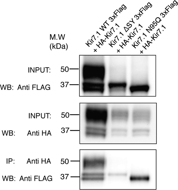
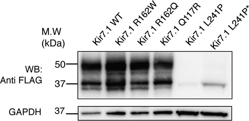
Kir7.1 is an inwardly rectifying potassium channel with important roles in the regulation of the membrane potential in retinal pigment epithelium, uterine smooth muscle, and hypothalamic neurons. Regulation of G protein-coupled inwardly rectifying potassium (GIRK) channels by G protein-coupled receptors (GPCRs) via the G protein βγ subunits has been well characterized. However, how Kir channels are regulated is incompletely understood. We report here that Kir7.1 is also regulated by GPCRs, but through a different mechanism. Using Western blotting analysis, we observed that multiple GPCRs tested caused a striking reduction in the complex glycosylation of Kir7.1. Further, GPCR-mediated reduction of Kir7.1 glycosylation in HEK293T cells did not alter its expression at the cell surface but decreased channel activity. Of note, mutagenesis of the sole Kir7.1 glycosylation site reduced conductance and open probability, as indicated by single-channel recording. Additionally, we report that the L241P mutation of Kir7.1 associated with Lebers congenital amaurosis (LCA), an inherited retinal degenerative disease, has significantly reduced complex glycosylation. Collectively, these results suggest that Kir7.1 channel glycosylation is essential for function, and this activity within cells is suppressed by most GPCRs. The melanocortin-4 receptor (MC4R), a GPCR previously reported to induce ligand-regulated activity of this channel, is the only GPCR tested that does not have this effect on Kir7.1.

Keywords: G protein–coupled receptor (GPCR); glycosylation; ion channel; potassium channel; signal transduction.


Conflict of interest statement

The authors declare that they have no conflicts of interest with the contents of this article

Figures

Figure 1.
Figure 1.
Inhibition of complex glycosylation of Kir7.1 by the β2AR. A, Western blotting analysis (WB) from whole-cell lysates of transiently transfected HEK293T cells with 3× FLAG-tagged Kir7.1 alone or co-expressed with either 3× HA–tagged β2AR or the MC4R. Transfections in A and B were performed using 0.25 μg of expression vector for the channel and the β2AR. 2.5 μg of expression vector for the MC4R was used to achieve receptor expression levels comparable with that seen with the β2AR (see Fig. 2). B, enzymatic digestion of whole-cell Kir7.1 lysates confirmed the 50-kDa Endo H–resistant doublet as the complex or mature glycosylated form, the 37-kDa band as the core glycan form, and the lowest band in lane 3 as the unglycosylated PNGase F–cleaved form. C, densitometric quantification of the blot shown in A, showing that MC4R had a modest increase in the ratio of mature to immature forms of kir7.1 p < 0.05, whereas β2AR significantly reduced the ratio of mature forms of Kir7.1 p < 0.005, one-way ANOVA representative of three independent experiments. The error bars represent S.D. M.W, molecular mass.
Figure 2.
Figure 2.
Increasing amounts of the β2AR increase the amounts of unglycosylated and core-glycosylated forms of Kir7.1, whereas the melanocortin-4 receptor does not. A and B, HEK293T cells were transiently transfected with a fixed amount of 3× FLAG-tagged Kir7.1 (0.25 μg) in the presence of increasing concentrations of 3× HA–β2AR (0.05, 0.08, 0.125, or 0.25 μg) or 3× HA–MC4R plasmid DNA (0.5, 0.8, 1.25, or 2.5 μg). Whole-cell lysates were analyzed by Western blotting analysis (WB). To assess receptor expression levels, the samples were rerun on a separate blot and imaged with anti-HA. This blot was stripped and reprobed for the loading control GAPDH to confirm plasmid concentration-based increase in receptor protein levels. C, densitometric analysis of the anti-FLAG blot to determine the ratio of mature to complex glycosylation. Densitometric analysis was also performed on the receptor blot and normalized to the GAPDH loading control for both β2AR expression and MC4R expression. The ratio of mature to immature Kir7.1 glycosylation was plotted against Log2 receptor intensity values, and a linear regression line was fitted to the data points. The data are representative of at least three experiments. D–F, comparable increases in receptor expression do not alter VSVG protein glycosylation. Whole-cell HEK293T lysates transiently expressing VSVG–GFP (0.25 μg) with Kir7.1 (0.25 μg) or increasing concentrations of β2AR (0.08, 0.125, or 0.25 μg) or MC4R (0.8, 1.25, or 2.5 μg) were treated ± Endo H. As a control, VSVG–GFP only expressing cells were treated with a transport inhibitor (brefeldin A (BFA)/monesin) and compared with untreated cell lysate. Anti-HA blots show receptor expression levels. **, Endo H–resistant; ***, Endo H–sensitive. VSVG–GFP+3× HA–β2AR OE wells show that a 3-fold overexpression of β2AR (0.75 μg) can affect VSVG–GFP glycosylation. The data are representative of three independent experiments. M.W, molecular mass.
Figure 3.
Figure 3.
Co-expression of the β2AR reduces total Kir7.1-mediated whole-cell currents. A, whole-cell patch-clamp recordings from HEK293T cells transiently expressing Kir7.1-M125R only versus Kir7.1-M125R co-expressed with MC4R (n = 5). B, a representative current-voltage (I–V) relationship is shown. The Kir7.1-M125R mutant has a methionine in the pore of the channel mutated to an arginine typically found at the same site in most other Kir channels. This mutation significantly increases the amount of detectable currents from the channel, with increased sensitivity to barium. Therefore, the amount of current inhibited by barium at the end of the experiment was recorded as the total current of Kir7.1-M125R from HEK293T cells. Mock-transfected cells did not exhibit barium-sensitive currents (data not shown). C, Kir7.1-M125R co-expressed with β2AR showed a significant reduction in whole-cell current in pA/pF, p0.0001 (n = 7 cells). The error bars represent S.E. D, representative current-voltage (I–V) relationship of Kir7.1-M125R (black) in the presence or absence of β2AR (red). NS, not significant.
Figure 4.
Figure 4.
Co-expression of the β2AR does not reduce the total amount of Kir7.1 at the plasma membrane but alters the ratio of mature glycosylated forms of Kir7.1 at the surface. Transfection levels were as in Fig. 1. A, Western blotting analysis (WB) of surface biotinylation of cells transiently transfected with 3× FLAG–tagged Kir7.1 co-expressed with the 3× HA–β2AR or 3× HA–MC4R. The experiment was performed as described by Chandrasekhar et al. (39). The cells were labeled with sulfo-NHS-SS-biotin, and surface proteins were immunoprecipitated with streptavidin–agarose beads. At least 45 μg of prequantified total protein was immunoprecipitated and loaded per lane. 15 μg (one-fifth) of total input protein was loaded for comparison. B, GAPDH control indicated that cells remained intact throughout biotinylation. Surface quantification was determined by densitometry analysis of the mature/complex doublet versus immature. The percentage of surface biotinylation was determined by dividing the values in the surface lanes by the input lane values multiplied by five. Percentages of complex or immature Kir7.1 at the surface were compared. The error bars represent S.D. with no significance observed between conditions determined by one-way ANOVA. C, comparison of the ratio of mature to immature forms of Kir7.1 in whole-cell lysates versus cell-surface biotinylated proteins. The results are representative of three to five independent experiments (p < 0.05; p < 0.005). The error bars represent S.D. MW, molecular mass; NS, not significant.
Figure 5.
Figure 5.
Creation of an unglycosylated mutant of Kir7.1. A, Lasergene MegAlign software was used to confirm Kir7.1 conserved glycosylation site of Asn-95 compared with other known glycosylated Kir channels, ROMK(Kir1.1) Asn-117 (23) and GIRK1 Asn-119 (22). B, the Asn-95 site of Kir7.1 was mutated to glutamine N95Q, and the whole-cell lysates of the 3× FLAG-tagged N95Q mutant and the similarly tagged WT channel were compared. No higher migrating forms of Kir7.1 were observed in the N95Q mutant. C, homology modeling of Kir7.1 based on Kir2.2 (PDB code 5KUK) revealed the location of the N95 site in the outer loops that gate the channel pore. This suggested a possible role for glycosylation in channel function. Modeling was performed using SWISS-MODEL, and the image was generated with PyMOL. Transfection levels were as shown in Fig. 1. M.W, molecular mass; WB, Western blotting analysis.
Figure 6.
Figure 6.
A and B, representative single-channel current traces from cell-attached patches of WT Kir7.1 (A) and Kir7.1N95Q (B) channels recorded from HEK293T cells. Patches were voltage-clamped at 130 mV. Openings are downward, and each representative trace was a continuous 5000-ms recording. C, average time histograms of openings (O1, O2, and O3) events are shown with the time constants (ms) and the relative contribution (%) of open events. D–F, bar graphs summarize the effects of Kir7.1N95Q on amplitude (D), mean open probability (NPo) (E), and mean open time of channels (F). The values are expressed as means ± S.E. (WT, n = 6; NQ, n = 7). Statistical differences were determined using unpaired t test relative to WT. C and O refer to the closed and open states, respectively.
Figure 7.
Figure 7.
Multiple GPCRs inhibit Kir7.1 glycosylation. A, Western blotting analysis (WB) of HEK293T whole-cell lysates transiently transfected with 3× FLAG–tagged Kir7.1, co-expressed with different 3× HA–tagged β-adrenergic receptors in the absence of ligands. Transfection levels were as in Fig. 1. Receptor expression was analyzed on a separate blot, with the loading control GAPDH shown. B, densitometric analysis of the anti-FLAG immunoblot of Kir7.1 shown in A to quantify the ratio of mature to immature glycosylation p < 0.005. The data from three independent experiments were analyzed by one-way ANOVA in GraphPad Prism, with Tukey post-test for multiple comparisons with S.D. M.W, molecular mass.
Figure 8.
Figure 8.
A conserved Golgi transport signal is required for complex glycosylation of Kir7.1. A, schematic showing a two-pore domain Kir channel subunit with two transmembrane regions, M1 and M2. The consensus N- and C-terminal Golgi export patch residues are shown, with known essential residues shown in red. Clustal alignment of the N- and C-terminal regions of the Golgi export patch of other closely related Kir channels that have been previously identified (28). Kir7.1 shares a highly conserved patch, except for a C-terminal leucine residue where other channels have a tryptophan. B, SWISS-MODEL of Kir7.1 structure based on the crystal structure of Kir2.2 (PDB code 5KUK). Regions highlighted in blue show the glycosylation site in each monomer of the channel. The N- and C-terminal portions of the Golgi export patch are highlighted in green and red, respectively. C, deletion of the serine and tyrosine residues in the Golgi export patch of Kir7.1 led to a loss of complex glycosylation compared with the WT channel determined by Western blotting analysis (WB) in Cos1 cells. D, compared with the WT channel, essentially no complex glycans were observed indicated by the significantly low ratio of 0.1 p < 0.005, (unpaired t test). E, enzymatic digestion of whole-cell lysates of the WT versus Kir7.1 SYΔ mutant from HEK293T cells confirmed that only the immature forms of Kir7.1 are expressed. F, β2AR also reduces the ratio of mature to immature glycosylated Kir7.1 in Cos1 cells. Western blotting analysis of whole-cell lysates from Cos1 cells co-expressing Kir7.1–3× FLAG with either 3× HA–β2AR or 3× HA–MC4R. 3× HA–β2AR also reduced Kir7.1 mature glycosylation in Cos1 cells as shown in the quantification by densitometric analysis in G (p < 0.05). Transfection levels in C, E, and F were as in Fig. 1. The data analyzed from four independent experiments were analyzed in GraphPad Prism with one-way ANOVA. The error bars for all data represent S.E. M.W, molecular mass.
Figure 9.
Figure 9.
Kir7.1 forms heterotetramers of immature and unglycosylated subunits. HEK293T cells were transiently transfected with HA-Kir7.1 with either FLAG-tagged Kir7.1 N95Q, Kir7.1 ΔSY, or Kir7.1 WT as a control; transfection levels were as in Fig. 1. Cell lysates were immunoprecipitated with an HA antibody and immunoblotted with anti-FLAG. The blot is representative of three independent experiments. M.W, molecular mass; WB, Western blotting analysis.
Figure 10.
Figure 10.
A retinopathy-associated variant of Kir7.1 has altered complex glycosylation. Shown is Western blotting analysis (WB) of three known variants of Kir7.1, following transfection of Kir7.1 mutants indicated; transfection levels were as in Fig. 1. Whole-cell lysates of HEK293T cells expressing a known variant of Kir7.1 were analyzed for changes in glycosylation. Kir7.1 L241P in lane 5 not only had lower expression but also had an absence of complex glycosylation. *, 15 μg of protein were loaded versus 7.5 μg in all other lanes. The blot is representative of four independent experiments. M.W, molecular mass.

Similar articles

Cited by

References

    1. Hibino H., Inanobe A., Furutani K., Murakami S., Findlay I., and Kurachi Y. (2010) Inwardly rectifying potassium channels: their structure, function, and physiological roles. Physiol. Rev. 90, 291–366 10.1152/physrev.00021.2009 - DOI - PubMed
    1. Döring F., Derst C., Wischmeyer E., Karschin C., Schneggenburger R., Daut J., and Karschin A. (1998) The epithelial inward rectifier channel Kir7.1 displays unusual K+ permeation properties. J. Neurosci. 18, 8625–8636 10.1523/JNEUROSCI.18-21-08625.1998 - DOI - PMC - PubMed
    1. Nakamura N., Suzuki Y., Sakuta H., Ookata K., Kawahara K., and Hirose S. (1999) Inwardly rectifying K+ channel Kir7.1 is highly expressed in thyroid follicular cells, intestinal epithelial cells and choroid plexus epithelial cells: implication for a functional coupling with Na+,K+-ATPase. Biochem. J. 342, 329–336 10.1042/bj3420329, 10.1042/0264-6021:3420329 - DOI - PMC - PubMed
    1. Yang D., Pan A., Swaminathan A., Kumar G., and Hughes B. A. (2003) Expression and localization of the inwardly rectifying potassium channel Kir7.1 in native bovine retinal pigment epithelium. Invest. Opthalmol. Vis. Sci. 44, 3178–3185 10.1167/iovs.02-1189 - DOI - PubMed
    1. Pattnaik B. R., Tokarz S., Asuma M. P., Schroeder T., Sharma A., Mitchell J. C., Edwards A. O., and Pillers D. A. (2013) Snowflake vitreoretinal degeneration (SVD) mutation R162W provides new insights into Kir7.1 ion channel structure and function. PLoS One 8, e71744 10.1371/journal.pone.0071744 - DOI - PMC - PubMed

Publication types

MeSH terms

Substances

Associated data

LinkOut - more resources