. 2019 Jan;18(1):65-85.
doi: 10.1074/mcp.RA118.001046. Epub 2018 Sep 26.

Identification of Novel Natural Substrates of Fibroblast Activation Protein-alpha by Differential Degradomics and Proteomics

Affiliations

Identification of Novel Natural Substrates of Fibroblast Activation Protein-alpha by Differential Degradomics and Proteomics

Hui Emma Zhang et al. Mol Cell Proteomics. 2019 Jan.

Abstract

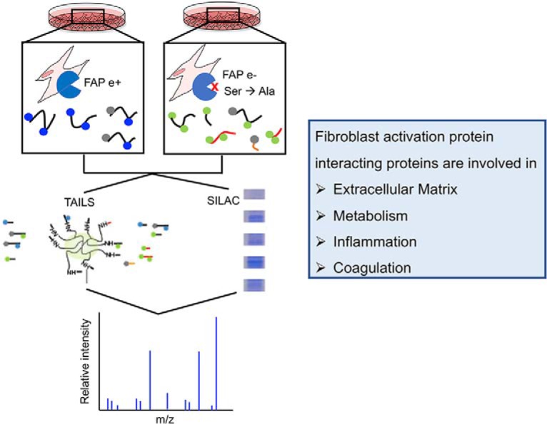
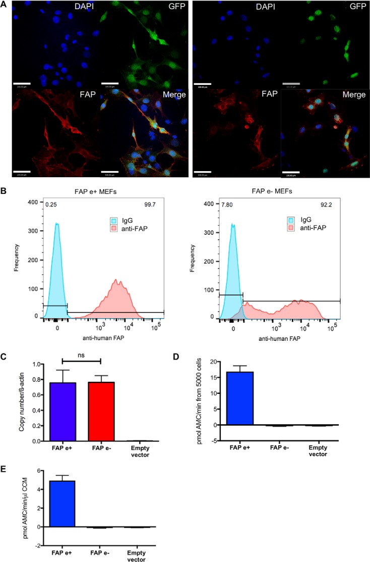
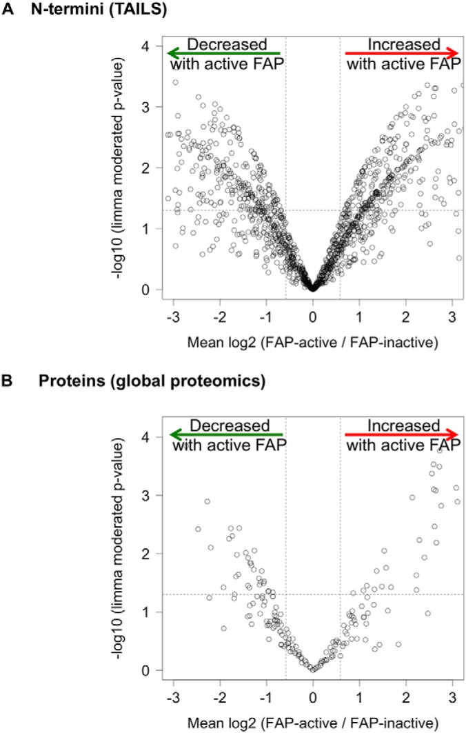
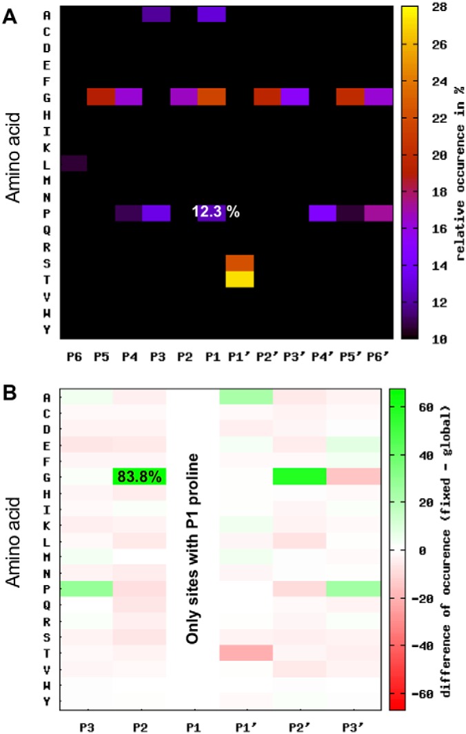
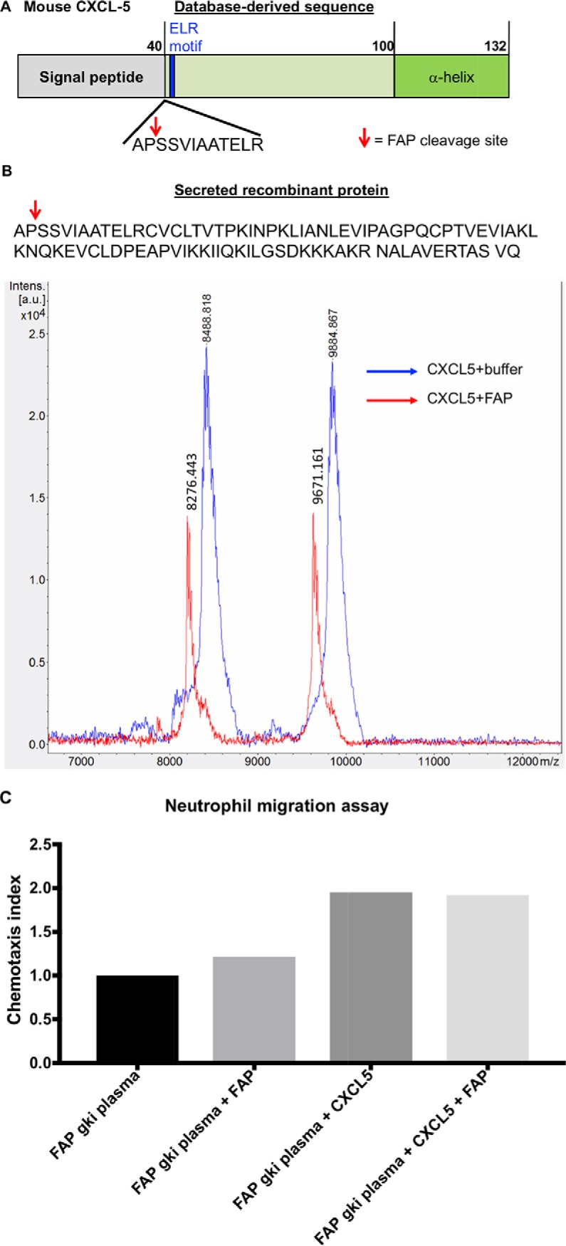
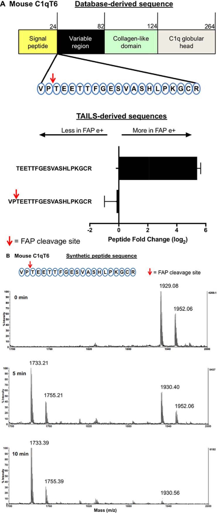
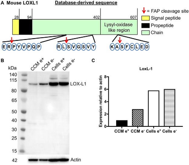
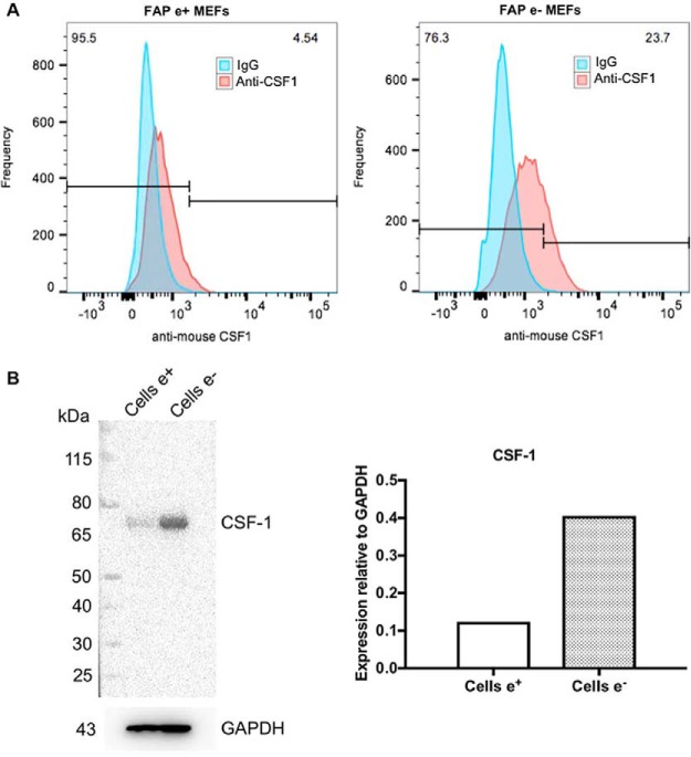
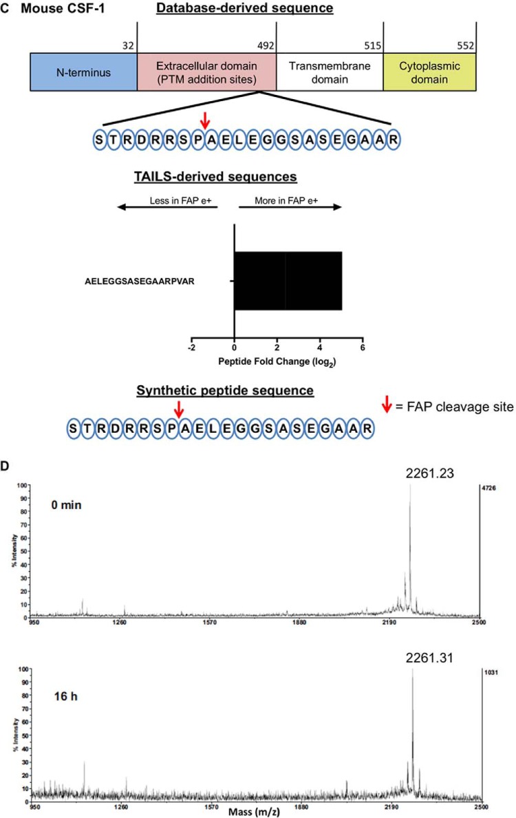
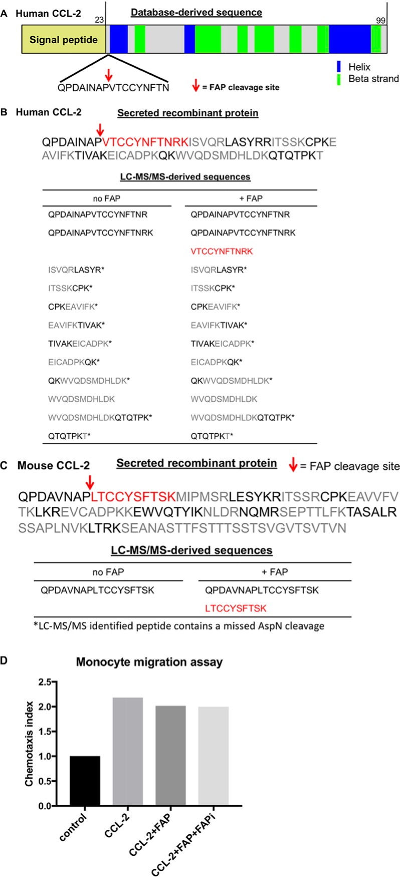
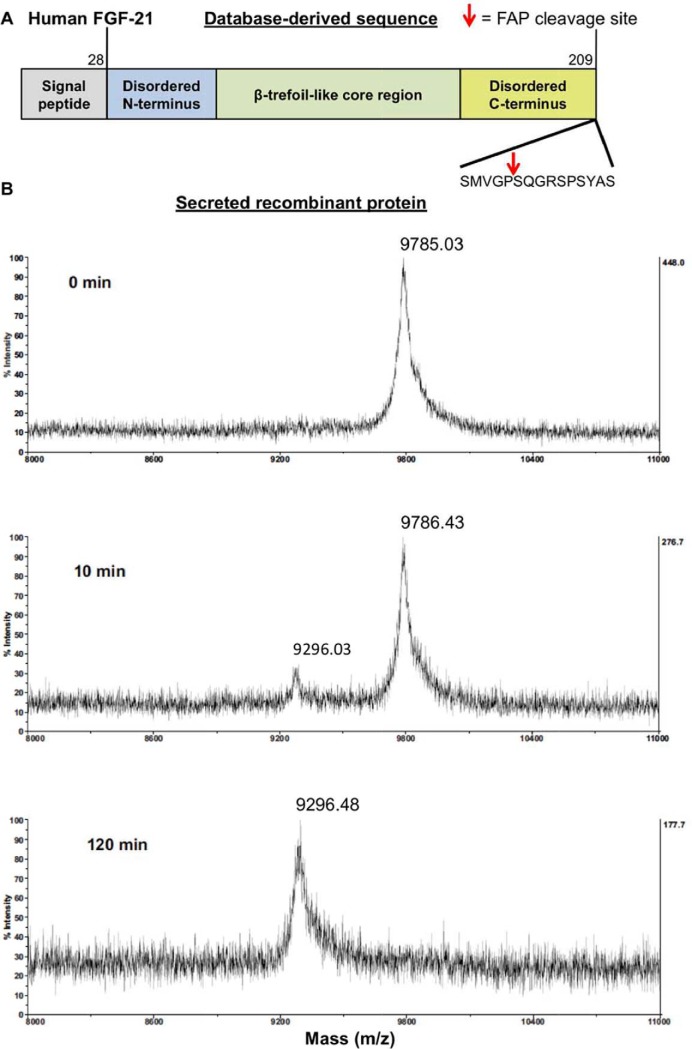
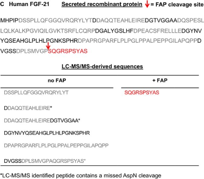
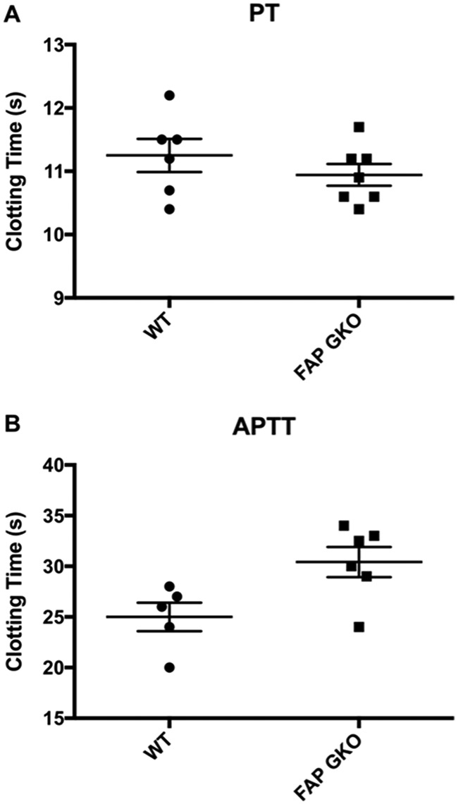
Fibroblast activation protein-alpha (FAP) is a cell-surface transmembrane-anchored dimeric protease. This unique, constitutively active serine protease has both dipeptidyl aminopeptidase and endopeptidase activities and can hydrolyze the post-proline bond. FAP expression is very low in adult organs but is upregulated by activated fibroblasts in sites of tissue remodeling, including fibrosis, atherosclerosis, arthritis and tumors. To identify the endogenous substrates of FAP, we immortalized primary mouse embryonic fibroblasts (MEFs) from FAP gene knockout embryos and then stably transduced them to express either enzymatically active or inactive FAP. The MEF secretomes were then analyzed using degradomic and proteomic techniques. Terminal amine isotopic labeling of substrates (TAILS)-based degradomics identified cleavage sites in collagens, many other extracellular matrix (ECM) and associated proteins, and lysyl oxidase-like-1, CXCL-5, CSF-1, and C1qT6, that were confirmed in vitro In addition, differential metabolic labeling coupled with quantitative proteomic analysis also implicated FAP in ECM-cell interactions, as well as with coagulation, metabolism and wound healing associated proteins. Plasma from FAP-deficient mice exhibited slower than wild-type clotting times. This study provides a significant expansion of the substrate repertoire of FAP and provides insight into the physiological and potential pathological roles of this enigmatic protease.

Keywords: Coagulation; Cytokines; Degradomics; Endopeptidases; Extracellular Matrix; Fibroblast Activation Protein; Fibroblasts; Proteases; SILAC; Substrate Identification.


Conflict of interest statement

The authors declare no competing interests

Figures

None
Graphical abstract
Fig. 1.
Fig. 1.
Validation of FAP expression in FAP e+ and FAP e− MEFs. A, FAP e+ (left) and FAP e− (right) transduced MEFs stained with the F19 antibody that binds to human FAP (red). Cell nuclei were counterstained with DAPI (blue). Scale bars: 100 μm. Confocal microscopy images are representative of three replicate cell lines. B, F19 antibody stained cell surface FAP on FAP e+ (left; 99.7%) and FAP e− (right; 92.2%) MEFs by flow cytometry. Mouse IgG was used as a negative control. C, qPCR of FAP mRNA expression in FAP e+, FAP e− and empty vector control MEFs, normalized to the house keeping gene β-actin. Mean ± S.D.; n = 3. D, E, FAP enzyme activity of FAP e+, FAP e− and empty vector control MEFs was measured in a FAP-specific enzyme assay on whole live cells in PBS (D) and cell conditioned medium (CCM) harvested from MEFs after 16 h serum starvation (E). Mean ± S.E.; n = 3; ns = not significant.
Fig. 2.
Fig. 2.
Proteomics and degradomics data. Volcano plot representation of the comparisons of FAP-active and FAP-inactive MEF secretomes following limma analyses of TAILS (A) and SILAC (B; global proteomics).
Fig. 3.
Fig. 3.
Understanding the cleavage site specificity of FAP. Graphical representation of the FAP e+ secretome specificity profile of peptides following limma analysis of TAILS. A, A heatmap of the occurrence of amino acids in each position, P6-P6′, relative to the natural abundance levels of amino acids in the mouse (93). The cleavage site specificity of FAP shows preferences for proline (P) in P1 (12.3%). B, Dependence plot displaying the amino acid occurrence (%) at positions P3-P3′ when proline is fixed at position P1. There was an 83.8% likelihood of glycine in position P2 when proline is at P1.
Fig. 4.
Fig. 4.
In vitro examination of candidate substrate CXCL-5. A, Schematic of the genome - encoded primary structure of mouse CXCL-5. The Uniprot-annotated N-terminal sequence is shown with the proposed FAP cleavage site indicated by a red arrow after Pro42. B, MALDI-TOF-MS spectra showing removal of Ala-Pro from the N terminus of mCXCL-5. C, Neutrophil migration assay using mCXCL-5 pre-incubated with FAPgki plasma, with rhFAP or buffer (control), at 37 °C for 24 h. Data are representative of two independent experiments.
Fig. 5.
Fig. 5.
In vitro examination of candidate substrate C1qT6. A, Schematic of the primary structure of mouse C1qT6. The Uniprot-annotated N terminus sequence is shown with proposed FAP cleavage site indicated by a red arrow after Pro26. The peptide identified from TAILS analysis and the corresponding fold changes are shown. Mean ± S.D.; n = 2–3. B, MALDI-TOF-MS spectra showing removal of Val-Pro dipeptide from the N terminus of synthetic recombinant peptide, consistent with a peak shift of 203. The peptide was almost completely cleaved at 10 min. The synthetic peptide had two forms, which differed in mass by ∼22 daltons as indicated by the presence of two peaks. Both forms were hydrolyzed by FAP.
Fig. 6.
Fig. 6.
In vitro examination of candidate substrate LOX-L1. A, Schematic of the primary structure of mouse LOX-L1. The Uniprot-annotated protein sequence is shown with the proposed FAP cleavage site indicated by red arrows. B, LOX-L1 immunoblotting of whole cell lysates and of cell conditioned medium (CCM) from the FAP e+ and e− MEFs. C, Densitometry was performed using beta-actin as loading control. The data are representative of three independent experiments.
Fig. 7.
Fig. 7.
In vitro examination of candidate substrate CSF-1. A, CSF-1 primary antibody paired with a secondary anti-goat Alexa Fluor 647 stained the N terminus of CSF-1 in the FAP e+ and e− MEFs after permeabilization with 0.05% (w/v) saponin. 23.7% of FAP e− MEFs were immunopositive compared with only 4.5% of the FAP e+ MEFs. Goat IgG was used as a negative control. B, Immunoblotting of CSF-1 in whole cell lysates revealed more cellular CSF-1 present in FAP e− MEFs than FAP e+ MEFs. Densitometry was performed using GAPDH as a loading control. (A, B) Representative of two independent experiments. C, Schematic of the primary structure of CSF-1. The Uniprot-annotated N terminus sequence is shown with the proposed FAP cleavage site indicated by a red arrow after Pro449. This cleavage site identified in TAILS is in the extracellular region, near the transmembrane domain. Identified peptide from TAILS analysis and its corresponding fold change are shown. Synthetic recombinant peptide consists of Ser442 to Arg463, with the proposed FAP cleavage site indicated by a red arrow. D, MALDI-TOF-MS showing no cleavage of the synthetic recombinant CSF-1 peptide after 16 h incubation with rhFAP at 37 °C.
Fig. 7.
Fig. 7.
In vitro examination of candidate substrate CSF-1. A, CSF-1 primary antibody paired with a secondary anti-goat Alexa Fluor 647 stained the N terminus of CSF-1 in the FAP e+ and e− MEFs after permeabilization with 0.05% (w/v) saponin. 23.7% of FAP e− MEFs were immunopositive compared with only 4.5% of the FAP e+ MEFs. Goat IgG was used as a negative control. B, Immunoblotting of CSF-1 in whole cell lysates revealed more cellular CSF-1 present in FAP e− MEFs than FAP e+ MEFs. Densitometry was performed using GAPDH as a loading control. (A, B) Representative of two independent experiments. C, Schematic of the primary structure of CSF-1. The Uniprot-annotated N terminus sequence is shown with the proposed FAP cleavage site indicated by a red arrow after Pro449. This cleavage site identified in TAILS is in the extracellular region, near the transmembrane domain. Identified peptide from TAILS analysis and its corresponding fold change are shown. Synthetic recombinant peptide consists of Ser442 to Arg463, with the proposed FAP cleavage site indicated by a red arrow. D, MALDI-TOF-MS showing no cleavage of the synthetic recombinant CSF-1 peptide after 16 h incubation with rhFAP at 37 °C.
Fig. 8.
Fig. 8.
In vitro examination of candidate substrate human CCL-2 (hCCL-2) and mouse CCL-2 (mCCL-2) using LC-MS/MS. A, Schematic of the primary structure of hCCL-2. The Uniprot-annotated N terminus sequence is shown with the proposed FAP cleavage site indicated by a red arrow after Pro31. B, C, Database-derived amino acid sequences of recombinant hCCL-2 (B) and mCCL-2 (C) are shown with red arrows indicating the FAP cleavage sites. The peptide sequence that is colored red corresponds to a peptide produced by FAP-mediated cleavage. Black and gray alternating peptides represent the peptides potentially produced by digestion with trypsin. Recombinant hCCL-2 and mCCL-2 were incubated with rhFAP (+ FAP) or a buffer control (no FAP) for 4 h at 37 °C. Samples were then treated with trypsin and analyzed by LC-MS/MS. 98% sequence coverage of hCCL-2 was observed in both samples, whereas 14% sequence coverage of mCCL-2 was seen, and the truncated peptides matching to the FAP-cleavage products were identified only in the samples that had been treated with FAP. Some trypsin cleavages were missed (*). Representative data from two technical replicates. D, Monocyte migration assay using mCCL-2 pre-incubated at 37 °C for 24 h with buffer (control), rhFAP, or rhFAP with a specific FAP inhibitor (FAPi). Data are representative of two independent experiments.
Fig. 9.
Fig. 9.
In vitro examination of candidate substrate human FGF21 (hFGF21). A, Schematic of the primary structure of hFGF21. The Uniprot-annotated N terminus sequence is shown with proposed FAP cleavage site indicated by a red arrow after Pro199. B, MALDI-TOF-MS spectra showing cleavage after Gly-Pro199. The ∼9786 Da peak corresponds to the doubly charged full-length protein. After 10 min incubation with rhFAP at 37 °C, cleavage resulted in the ∼9296 Da peak corresponding to the full-length protein lacking the last 10 amino acids. FGF-21 was fully digested at 120 min incubation with rhFAP. C, Database-derived amino acid sequence of rhFGF21, with a red arrow at the FAP cleavage site and the red peptide corresponds to the peptide produced from FAP cleavage. Black and gray alternating peptides represent the peptides produced after digestion with AspN. Recombinant hFGF21 was incubated with rhFAP (+ FAP) or a buffer control (no FAP) for 30 min at 37 °C, digested with AspN and analyzed by LC-MS/MS. The peptides identified in each sample are listed; 33.7% sequence coverage of rhFGF21 was observed in the untreated sample whereas only 5.5% sequence coverage was observed in the rhFAP-treated sample, and the truncated peptide matching to the FAP-cleavage product was identified only in the sample that was treated with rhFAP. Representative of two technical replicates.
Fig. 9.
Fig. 9.
In vitro examination of candidate substrate human FGF21 (hFGF21). A, Schematic of the primary structure of hFGF21. The Uniprot-annotated N terminus sequence is shown with proposed FAP cleavage site indicated by a red arrow after Pro199. B, MALDI-TOF-MS spectra showing cleavage after Gly-Pro199. The ∼9786 Da peak corresponds to the doubly charged full-length protein. After 10 min incubation with rhFAP at 37 °C, cleavage resulted in the ∼9296 Da peak corresponding to the full-length protein lacking the last 10 amino acids. FGF-21 was fully digested at 120 min incubation with rhFAP. C, Database-derived amino acid sequence of rhFGF21, with a red arrow at the FAP cleavage site and the red peptide corresponds to the peptide produced from FAP cleavage. Black and gray alternating peptides represent the peptides produced after digestion with AspN. Recombinant hFGF21 was incubated with rhFAP (+ FAP) or a buffer control (no FAP) for 30 min at 37 °C, digested with AspN and analyzed by LC-MS/MS. The peptides identified in each sample are listed; 33.7% sequence coverage of rhFGF21 was observed in the untreated sample whereas only 5.5% sequence coverage was observed in the rhFAP-treated sample, and the truncated peptide matching to the FAP-cleavage product was identified only in the sample that was treated with rhFAP. Representative of two technical replicates.
Fig. 10.
Fig. 10.
Influence of FAP on the extrinsic and intrinsic coagulation pathways. Plasma was collected from wild type (WT) and FAP gene knockout (GKO) mice and prothrombin times (PT) and activated partial thromboplastin times (aPTT) were measured. A, No significant difference in prothrombin times between WT and FAP GKO mouse plasma was observed. B, A significant increase in activated partial prothrombin times was observed in the FAP GKO plasma compared with WT plasma (p = 0.0325). Mean ± S.E., n = 5–7.

Similar articles

Cited by

References

    1. Park J. E., Lenter M. C., Zimmermann R. N., Garin-Chesa P., Old L. J., and Rettig W. J. (1999) Fibroblast activation protein: A dual-specificity serine protease expressed in reactive human tumor stromal fibroblasts. J. Biol. Chem. 274, 36505–36512 - PubMed
    1. Aertgeerts K., Levin I., Shi L., Snell G. P., Jennings A., Prasad G. S., Zhang Y., Kraus M. L., Salakian S., Sridhar V., Wijnands R., and Tennant M. G. (2005) Structural and kinetic analysis of the substrate specificity of human fibroblast activation protein {alpha}. J. Biol. Chem. 280, 19441–19444 - PubMed
    1. Lee K. N., Jackson K. W., Christiansen V. J., Lee C. S., Chun J. G., and McKee P. A. (2006) Antiplasmin-cleaving enzyme is a soluble form of fibroblast activation protein. Blood 107, 1397–1404 - PubMed
    1. Hamson E. J., Keane F. M., Tholen S., Schilling O., and Gorrell M. D. (2014) Understanding Fibroblast Activation Protein (FAP): substrates, activities, expression and targeting for cancer therapy. Proteomics Clin. Appl. 8, 454–463 - PubMed
    1. Polak N., and Gorrell M. D. (2018) Fibroblast activation protein; FAP. In: Choi S., ed. Encyclopedia of Signaling Molecules, 2nd Ed., pp. 1676–1681, Springer International Publishing, Cham

Publication types

MeSH terms