. 2018 Oct 9;115(41):10357-10362.
doi: 10.1073/pnas.1721678115. Epub 2018 Sep 26.

PAX5-ELN oncoprotein promotes multistep B-cell acute lymphoblastic leukemia in mice

Affiliations

PAX5-ELN oncoprotein promotes multistep B-cell acute lymphoblastic leukemia in mice

Laura Jamrog et al. Proc Natl Acad Sci U S A. .

Abstract

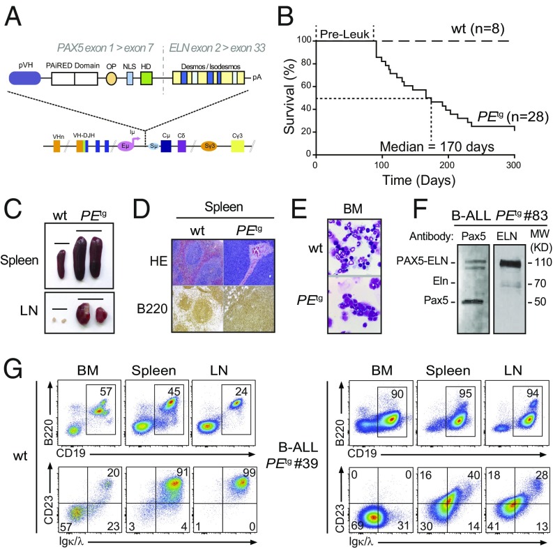
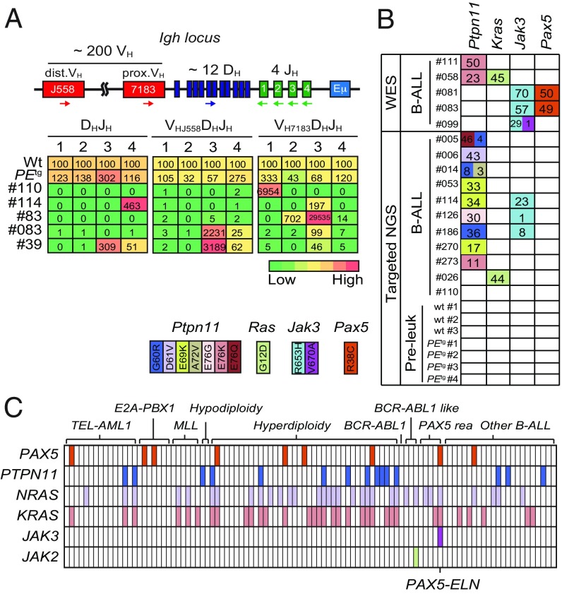
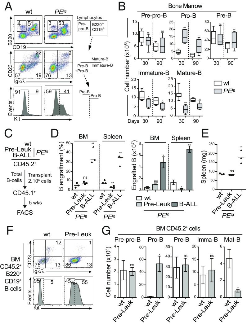
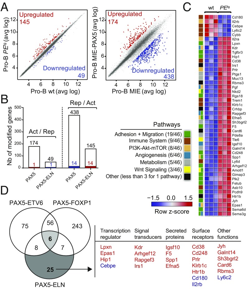
PAX5 is a well-known haploinsufficient tumor suppressor gene in human B-cell precursor acute lymphoblastic leukemia (B-ALL) and is involved in various chromosomal translocations that fuse a part of PAX5 with other partners. However, the role of PAX5 fusion proteins in B-ALL initiation and transformation is ill-known. We previously reported a new recurrent t(7;9)(q11;p13) chromosomal translocation in human B-ALL that juxtaposed PAX5 to the coding sequence of elastin (ELN). To study the function of the resulting PAX5-ELN fusion protein in B-ALL development, we generated a knockin mouse model in which the PAX5-ELN transgene is expressed specifically in B cells. PAX5-ELN-expressing mice efficiently developed B-ALL with an incidence of 80%. Leukemic transformation was associated with recurrent secondary mutations on Ptpn11, Kras, Pax5, and Jak3 genes affecting key signaling pathways required for cell proliferation. Our functional studies demonstrate that PAX5-ELN affected B-cell development in vitro and in vivo featuring an aberrant expansion of the pro-B cell compartment at the preleukemic stage. Finally, our molecular and computational approaches identified PAX5-ELN-regulated gene candidates that establish the molecular bases of the preleukemic state to drive B-ALL initiation. Hence, our study provides a new in vivo model of human B-ALL and strongly implicates PAX5 fusion proteins as potent oncoproteins in leukemia development.

Keywords: B-cell acute lymphoblastic leukemia; PAX5 fusion proteins; engineered mouse models; leukemia initiation; oncogenic transformation.


Conflict of interest statement

The authors declare no conflict of interest.

Figures

Fig. 1.
Fig. 1.
Human PAX5-ELN expression induces efficient B-ALL development. (A) Generation of knockin mouse model expressing PAX5-ELN fusion protein. The sequence encoding the human PAX5-ELN fusion protein was inserted downstream of Eμ enhancer. Desmos, Desmosine; HD, homeodomain; OP, octapeptide; pVH, VH gene promoter. (B) Kaplan–Meier curves of the time to leukemia for cohorts of PEtg mice (n = 28). WT mice (n = 8) were used as controls. Pre-Leuk, preleukemic time. (CE) PAX5-ELN induces B-ALL development characterized by leukemic cell invasion in the bone marrow, spleen, and lymph nodes. (C) Pictures of spleens (Upper) and LNs (Lower) from WT and leukemic PEtg mice are shown. (D) Staining with hematoxylin and eosin (HE) and immunohistochemistry of B220 are shown of spleens from WT and leukemic PEtg mice. (E) Pictures of May-Grünwald–Giemsa–stained cytospin of BM cells from WT and leukemic PEtg mice. (F) Protein extract of leukemic cells from a B-ALL PEtg mouse (no. 83) was subjected to immunoblotting with anti–N-terminal Pax5 (N19) antibody for the detection of PAX5-ELN and endogenous Pax5 and with anti-ELN antibody for the detection of PAX5-ELN and endogenous Eln. (G) Total cells from the BM, spleen, and LNs of WT (Left) and leukemic PEtg (Right) mouse (no. 39) were immunophenotyped using the B220, CD19, CD23, and Igκ/λ markers.
Fig. 2.
Fig. 2.
Clonal selection in PAX5-ELN–induced B-ALL. (A) Schematic of the mouse IgH locus (Upper). The locus is composed of ∼200 VH (red boxes), ∼12 DH (blue), and 4 JH (green) segments that undergo recombination in B-cell precursors to produce a functional VHDHJH unit. Genomic DNAs were prepared from purified PEtg pro-B cells and five leukemic PEtg mice and subjected to quantitative PCR to quantify DHJH and VHDHJH rearrangements using primers that bind the indicated gene segments. dVH, distal VH; pVH, proximal VH. PCR of the HS5 element downstream of the 3′ regulatory region was performed for normalization of DNA input. PCR was performed in triplicate. Quantification of DHJH and VHDHJH rearrangements is represented as a heat map (Lower). DNA from purified WT pro-B cells was used as a control and normalization (100% of the signal) for each gene rearrangement ranked in each column. (B) Recurrent mutations in B-ALL blasts induced by PAX5-ELN. Whole-exome sequencing (WES) was performed on BM cells from five leukemic PEtg mice. Mutations found on Ptpn11, Kras, Pax5, and Jak3 genes were verified and screened for recurrence by targeted next-generation sequencing (NGS) on BM cells from 11 leukemic, 3 WT, and 4 preleukemic 30-d-old PEtg mice. Each row represents a leukemia sample, and each column represents a genetic alteration. Colors indicate the position of the mutation, and numbers represent the variant allele frequency of each mutation. (C) Mutations on PTPN11, KRAS, NRAS, JAK3, JAK2, and PAX5 genes were screened for recurrence on 101 B-ALL patient samples. Each column represents a B-ALL sample classified according to the indicated oncogenic subtypes, and each row represents a genetic alteration. Colored boxes indicate the presence of a mutation, listed in Dataset S1.
Fig. 3.
Fig. 3.
Pro-B cells are expanded by PAX5-ELN at a preleukemic phase. (A and B) Preleukemic PEtg pro-B cells exhibit an aberrant expansion potential in vivo. Immunophenotyping of BM cells from 30-d-old WT and PEtg mice was performed using B220, CD19, CD23, Igκ/λ, and Kit markers (A, Left), and a gating strategy was applied to discriminate B-cell subpopulations (A, Right and SI Appendix, Fig. S2). Absolute numbers of pre-pro-B, pro-B, pre-B, immature B, and mature B cells from the BM of 30- and 90-d-old WT and PEtg mice were calculated (B; n = 5 to 8 mice per condition). (CE) Preleukemic and leukemic PEtg B cells exhibit different engraftment potentials. Total B cells from the BM of preleukemic and B-ALL PEtg mice (CD45.2+) were purified and i.v. transplanted in recipient mice (CD45.1+, n = 3, 2.106 cells per mouse) pretreated with 30 mg/kg busulfan (C). The same number of total B cells from the BM of WT mice was transplanted in parallel as control (C; n = 3). The proportion (D, Left) and absolute numbers (D, Right) of donor-derived (CD45.2+) B cells in the recipient BM were calculated 5 wk after transplantation. Spleen weights of engrafted mice were compared (E). (F and G) Preleukemic PEtg pro-B cells expand after transplantation. Immunophenotyping of engrafted B cells (CD45.2+B220+CD19+) in the BM was performed (F), and absolute numbers of donor-derived (CD45.2+) preleukemic pre-pro-B, pre-B, pro-B, immature B, and mature B cells in the recipient BM were calculated (G). Error bars represent standard deviations, ***P < 0.0005, **P < 0.005, and *P < 0.05; ns, nonsignificant.
Fig. 4.
Fig. 4.
(A) Scatter plot of gene expression differences between in vivo purified WT and PEtg preleukemic pro-B cells (Left) and between ex vivo E17.5 fetal liver Pax5−/− pro-B cells transduced with either MIE-PAX5 or MIE retroviral vectors (Right) based on three independent microarray experiments. The normalized expression data of individual coding genes (indicated by dots) were plotted as the average log ratio (avg log). Up- and down-regulated genes with an expression difference of >1.5- and >3-fold, and an adjusted P value of <0.05, are colored in red or blue, respectively. (B) Absence of a general dominant-negative effect of PAX5-ELN on ex vivo PAX5-regulated genes in pro-B cells. Comparison of PAX5-activated and PAX5-ELN–repressed genes (Left) and of PAX5-repressed and PAX5-ELN–activated genes (Right) in preleukemic pro-B cells. Overlap indicates that one gene was activated by PAX5 and repressed by PAX5-ELN and 14 genes of the PAX5-repressed genes were activated by PAX5-ELN as represented by colored bars. (C) Heat map displaying the differential expression of PAX5-ELN–activated (red) and –repressed (blue) genes in WT (n = 3) and PEtg (n = 3) pro-B cells. The 46 PAX5-ELN–modified genes were selected on the basis of an expression difference of >2-fold (P < 0.05) and for encoding a protein implicated in one of the indicated pathways. The expression value of each gene is visualized according to the indicated scale. The pathway annotation is shown (Left). (D) Venn diagram indicating the overlap between PAX5-ETV6–, PAX5-FOXP1– (15), and PAX5-ELN–modified genes in preleukemic pro-B cells, selected for an expression difference of >3-, 3-, and 2-fold, respectively (Left). PAX5-ELN–modified genes that were included neither in PAX5-ETV6 nor in PAX5-FOXP1 signatures are listed according to their biological functions as transcriptional regulators, signal transducers, secreting proteins, and surface receptors. PAX5-ELN–activated and –repressed genes are indicated in red and blue, respectively.

Similar articles

Cited by

References

    1. Pui CH, Carroll WL, Meshinchi S, Arceci RJ. Biology, risk stratification, and therapy of pediatric acute leukemias: An update. J Clin Oncol. 2011;29:551–565. - PMC - PubMed
    1. Armstrong SA, Look AT. Molecular genetics of acute lymphoblastic leukemia. J Clin Oncol. 2005;23:6306–6315. - PubMed
    1. Hardy RR, et al. B-cell commitment, development and selection. Immunol Rev. 2000;175:23–32. - PubMed
    1. Adams B, et al. Pax-5 encodes the transcription factor BSAP and is expressed in B lymphocytes, the developing CNS, and adult testis. Genes Dev. 1992;6:1589–1607. - PubMed
    1. Urbánek P, Wang ZQ, Fetka I, Wagner EF, Busslinger M. Complete block of early B cell differentiation and altered patterning of the posterior midbrain in mice lacking Pax5/BSAP. Cell. 1994;79:901–912. - PubMed

Publication types

MeSH terms

LinkOut - more resources