. 2018 Oct 9;115(41):E9560-E9569.
doi: 10.1073/pnas.1810648115. Epub 2018 Sep 26.

ATP hydrolysis-coupled peptide translocation mechanism of Mycobacterium tuberculosis ClpB

Affiliations

ATP hydrolysis-coupled peptide translocation mechanism of Mycobacterium tuberculosis ClpB

Hongjun Yu et al. Proc Natl Acad Sci U S A. .

Abstract

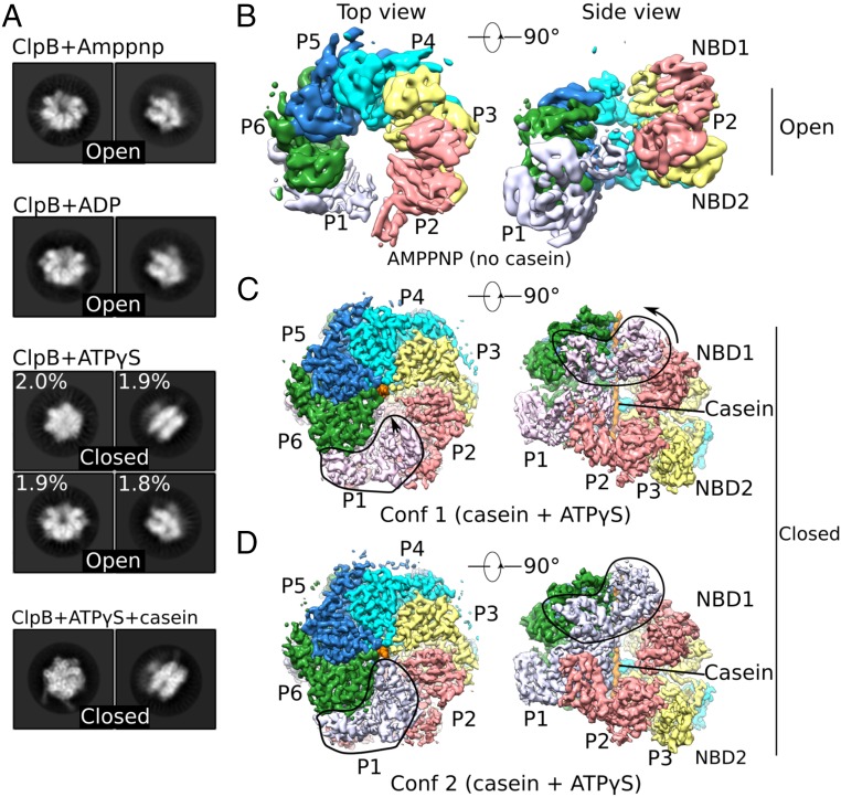
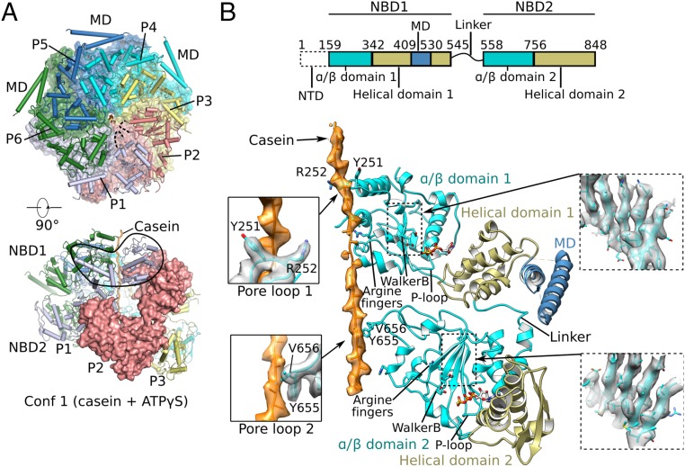
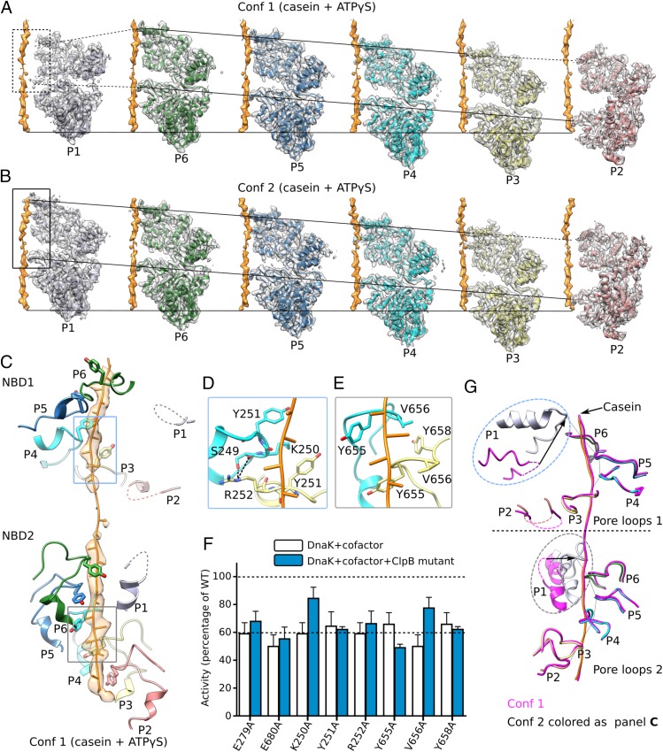
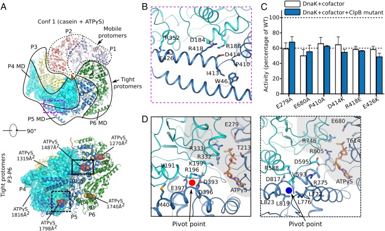
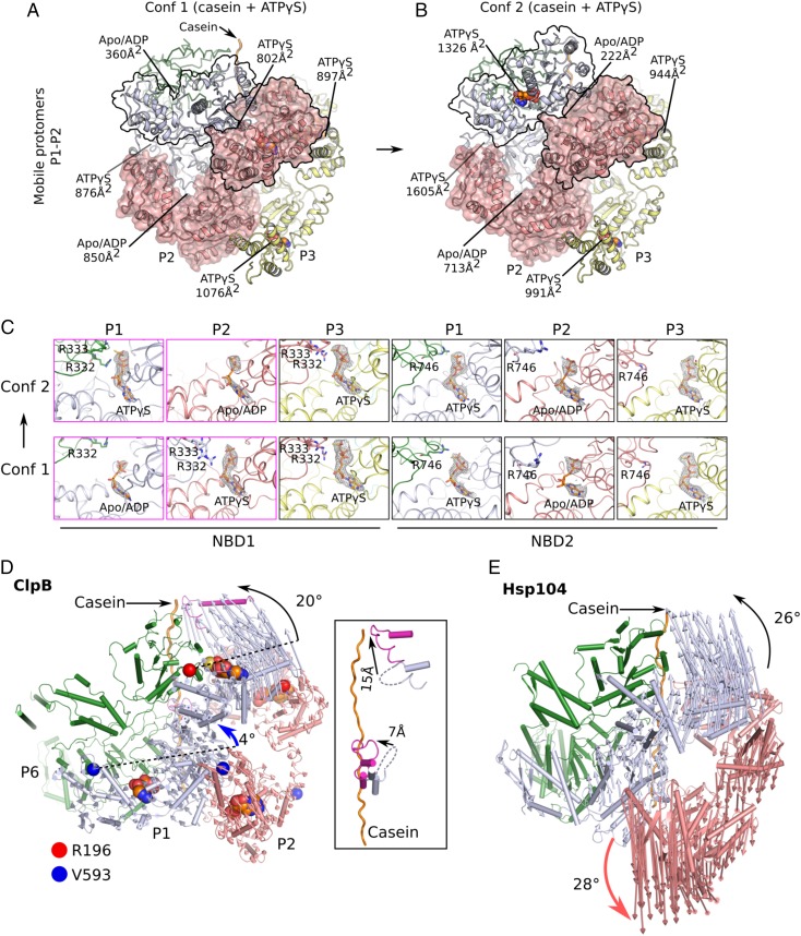
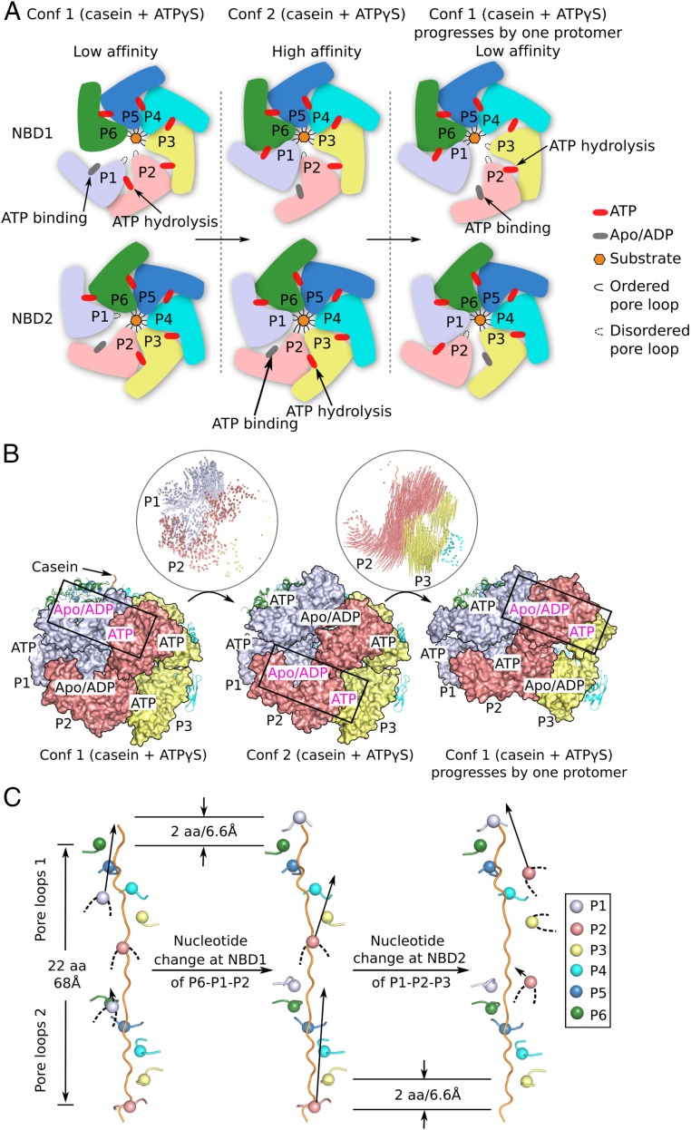
The protein disaggregase ClpB hexamer is conserved across evolution and has two AAA+-type nucleotide-binding domains, NBD1 and NBD2, in each protomer. In M. tuberculosis (Mtb), ClpB facilitates asymmetric distribution of protein aggregates during cell division to help the pathogen survive and persist within the host, but a mechanistic understanding has been lacking. Here we report cryo-EM structures at 3.8- to 3.9-Å resolution of Mtb ClpB bound to a model substrate, casein, in the presence of the weakly hydrolyzable ATP mimic adenosine 5'-[γ-thio]triphosphate. Mtb ClpB existed in solution in two closed-ring conformations, conformers 1 and 2. In both conformers, the 12 pore-loops on the 12 NTDs of the six protomers (P1-P6) were arranged similarly to a staircase around the bound peptide. Conformer 1 is a low-affinity state in which three of the 12 pore-loops (the protomer P1 NBD1 and NBD2 loops and the protomer P2 NBD1 loop) are not engaged with peptide. Conformer 2 is a high-affinity state because only one pore-loop (the protomer P2 NBD1 loop) is not engaged with the peptide. The resolution of the two conformations, along with their bound substrate peptides and nucleotides, enabled us to propose a nucleotide-driven peptide translocation mechanism of a bacterial ClpB that is largely consistent with several recent unfoldase structures, in particular with the eukaryotic Hsp104. However, whereas Hsp104's two NBDs move in opposing directions during one step of peptide translocation, in Mtb ClpB the two NBDs move only in the direction of translocation.

Keywords: AAA-ATPase; Mycobacterium tuberculosis; cryo-EM; disaggregase; proteostasis.


Conflict of interest statement

The authors declare no conflict of interest.

Figures

Fig. 1.
Fig. 1.
Cryo-EM analyses of multiple states of Mtb ClpB disaggregase. (A) Representative 2D images of the ClpB asymmetric hexamer (Left: top view; Right: side view) in the presence of different nucleotides with or without casein substrate. (BD) Cryo-EM maps of different trapped conformations of ClpB, segmented by individual protomers. B shows an open left-handed spiral structure of ClpB in the presence of AMP-PNP. C and D show two closed conformations of ClpB in a complex with casein and ATPγS. Outlined regions exhibit major conformational differences between these two conformations. Conf 1, conformer 1; Conf2, conformer 2.
Fig. 2.
Fig. 2.
Cryo-EM structure of conformer 1 of Mtb ClpB complexed with ATPγS and casein. (A) Two orthogonal views of the atomic model of conformer 1 (corresponding to Fig. 1C). (Upper) The NBD2 ring is shown, overlapped with a semitransparent surface view. (Lower) Side view of conformer 1 with one protomer (P2) shown in surface view. (B, Upper) The domain architecture of ClpB. (Lower) Detailed structural elements of protomer P4 are shown in cartoon representation. The two α/β subdomains are shown in cyan, two helical subdomains in olive, MD domain in dark blue, and casein peptide in orange with electron density superimposed in semitransparent surface. Key features such as arginine fingers, Walker B motifs, P loops, and the linker between NBD1 and NBD2 are labeled. (Left Insets) Enlarged views of the interactions between the casein peptide and pore-loop 1 (Upper) and pore-loop 2 (Lower). (Right Insets) Electron density of the middle β-sheet of NBD1 (Upper) and NBD2 (Lower).
Fig. 3.
Fig. 3.
Interactions between ClpB and bound substrate mimic casein. (A and B) Individual protomers within conformer 1 (Conf 1) (A) and conformer 2 (Conf 2) (B) are shown separately with a 60° rotation around the bound casein, superposed with their corresponding cryo-EM densities. The casein peptide was aligned across panels (horizontal solid lines) to show the relative height of each protomer in the hexamer. The two slanted lines in each panel connect the pore-loops 1 and 2 across individual protomers, respectively. The dashed lines connect to disordered loops. In both conformations, protomers P3–P6 were fully engaged with the casein through their respective pore-loops 1 and 2, and the tyrosine sidechains of all eight pore-loops had good densities (Fig. 2B). Furthermore, protomer P2 contacted casein via pore-loop 2, but its pore-loop 1 was disordered, so P2 was partially engaged with the peptide. Both pore-loops of P1 were disordered or unengaged in conformer 1 but become fully engaged in conformer 2. (C) In conformer 1, the pore-loops inside the central chamber surrounded and bound the substrate casein in a right-handed spiral mode. This spiral was discontinued at pore-loops 1 of protomers P1 and P2 in the NBD1 ring and at pore-loop 2 of protomer P1 in the NBD2 ring; these pore-loops were flexible and did not contact the substrate. (D) Interactions among neighboring pore-loops 1 and the substrate. Y251 stacked with the casein main chain while R252 H-bonded with S249 of neighboring pore-loops to stabilize the right-handed spiral arrangement. (E) Interactions among neighboring pore-loops 2 and the substrate. Y655 and V656 stacked with the peptide while V656 stacked with Y658 of neighboring loops to stabilize their spiral arrangement. (F) Mutational analysis of the key residues in the pore-loops using a luciferase-based model protein aggregate reactivation assay. The Walker B mutations E279A and E680A were used as negative controls. Data were normalized to reactions containing wild-type ClpB, Hsp20, and DnaK along with cofactors DnaJ1, DnaJ2, and GrpE, which together reactivate denatured luciferase. The dashed line at 60% of wild-type activity defines the yield of reactions lacking ClpB (white bars), indicating the background refolding activity of DnaK and cofactors alone. Hence, reactions with ClpB mutants (blue bars) that show about 60% activity are defined as nonfunctional. (G) A comparison of pore-loops in conformer 1 (magenta) and conformer 2 (colored by protomers as in C). The two conformations were aligned by their respective caseins. Dashed curves mark disordered loops.
Fig. 4.
Fig. 4.
Protomer organization in conformer 1 (Conf 1) of Mtb ClpB bound to casein. (A) Mobile (protomers P1 and P2; dashed protomer boundary) vs. tightly packed protomers (protomers P3–P6; solid boundary) in conformer 1. (B) Three pairs of salt bridges between MD motif 1 of protomer P5 and NBD1 of the neighboring protomer P4 in the ClpB region marked by the purple dashed box in A. A hydrophobic region formed by P410, I413, and W463 at the right end of coiled-coil motif 1 might facilitate the relative rotation between motif 1 and motif 2 (not shown) of the MD domain. (C) Impact of the mutation of key residues in the MD domain on ClpB activity. The same Walker B mutations E279A and E680A shown in Fig. 3F were used as negative controls. Refer to Fig. 3F for a description of this assay. (D) Close-up views of the interfaces between the two NBD1 domains (Left) and two NBD2 domains (Right) of protomers P4 and P5 in regions marked by the solid-outlined and dashed-outlined boxes in A, Lower, respectively. The NBD1–NBD1 and NBD2–NBD2 interfaces were similar; both involved the ATP-binding pocket (shaded in gray) that interacted with the arginine finger from a neighboring protomer and a helix of the small helical domain that interacted with a loop from a neighboring protomer, either by salt bridges in NBD1 (R196 vs. D393/D396/E397) or by hydrophobic packing in NBD2 (V593 vs. L772/L776/L819/L823).
Fig. 5.
Fig. 5.
Nucleotide states and structural changes between conformers 1 and 2 of Mtb ClpB bound to casein. (A and B) The different arrangement of the mobile protomers P1 and P2 in conformer 1 (Conf 1) (A) and conformer 2 (Conf 2) (B). The contact areas of NBD1–NBD1 and NBD2–NBD2 and the bound-nucleotide states are labeled. (C) A comparison of the bound nucleotides in NBD1 and NBD2 of each protomer between conformers 1 and 2, with all of the densities displayed at the same threshold. The nucleotide states in protomers P4–P6 which bound ATPγS in both NBD1 and NBD2 are shown in SI Appendix, Fig. S5. (D) Structural changes from conformer 1 (cartoon view) to conformer 2 (hidden) are revealed by aligning their respective bound peptides. Vectors connect the same structural elements in the two conformers. Changes occurred primarily in protomer P1 (light blue), much less in protomer P2 (salmon), and were negligible in protomers P3–P6. Changes in protomers P1 and P2 between conformers 1 and 2 were described as rigid-body rotations of NBD1 and NBD2 around their respective pivotal points. The pivot point residues are marked by a red or a blue circle, respectively, as in Fig. 4D. The Inset shows the pore-loop positions in the two conformers. (E) Structural changes of the eukaryotic Hsp104 from the closed state (PDB ID code 5VJH; shown in cartoon) to the extended state (PDB ID code 5VYA) are illustrated by the vectors connecting the same residues in the two states. The bidirectional movement of the two neighboring protomers, as highlighted by the curved black and red arrows, creates a transient gap stage in both the NBD1 and NBD2 rings in Hsp104.
Fig. 6.
Fig. 6.
A putative three-stage, rotary sequential nucleotide-driven substrate translocation mechanism of Mtb ClpB. (A) A sketch of the proposed mechanism in which the hexamer progresses from stage 1 (conformer 1) to stage 2 (conformer 2) and finally to stage 3. Stage progression is driven by ATP binding and hydrolysis in NBD1 and NBD2 of the two mobile protomers. (B) Atomic trajectories of the progressive conformational changes between stage 1 and 2 and between stage 2 and 3 as illustrated in A. To generate the model for stage 3, conformer 1 was superposed with conformer 2 by aligning protomer P5 in conformer 1 onto protomer P6 in conformer 2. (C) Unidirectional and progressive rearrangements of the 12 pore-loops as the ClpB hexamer goes through the three stages. From stage 1 (Left) to stage 2 (Middle), protomer P1 transits from the unengaged state with none of pore-loops 1 and 2 contacting casein to the fully engaged state with both pore-loops 1 and 2 contacting casein. This is caused by the ATP rebinding to NBD1 of protomer P1 and ATP hydrolysis by NBD1 of protomer P2. From stage 2 (Middle) to stage 3 (Right), protomer P2 transits from a partially engaged state with only pore-loop 2 contacting casein to an unengaged state with neither pore-loop 1 nor 2 contacting casein, and protomer P3 transits from a fully engaged state to a partially engaged state. This step may be caused by ATP rebinding to NBD2 of P2 and ATP hydrolysis by NBD2 of P3. The net effect of going through these three stages is a counterclockwise propagation of conformer 1 by one protomer. Continued cycling through the three stages leads to a rotary sequential model for substrate translocation in Mtb ClpB. Conf 1, conformer 1; Conf 2, conformer 2.

Similar articles

Cited by

References

    1. Doyle SM, Genest O, Wickner S. Protein rescue from aggregates by powerful molecular chaperone machines. Nat Rev Mol Cell Biol. 2013;14:617–629. - PubMed
    1. Mogk A, et al. Identification of thermolabile Escherichia coli proteins: Prevention and reversion of aggregation by DnaK and ClpB. EMBO J. 1999;18:6934–6949. - PMC - PubMed
    1. Doyle SM, Wickner S. Hsp104 and ClpB: Protein disaggregating machines. Trends Biochem Sci. 2009;34:40–48. - PubMed
    1. Lupoli TJ, Fay A, Adura C, Glickman MS, Nathan CF. Reconstitution of a Mycobacterium tuberculosis proteostasis network highlights essential cofactor interactions with chaperone DnaK. Proc Natl Acad Sci USA. 2016;113:E7947–E7956. - PMC - PubMed
    1. Weibezahn J, et al. Thermotolerance requires refolding of aggregated proteins by substrate translocation through the central pore of ClpB. Cell. 2004;119:653–665. - PubMed

Publication types

MeSH terms