. 2019 Jan;17(1):289-298.
doi: 10.1158/1541-7786.MCR-18-0364. Epub 2018 Sep 26.

Targeting of SGK1 by miR-576-3p Inhibits Lung Adenocarcinoma Migration and Invasion

Affiliations

Targeting of SGK1 by miR-576-3p Inhibits Lung Adenocarcinoma Migration and Invasion

Evan J Greenawalt et al. Mol Cancer Res. 2019 Jan.

Abstract

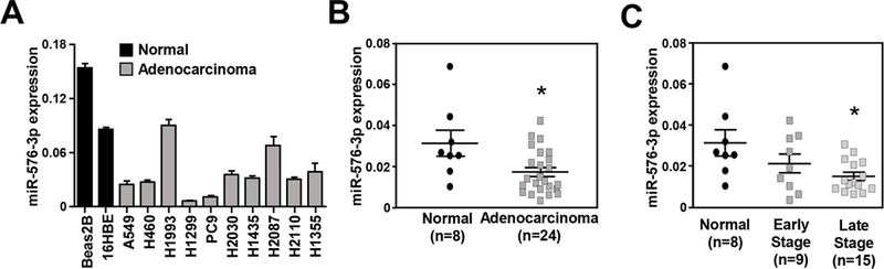
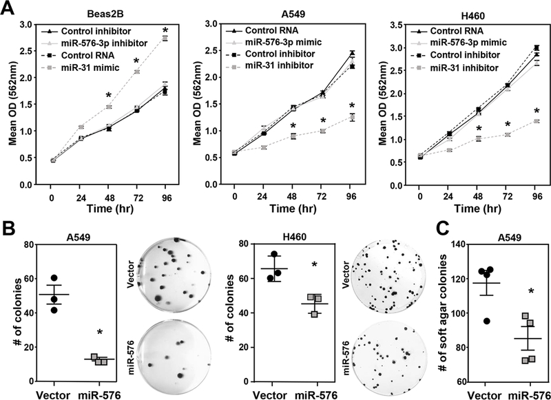
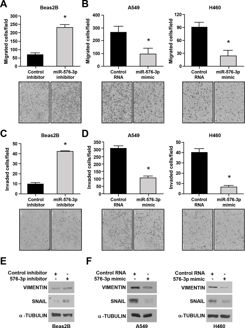
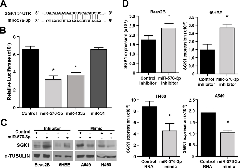
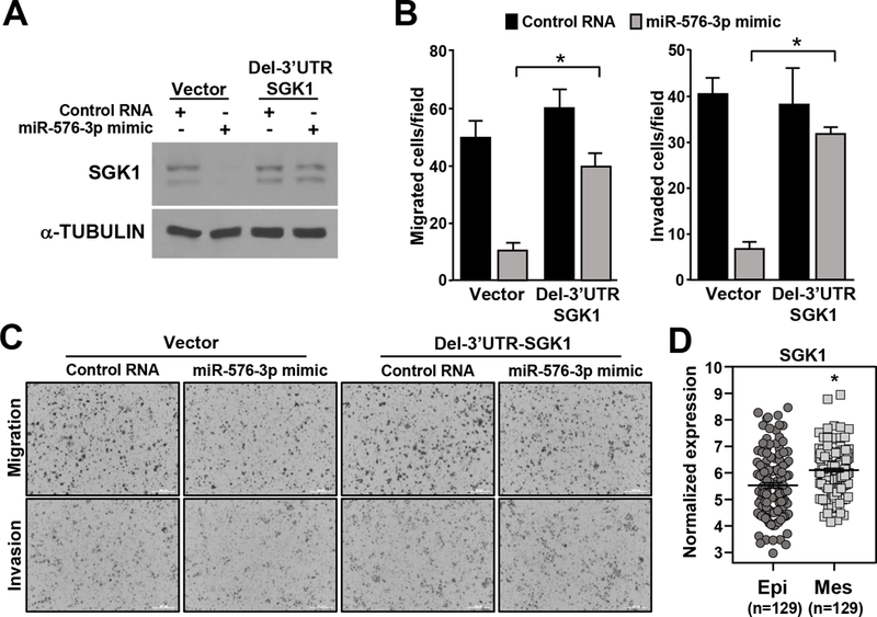
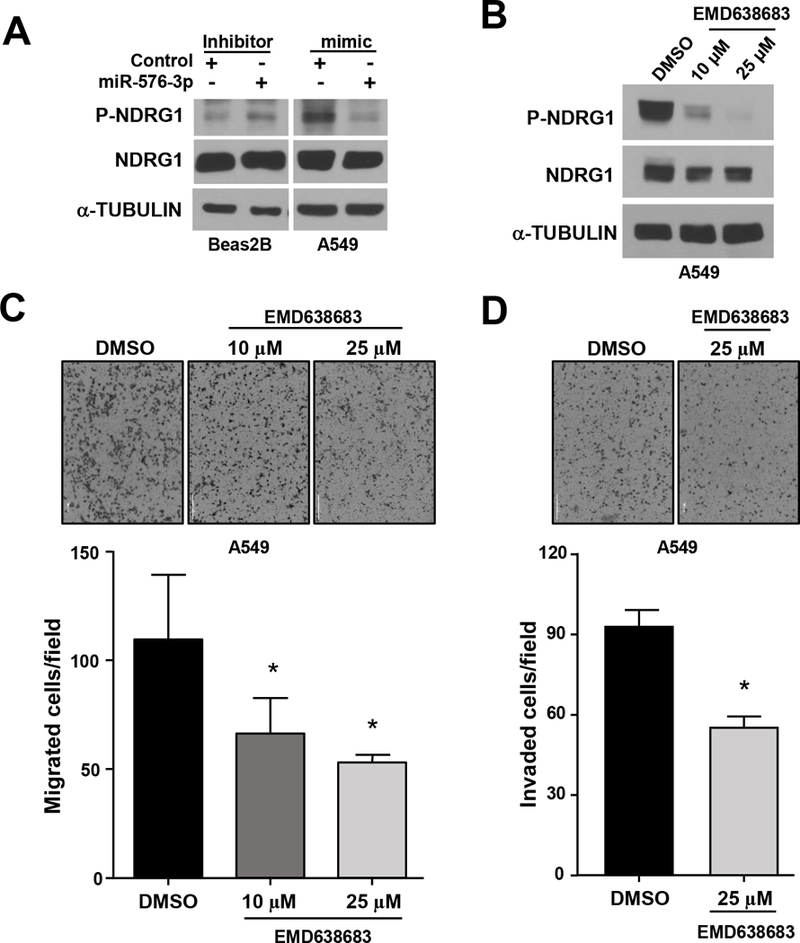
Metastatic lung cancer is common in patients with lung adenocarcinoma, but the molecular mechanisms of metastasis remain incompletely resolved. miRNA regulate gene expression and contribute to cancer development and progression. This report identifies miR-576-3p and its mechanism of action in lung cancer progression. miR-576-3p was determined to be significantly decreased in clinical specimens of late-stage lung adenocarcinoma. Overexpression of miR-576-3p in lung adenocarcinoma cells decreased mesenchymal marker expression and inhibited migration and invasion. Inhibition of miR-576-3p in nonmalignant lung epithelial cells increased migration and invasion as well as mesenchymal markers. Serum/glucocorticoid-regulated kinase 1 (SGK1) was a direct target of miR-576-3p, and modulation of miR-576-3p levels led to alterations in SGK1 protein and mRNA as well as changes in activation of its downstream target linked to metastasis, N-myc downstream regulated 1 (NDRG1). Loss of the ability of miR-576-3p to bind the 3'-UTR of SGK1 rescued the inhibition in migration and invasion observed with miR-576-3p overexpression. In addition, increased SGK1 levels were detected in lung adenocarcinoma patient samples expressing mesenchymal markers, and pharmacologic inhibition of SGK1 resulted in a similar inhibition of migration and invasion of lung adenocarcinoma cells as observed with miR-576-3p overexpression. Together, these results reveal miR-576-3p downregulation is selected for in late-stage lung adenocarcinoma due to its ability to inhibit migration and invasion by targeting SGK1. Furthermore, these results also support targeting SGK1 as a potential therapeutic for lung adenocarcinoma. IMPLICATIONS: This study reveals SGK1 inhibition with miR-576-3p or pharmacologically inhibits migration and invasion of lung adenocarcinoma, providing mechanistic insights into late-stage lung adenocarcinoma and a potential new treatment avenue.


Conflict of interest statement

Conflict of interest: The authors declare no conflicts of interest.

Figures

Figure 1.
Figure 1.. miR-576–3p expression is downregulated in human lung adenocarcinoma.
(A) qRT-PCR analysis of miR-576–3p in two bronchial epithelial cell lines and a panel of lung adenocarcinoma cell lines; (triplicates) SEM. (B) qRT-PCR analysis of normal human lung and lung adenocarcinoma patient samples; (triplicates) SEM; *P=5.8×10⁻³, two-tailed t-test. (C, D) qRT-PCR data from B grouped as early-stage (I & II) and late-stage (III & IV) (C) or separated into tumor stage (D); (triplicates) SEM; late-stage, C, *P=3×10⁻³; Stage III, D, *P=8.3×10⁻³; Stage IV, D, *P=0.038, one-tailed t-test. All data are relative to the endogenous control RNU6B small RNA.
Figure 2.
Figure 2.. miR-576–3p does not affect proliferation, but inhibits colony formation.
(A) The indicated cell lines were transfected with miR-576–3p mimic, miR-576–3p inhibitor, or controls, including a miR-31 mimic and inhibitor as a positive control. MTT assays (quadruplicate) were performed at the indicated intervals; SEM, *P<0.001, two-tailed t-test. (B, C) Colony formation (B) and soft agar (C) assays (both triplicates) were performed with the indicated lung adenocarcinoma cells retrovirally overexpressing miR-576 or scrambled control. Representative images (10×) shown. Data are representative of three independent experiments; SEM; B, A549 *P=7.0×10⁻³; H460 *P=1.9×10⁻²; C, *P<0.02, two-tailed t-test.
Figure 3.
Figure 3.. miR-576–3p alters mesenchymal marker expression and inhibits migration and invasion.
The indicated cell lines were transfected with the miR-576–3p inhibitor (A, C, E), miR-576–3p mimic (B, D, F), or control. (A-D) Transwell migration and invasion assays were performed; SEM; A, *P=9.7×10⁻³; B, A549, *P=0.028; H460, *P=2.0×10⁻³; C, *P=1.7×10⁻³; D, A549, *P=2.5×10⁻³; H460, *P=1.6×10⁻⁴; two-tailed t-test. Representative images shown (10×). (E, F) Western blots for the indicated proteins were performed. Data are representative of at least three independent experiments.
Figure 4.
Figure 4.. SGK1 is a direct target of miR-576–3p.
(A) Schematic of the SGK1 3’-UTR binding site for miR-576–3p. (B) Luciferase activity (triplicates) was measured after transfection of the indicated miRNA mimic or control RNA into 293T cells; SEM; miR-576–3p, *P=8.8×10⁻⁴; miR-133b, *P=5.9×10⁻⁴, two-tailed t-test. miR-133b and miR-31 mimics served as positive and negative controls, respectively. (C, D) Western blots (C) and qRT-PCR (triplicate, D) were performed with cells transfected with miR-576–3p mimic, miR-576–3p inhibitor, or controls; SEM; Beas2B, *P=0.021; 16HBE, *P=8.7×10⁻³; H460, *P=8.3×10⁻³; A549, *P=2.4×10⁻³, two-tailed t-test. Data are representative of three independent experiments.
Figure 5.
Figure 5.. Deletion of the miR-576–3p binding site in SGK1 rescues the inhibition of migration and invasion from miR-576–3p.
H460 cells infected with an empty retrovirus or a retrovirus expressing SGK1 lacking its 3’-UTR (Del-3’UTR SGK1) were transfected with miR-576–3p mimic or control RNA. (A) Western blots were performed for the proteins indicated. (B) Transwell migration and invasion assays were performed; SEM; migration, *P=8.9×10⁻⁶; invasion, *P=1.6×10⁻⁷ two-tailed t-test. (C) Representative images of B (10×). Data are representative of three independent experiments. (D) SGK1 expression in TCGA RNA-seq data divided into mesenchymal and epithelial subtypes, *P<1.43 ×10⁻⁵.
Figure 6.
Figure 6.. Pharmacological SGK inhibition prevents downstream target phosphorylation and cell migration and invasion.
(A) The indicated cells were transfected with miR-576–3p mimic, inhibitor, or controls. Phospho-NDRG1 (Thr346) and total NDRG1 levels were determined by Western blot. (B-D) A549 cells were treated with vehicle control (DMSO) or with EMD638683 at the concentrations indicated. Western blots (B) and transwell migration (C) and invasion (D) assays performed (representative images shown, 4×); SEM; C, 10μM *P=0.045, 25μM *P=0.025; D, *P=5.4×10⁻⁵, two-tailed t-test. Data are representative of three independent experiments.

Similar articles

Cited by

References

    1. Siegel RL, Miller KD, and Jemal A, Cancer statistics, 2018. CA Cancer J Clin 2018;68:7–30. - PubMed
    1. Peng Y and Croce CM, The role of MicroRNAs in human cancer. Signal Transduct Target Ther 2016;1:15004. - PMC - PubMed
    1. Inamura K and Ishikawa Y, MicroRNA In Lung Cancer: Novel Biomarkers and Potential Tools for Treatment. J Clin Med 2016;5. - PMC - PubMed
    1. Legras A, Pecuchet N, Imbeaud S, Pallier K, Didelot A, Roussel H, et al., Epithelial-to-Mesenchymal Transition and MicroRNAs in Lung Cancer. Cancers (Basel) 2017;9. - PMC - PubMed
    1. Edmonds MD, Boyd KL, Moyo T, Mitra R, Duszynski R, Arrate MP, et al., MicroRNA-31 initiates lung tumorigenesis and promotes mutant KRAS-driven lung cancer. J Clin Invest 2016;126:349–64. - PMC - PubMed

Publication types

MeSH terms

Substances