. 2019 Jan;17(1):277-288.
doi: 10.1158/1541-7786.MCR-18-0558. Epub 2018 Sep 26.

TGFβ1 Cell Cycle Arrest Is Mediated by Inhibition of MCM Assembly in Rb-Deficient Conditions

Affiliations

TGFβ1 Cell Cycle Arrest Is Mediated by Inhibition of MCM Assembly in Rb-Deficient Conditions

Brook S Nepon-Sixt et al. Mol Cancer Res. 2019 Jan.

Abstract

Transforming growth factor β1 (TGFβ1) is a potent inhibitor of cell growth that targets gene-regulatory events, but also inhibits the function of CDC45-MCM-GINS helicases (CMG; MCM, Mini-Chromosome Maintenance; GINS, Go-Ichi-Ni-San) through multiple mechanisms to achieve cell-cycle arrest. Early in G1, TGFβ1 blocks MCM subunit expression and suppresses Myc and Cyclin E/Cdk2 activity required for CMG assembly, should MCMs be expressed. Once CMGs are assembled in late-G1, TGFβ1 blocks CMG activation using a direct mechanism involving the retinoblastoma (Rb) tumor suppressor. Here, in cells lacking Rb, TGFβ1 does not suppress Myc, Cyclin E/Cdk2 activity, or MCM expression, yet growth arrest remains intact and Smad2/3/4-dependent. Such arrest occurs due to inhibition of MCM hexamer assembly by TGFβ1, which is not seen when Rb is present and MCM subunit expression is normally blocked by TGFβ1. Loss of Smad expression prevents TGFβ1 suppression of MCM assembly. Mechanistically, TGFβ1 blocks a Cyclin E-Mcm7 molecular interaction required for MCM hexamer assembly upstream of CDC10-dependent transcript-1 (CDT1) function. Accordingly, overexpression of CDT1 with an intact MCM-binding domain abrogates TGFβ1 arrest and rescues MCM assembly. The ability of CDT1 to restore MCM assembly and allow S-phase entry indicates that, in the absence of Rb and other canonical mediators, TGFβ1 relies on inhibition of Cyclin E-MCM7 and MCM assembly to achieve cell cycle arrest. IMPLICATIONS: These results demonstrate that the MCM assembly process is a pivotal target of TGFβ1 in eliciting cell cycle arrest, and provide evidence for a novel oncogenic role for CDT1 in abrogating TGFβ1 inhibition of MCM assembly.


Conflict of interest statement

The authors declare no financial or other conflicts of interest

Figures

Figure 1:
Figure 1:. TGFß1 inhibits cell cycle progression of Rb-deficient cells in a Smad-dependent manner.
(A) Diagram showing experimental design and timing of cell cycle events in MK cells. (B) EGF-synchronized wild type MK or MK(Rb-) cells were assessed for TGFß1-induced growth suppression when exposed to TGFß1 in early-G1. BrdU incorporation in early S-phase was used to determine nuclear labeling percentages. Results are means from triplicate fields of >200 nuclei analyzed, +/− 1 s.d., with similar results obtained in a second biological replicate experiment. Statistical analyses are two-sided Students t-tests; two stars denote p<0.01 (labeling used for statistics throughout report). (C) Asynchronous cells were assessed for total Rb protein levels by immunoblotting complete cell lysates. Actin was probed as a loading control. (D) Synchronized MK(Rb-) cells were transfected with siRNAs against Luciferase (Luc), Smad3, or Smad4 during the EGF withdrawal interval and assessed by immunoblots at time of release (G0) or at the G1/S transition (12 hr) for protein expression. Antibodies used are indicated at left. (E) Synchronized MK(Rb-) cells were assessed for their ability to enter S-phase after exposure to indicated siRNAs, and with or without TGFß1 addition in early-G1. Non-transfection control (no Tfx) was also compared. BrdU incorporation assays were performed in early S-phase. Results are means from triplicate fields of >200 cells, +/− 1 s.d, normalized to non-TGFß1-treated controls (assigned at 100%). Similar results were obtained in a second biological replicate experiment. (F) Immunofluorescent images of representative fields for the data collected in panel E. (G) Synchronized MK(Rb-) cells were transfected with siRNAs against Luciferase or Smad2 during EGF withdrawal, and assessed by immunoblots at time of release (G0) or at the G1/S transition (12 hr) for protein expression in total lysates. Antibodies used are at right. (H) Cells treated as in panel G were assessed for their ability to enter S-phase, with or without TGFß1 added in early-G1. Non-transfection control (No Tfx) was also compared. BrdU incorporation assays were performed in early S-phase. Results are means from triplicate fields of >200 cells, +/− 1 s.d, normalized to non-TGFß1-treated controls (assigned at 100%). Similar results were obtained in a second biological replicate experiment.
Figure 2:
Figure 2:. TGFß1-induced growth arrest of MK(Rb-) cells is not due to inhibition of c-Myc, CycE/Cdk2, or MCM expression.
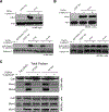
(A) Synchronized wild type MK cells exposed to TGFß1 in early-G1 were assessed at time of release and in late-G1 for c-Myc protein expression (top, total protein immunoblots) and Cyclin E/Cdk2 kinase activity (bottom, Histone H1 substrate). Input Histone H1 was stained with Coomassie blue as a loading control. Rabbit IgG was used as a negative control for the immunoprecipitation. (B) Synchronized MK(Rb-) cells were assessed by the same experimental procedure as in panel A. Similar results were obtained in three independent experiments for panels A&B. (C) Synchronized MK or MK(Rb-) cells were exposed to TGFß1 in early-G1 and assessed by immunoblotting for the expression of MCM subunits at time of release and in late-G1. Cdt1 was unaltered and used as a loading control. Results are representative of three independent experiments with similar findings.
Figure 3:
Figure 3:. TGFß1 induces cell cycle arrest in Rb-deficient cells by inhibiting MCM assembly and Cyclin E-Mcm7 interactions.
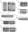
(A) Synchronized MK(Rb-) cells were exposed to TGFß1 in early-G1 and assessed in late-G1 by immunoblotting for protein expression, chromatin recruitment, and DNA loading status of Mcm2–7, Cdt1, and Cdc6. Lysates from equal cell numbers are loaded in pairs. Antibodies used in blots are indicated at left. Results are representative of three independent experiments with similar findings. (B) Samples from panel A were used in a titration experiment to determine the approximate amount of Mcm2/7 bound to chromatin in TGFß1-treated MK(Rb-) cells. Control (no TGFß1) samples were diluted as indicated and immunoblotted against non-diluted samples treated with TGFß1. 100% indicates no dilution. Note that the ~50–60% dilutions of Mcm2/7 protein roughly match the intensity of TGFß1-treated Mcm2/7 signals. (C) Synchronized MK(Rb-) cells were exposed to TGFß1 in early-G1 and assessed for effects of Smad4 loss on MCM subunit expression and chromatin recruitment status. Total protein lysates or chromatin-bound fractions were collected in late-G1 (10 hr) for immunoblotting. siRNAs were transfected during the synchronization in EGF depleted medium. Antibodies used are at left. (D) MK(Rb-) cells were subjected to the same experimental conditions and immunoblotting as in panel C, but using si-Smad3. Histone H4 serves as a loading control. (E) Synchronized MK(Rb-) cells were exposed to TGFß1 in early-G1 and analyzed for chromatin-bound proteins in late-G1 by immunoblotting. Antibodies for analysis are at left. (F) Synchronized MK(Rb-) cells treated with TGFß1 in early-G1 were assessed in late-G1 for the interaction between Cyclin E and Mcm7. Anti-Cyclin E was used in the immunoprecipitation step (IP), followed by immunoblotting (IB) for associated Mcm7 or Cdk2. Input Mcm7 levels in total protein lysates are shown at the top. Rabbit IgG was used in the IP as a negative control. Similar results were found in an independent experiment. (G) MK(Rb-) cells were analyzed by immunoblotting in late-G1 for the effects of TGFß1 on the phosphorylation status of Cdc6. An antibody specific for phospho-serine residue 54 on Cdc6 was used on total cell lysates. Actin probing served as a loading control. Results are representative of three independent experiments with similar outcomes.
Figure 4:
Figure 4:. Cdt1 overexpression in Rb-depleted cells abrogates TGFß1 cell cycle arrest.
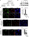
(A) Experimental design for the synchronization-transfection approach. MK(Rb-) cells were used in panels B-E. MK wild type cells were used in panels F&G. (B) Immunoblot showing kinetics of wild type HA-Cdt1 protein expression at indicated time points following release into G1 and S-phase. Anti-HA antibody was used to analyze ectopic Cdt1 levels against Actin probing as a loading control. (C) Cells were synchronized and transfected with HA-Cdt1 or HA-PSF(splice), and treated with TGFß1 in early-G1. The ability of cells to enter S-phase under each condition was assessed using BrdU incorporation assays 15 hr after release. DAPI was used to mark locations of all nuclei. Arrows denote selected HA-positive (transfected) cells as references. (D) Quantitation of the results from the experiment in panel C. BrdU-labeled nuclei for MK(Rb-) cells expressing indicated ectopic proteins were compared to non-transfected cells. Results in graph are means of triplicate counts of ~150 cells per field, +/− 1 s.d. Similar results were obtained in a second biological replicate experiment. (E) Larger representative fields showing HA-Cdt1-transfected cells exposed to TGFß1 from the experiment performed in panel C. The inset box is the area enlarged in panel C. (F) Wild type MK cells synchronized and transfected with HA-Cdt1, and exposed to TGFß1 in early-G1. BrdU and anti-HA analyses were performed as in panel C. Note that HA-Cdt1 does not override TGFß1 in wild type MK cells. (G) Quantitation of the results for the experiment in panel F. BrdU analysis was performed as described for panel D. Similar results were obtained in a second biological replicate experiment.
Figure 5:
Figure 5:. The MCM-interacting domain of Cdt1 is required to rescue MCM recruitment and abrogate TGFß1 cell cycle arrest.
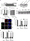
(A) Diagram of HA-tagged Cdt1 truncation mutants tested. (B) Immunoblot using anti-HA to verify similar expression of each HA-Cdt1 truncation mutant relative to wild type HA-Cdt1. Transfection of MK(Rb-) cells occurred during synchronization, and protein expression was assessed after release in late-G1 (10 hr). Separate images are shown, but are derived from the same immunoblot exposure. (C) Synchronized MK(Rb-) cells were transfected during EGF deprivation with plasmids expressing indicated Cdt1 ectopic proteins, or not transfected as a control. Cells were released, and TGFß1 was added to the medium in early-G1. The ability of cells to enter S-phase was assessed using BrdU incorporation assays at 15 hr. HA-positive (transfected) cells were identified using anti-HA IF methods. Results in graph are means of triplicate counts of ~150 cells per field, +/− 1 s.d. Similar results were obtained in a second biological replicate experiment. (D) An experiment using the same design as in panel C, and done in parallel, was performed in the absence of TGFß1 treatment. Note that none of the HA-Cdt1 proteins act as a dominant-negative in suppressing entry into S-phase. (E) MK(Rb-) cells were transfected during synchronization with indicated constructs. Where indicated, cells were treated with TGFß1 in early-G1, and immunoblots performed on total protein lysates collected in late-G1. Antibodies used are shown at the left. (F) Synchronized MK(Rb-) cells were transfected during EGF deprivation with plasmids expressing indicated HA-Cdt1 proteins, and TGFß1 was added to the culture medium in early-G1. The levels of chromatin-bound (recruited) Mcm2 under each condition were assessed by IF methods in late-G1 (10 hr). Cells were pre-extracted in low-salt detergent buffer prior to fixation and staining with anti-HA and anti-Mcm2 antibodies. Arrows indicate select HA-positive (transfected) cells as a reference. Images were obtained using the same exposure settings across samples. (G) Quantitation of the experiment performed in panel F. Cells were scored for presence or absence of Mcm2 signal on chromatin for each condition. Transfected cells were HA-positive in IF analysis. ‘No HA signal’ samples were derived from non-transfected (HA-negative) cells in the same fields that had been exposed to TGFß1. Results are means of triplicate counts of ~50 cells per field, +/− 1 s.d. Similar results were obtained in a second biological replicate experiment. (H) An experiment designed identically to that in panels F&G was performed, except extraction conditions used a high-salt buffer to remove MCMs not loaded onto DNA.
Figure 6:
Figure 6:. Model for TGFß1-Induced Pathways Regulating Cell Growth in the Presence and Absence of Rb.
Canonical targets of TGFß1 are described for wild type cells (containing Rb; Rb+), with mechanisms differing for early-G1 or late-G1 TGFß1 exposure (left side). Findings from this report obtained from cells lacking the Rb protein (Rb-) are described on the right side. See text for detailed discussion.

Similar articles

Cited by

References

    1. Miyazawa K, Shinozaki M, Hara T, Furuya T, Miyazono K. Two major Smad pathways in TGF-beta superfamily signalling. Genes Cells. 2002;7(12):1191–204. - PubMed
    1. Hill CS. The Smads. Int J Biochem Cell Biol. 1999;31(11):1249–54. - PubMed
    1. Pietenpol JA, Holt JT, Stein RW, Moses HL. Transforming growth factor b-1 suppression of c-myc gene transcription: Role in inhibition of keratinocyte proliferation. ProcNatlAcadSciUSA. 1990;87:3758–62. - PMC - PubMed
    1. Seoane J, Pouponnot C, Staller P, Schader M, Eilers M, Massague J. TGFbeta influences Myc, Miz-1 and Smad to control the CDK inhibitor p15INK4b. Nat Cell Biol. 2001;3(4):400–8. - PubMed
    1. Warner BJ, Blain SW, Seoane J, Massague J. Myc downregulation by transforming growth factor beta required for activation of the p15Ink4b G1 arrest pathway. MolCellBiol. 1999;19:5913–22. - PMC - PubMed

Publication types

MeSH terms

Substances