. 2018 Nov 27;92(24):e00737-18.
doi: 10.1128/JVI.00737-18. Print 2018 Dec 15.

Characterization of a Threonine-Rich Cluster in Hepatitis C Virus Nonstructural Protein 5A and Its Contribution to Hyperphosphorylation

Affiliations

Characterization of a Threonine-Rich Cluster in Hepatitis C Virus Nonstructural Protein 5A and Its Contribution to Hyperphosphorylation

Christian Schenk et al. J Virol. .

Abstract

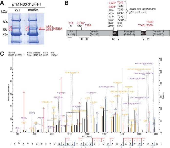
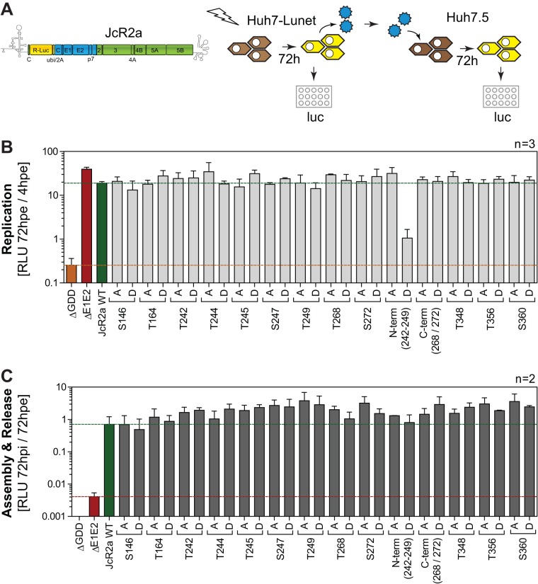
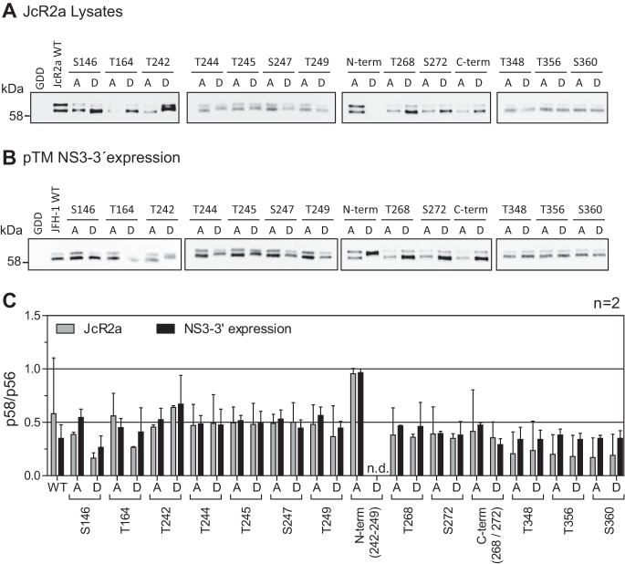
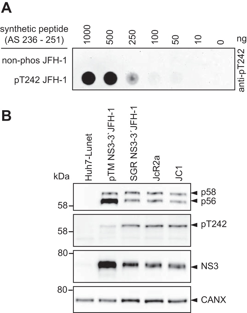
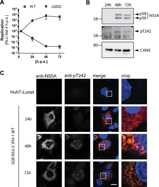
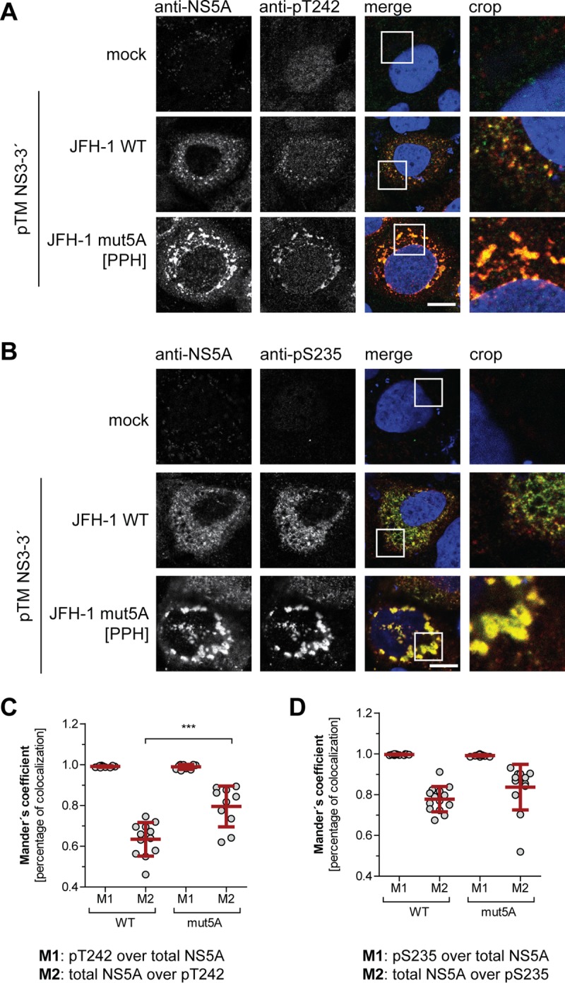
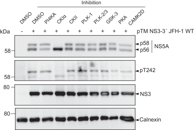
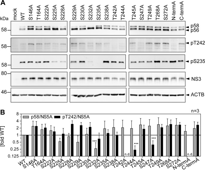
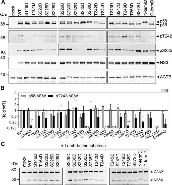
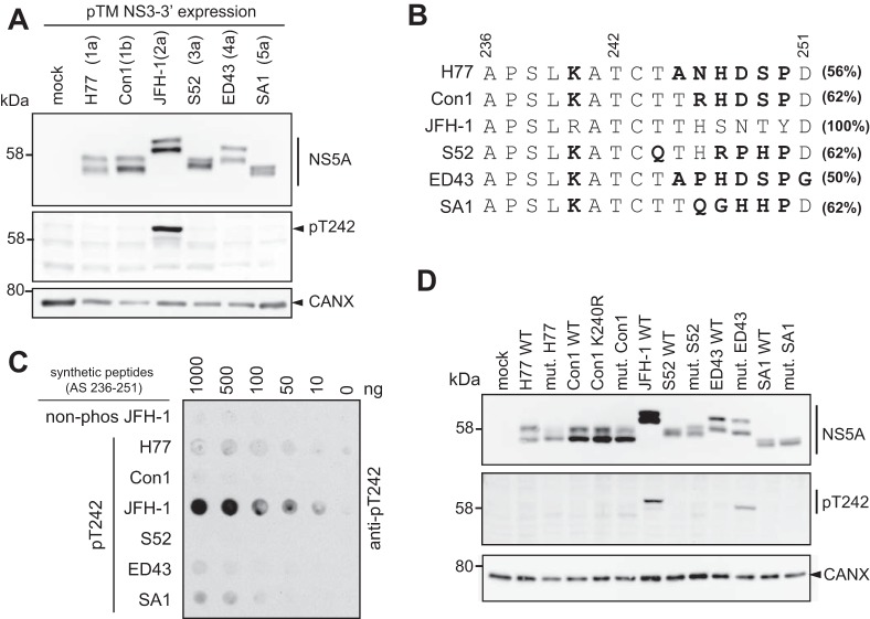
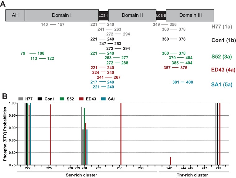
Hepatitis C virus (HCV) nonstructural protein 5A (NS5A) is a phosphoprotein with key functions in regulating viral RNA replication and assembly. Two phosphoisoforms are discriminated by their different apparent molecular weights: a basally phosphorylated (p56) and a hyperphosphorylated (p58) variant. The precise mechanisms governing p58 synthesis and specific functions of the isoforms are poorly understood. Our study aimed at a deeper understanding of determinants involved in p58 synthesis. We analyzed two variants of p56 and p58 of isolate JFH-1 separately by mass spectrometry using an expression model and thereby identified a threonine-rich phosphopeptide exclusively found in the hyperphosphorylated variant. Individual exchange of possible phosphoacceptor sites to phosphoablatant or -mimetic residues had little impact on HCV replication or assembly in cell culture. A phosphospecific antibody recognizing pT242 revealed that this position was indeed phosphorylated only in p58 and depended on casein kinase Iα. Importantly, phosphoablative mutations at positions T244 and S247 abrogated pT242 detection without substantial effects on global p58 levels, whereas mutations in the preceding serine-rich cluster dramatically reduced total p58 levels but had minor impact on pT242 levels, suggesting the existence of distinct subspecies of hyperphosphorylated NS5A. Mass spectrometry analyses of different genotypes showed variable phosphorylation patterns across NS5A and suggested that the threonine-rich region is also phosphorylated at T242 in gt4a and at S249 in gt1a, gt1b, and gt4a. Our data therefore indicate that p58 is not a single homogenously phosphorylated protein species but rather a population of various phosphoisoforms, with high variability between genotypes.IMPORTANCE Hepatitis C virus infections affect 71 million people worldwide and cause severe chronic liver disease. Recently, efficient antiviral therapies have been established, with inhibitors of nonstructural protein NS5A as a cornerstone. NS5A is a central regulator of HCV replication and assembly but is still enigmatic in its molecular functions. It exists in two phosphoisoforms, p56 and p58. We identified a phosphopeptide exclusively found in p58 and analyzed the determinants involved in phosphorylation of this region. We found evidence for very different phosphorylation patterns resulting in p58. These results challenge the concept of p58 being a homogenous species of NS5A molecules phosphorylated at the same positions and argues for at least two independently phosphorylated variants showing the same electrophoretic mobility, likely serving different functions.

Keywords: NS5A; PI4KA; PI4KIIIa; basal phosphorylation; hepatitis C virus; hyperphosphorylation; p56; p58; phosphorylation; positive-strand RNA virus.


Figures

FIG 1
FIG 1
Identification of NS5A phosphorylation sites. (A) Huh7-Lunet T7 cells were transfected with constructs encompassing NS3-5B JFH-1 with the NS5A WT or harboring a triple-alanine mutation in the PFIS, abrogating PI4KA interaction (mut5A [39]). Twenty-four h later, cells were lysed, NS5A was immunoprecipitated, and p58 and p56 bands were cut out after SDS-PAGE and subjected to mass spectrometry analysis to identify phosphorylated peptides. (B) Schematic of the different domains of NS5A. The location of the identified phosphopeptides is indicated below. All numberings on top refer to amino acid positions within NS5A JFH-1 (GenBank accession number KC164983.1). Residues shown in red are sites identified to be phosphorylated with high probability. Additional potential phosphoacceptor sites within each peptide that could not be identified as phosphorylated are shown in a smaller font size. Residues in italics of peptide V represent potential phosphoacceptor, where the exact site of phosphorylation could not be determined. Asterisks mark the residues already reported elsewhere: S146 (25), S222 (25, 41, 65), S225 (25), S232 (48), S235/238 (34), T249 (52), T348 (25), and T356 (40). (C) MSMS spectrum of peptide AT(ph)CTTHSNTYDVDMVDANLLM(ox)EGGVAQTEPESR with a mass of 3,723.5376 Da from NS3-3′ JFH-1 detected in sample C1. This peptide was identified with a MaxQuant score of 65.19. Annotated fragments are color coded. y ions, red; b ions, blue; internal fragments, purple; a, b, and y ions and internal fragments with additional loss of ammonia, water or phosphoric acid, yellow. MaxQuant protein database software annotated the threonine in position two as most likely to be phosphorylated by the relation of the nonphosphorylated singly charged internal fragment TTHSNTYDVDM(ox)VDANL (m/z = 1,793.76) and the phosphorylated internal fragment T(ph)CTTHSNTYD (m/z = 1,261.42).
FIG 2
FIG 2
Impact of phosphoablatant and -mimetic mutations at potential phosphoacceptor sites on replication and virus production. (A) Schematic representation of the experimental procedure. Huh7-Lunet cells were transfected with WT and mutant full-length monocistronic JcR2a reporter virus genomes encoding renilla luciferase, and RNA replication efficiency was determined by luciferase measurement. Supernatants of transfected cells were transferred to Huh7.5 cells to assess production of infectious virus, again by quantification of luciferase activity 72 h postinfection (hpi). hpe, hours postelectroporation. (B and C) Analysis of phosphoacceptor sites harboring alterations of serine and threonine residues to alanine (to A) or aspartate (to D). Mutant “N-term” comprises all five serine and threonine mutations from T242 to T249. Mutant “C-term” comprises substitutions at both positions T268 and S272. (B) Replication is represented in relative light units (RLU) measured 72 h posttransfection and normalized to 4 h posttransfection. A replication-deficient JcR2a variant harboring a deletion in NS5B was used as a negative control for replication (ΔGDD), indicated by the orange line. A green line highlights the replication level of JcR2a WT. The data shown are the mean values with standard deviations (SD) from three independent experiments with two technical replicates each. (C) Supernatants of transfected cells described for panel B were used for reinfection of naive Huh7.5 cells to assess production of infectious virus. Huh7.5 cells were lysed 72 h postinfection and analyzed for luciferase activity. Virus production efficiency is expressed in luciferase activity RLU 72 h postinfection relative to 72 h posttransfection in order to normalize for defects in RNA replication. An assembly-deficient JcR2a variant harboring a deletion in E1E2 proteins was used as a negative control for reinfection (ΔE1E2), indicated by the red line. A green line indicates the virus production of JcR2a WT. The data shown are the mean values with standard deviations from two independent experiments with two technical replicates each.
FIG 3
FIG 3
Comparison of electrophoretic mobility of NS5A phosphoisoforms upon virus replication and NS3-5B expression. Huh7-Lunet cells were electroporated with RNA encoding the full-length reporter virus JcR2a (A), or Huh7-Lunet T7 cells were transfected with pTM vectors expressing NS3-5B of isolate JFH-1 (B), either the WT or a mutant harboring the indicated phosphoablative alanine (A) or the phosphomimetic aspartic acid (D) mutations. Cells were lysed 72 h (A) or 24 h after transfection (B) and subjected to Western blotting using NS5A-specific antibody 9E10. The data shown are from one representative experiment (n = 2). (C) Quantification of the p58/p56 ratios comparing JcR2a (gray bars) and pTM expression (black bars) based on the Western blots shown in panels A and B. n.d., not done, either due to replication defects (JcR2a) or due to the lack of separation between p56 and p58. The quantifications shown are from two experiments (n = 2). Note for this and subsequent figures that the phosphoisoforms of NS5A of isolate JFH-1 have higher apparent molecular weights than 56 and 58 kDa but are still referred to as p56 and p58, respectively.
FIG 4
FIG 4
T242 is exclusively phosphorylated in the hyperphosphorylated form of NS5A. (A) Different concentrations of peptide either phosphorylated at position T242 or nonphosphorylated were titrated on PVDF membrane and incubated with purified, polyclonal anti-pT242 antibodies. (B) Huh7-Lunet T7 cells were either transfected with a pTM vector encoding JFH-1 NS3-3′ or electroporated with in vitro transcripts encoding a bicistronic reporter replicon (SGR JFH-1), a full-length reporter virus genome (JcR2a), or an unmodified full-length virus genome (JC1). The cells were lysed 24 h posttransfection or 72 h postelectroporation, respectively, and analyzed by 7.5% SDS-PAGE/Western blotting using anti-NS5A (monoclonal 9E10)-, anti-NS5A-pT242-, anti-NS3-, and anti-calnexin-specific antibodies. Shown is one representative experiment (n = 2). Note the discrepancy between the 58-kDa molecular weight (MW) marker band (left) and the band referred to as p58/pT242 (right), which is due to a higher apparent MW of the NS5A phosphoisoforms in the case of JFH-1.
FIG 5
FIG 5
NS5A phosphorylated at T242 has no distinct subcellular localization compared to total NS5A. Huh7-Lunet cells were transfected with in vitro transcripts encoding a JFH-1 reporter replicon (A to C) and analyzed for RNA replication (A), NS5A phosphorylation (B), and pT242 localization (C) at the indicated time points. (A) RNA replication of a WT replicon compared to a replication-deficient mutant (ΔGDD) is represented in relative light units (RLU) measured 24 h, 48 h, and 72 h posttransfection and shown as fold relative to values at 4 h posttransfection to normalize for transfection efficiency. (B) Transfected cells from panel A were lysed and analyzed by Western blotting for NS5A phosphorylation using anti-NS5A (monoclonal antibody 9E10)-, anti-NS5A-pT242-, and anti-calnexin (CAXN)-specific antibodies, as indicated on the right. (C) Cells were seeded on coverslips and fixed at the indicated time points postelectroporation. Total NS5A and NS5A phosphorylated at T242 were detected by immunofluorescence analysis using monoclonal antibody 9E10 (red) and anti-NS5A-pT242 (green), respectively. Nuclei were stained with DAPI. Scale bar, 10 µm. Shown is a representative experiment of two with comparable outcomes.
FIG 6
FIG 6
Subcellular localization of WT NS5A compared to NS5A mutant with impaired PI4KA activation. (A and B) Huh7-Lunet T7 cells were transfected with a pTM vector encoding NS3-5B of JFH-1 WT or mutPPH, seeded on coverslips, and fixed 24 h posttransfection. Total NS5A and NS5A phosphorylated at T242 (A) or S235 (B) was detected by immunofluorescence analysis using monoclonal antibody 9E10 (red) and anti-NS5A-pT242 or anti-NS5A-pS235 (both green), respectively. Nuclei were stained with DAPI. Scale bar, 10 µm. (C and D) Mander’s coefficient representing colocalization of pT242 with NS5A (M1) (C) or pS235 with NS5A (D) and vice versa (M2) was calculated for the pTM-transfected cells using Fiji. At least 10 cells per condition were analyzed. ***, P < 0.01 (homoscedastic, two-tailed t test).
FIG 7
FIG 7
Impact of kinase inhibitors on phosphorylation of T242. Huh7-Lunet T7 cells were transfected with a pTM vector encoding NS3-5B of JFH-1. Four hours after transfection the medium was replaced with new medium containing drugs targeting the indicated kinases that have been described to modulate NS5A phosphorylation. Cells were lysed 24 h after transfection and analyzed by Western blotting for NS5A phosphorylation using anti-NS5A (monoclonal antibody 9E10)-, anti-NS5A-pT242-, anti-NS3-, and anti-calnexin-specific antibodies as indicated. One representative of three experiments is shown.
FIG 8
FIG 8
Impact of phosphoablative mutations of potential phosphoacceptor sites on T242 phosphorylation. Huh7-Lunet T7 cells were transfected with pTM vectors expressing NS3-5B of isolate JFH-1, either WT or harboring the indicated phosphoablative alanine mutations. Mutant “N-termA” comprises all five serine and threonine mutations from T242 to T249. Mutant “C-termA” comprises both substitutions at positions T268 and S272. (A) Cells were lysed 24 h after transfection and analyzed by Western blotting using anti-NS5A (monoclonal antibody 9E10, upper)-, anti-NS5A-pT242-, anti-NS5A-pS235-, anti-NS3, and anti-β-actin (ACTB)-specific antibodies. (B) Ratio of p58 and pT242 to total NS5A. Intensities of the p58 bands, the pT242 bands, and the sum of p56 and p58 (total NS5A) as shown in panel A were quantified and used to calculate the indicated ratios. An additional staining of NS3 was performed subsequently on the same membranes previously used for detection of NS5A or pT242. These NS3 signals were used to obtain relative NS5A or pT242 levels for each membrane, which built the basis for pT242/NS5A levels. Bars represent mean values and SD from one representative experiment with three technical replicates. A second biological replicate was performed with a similar outcome (n = 2). n.d., not detectable. ***, P < 0.01 (homoscedastic, two-tailed t test).
FIG 9
FIG 9
Impact of phosphomimetic mutations of potential phosphoacceptor sites on T242 phosphorylation. Huh7-Lunet T7 cells were transfected with pTM vectors expressing NS3-5B of isolate JFH-1, either WT or harboring the indicated phosphomimetic aspartic acid mutations. Mutant “N-termD” comprises all five serine and threonine mutations from T242 to T249. Mutant “C-termD” comprises both substitutions at positions T268 and S272. (A) Cells were lysed 24 h after transfection and analyzed by Western blotting using anti-NS5A (monoclonal antibody 9E10, upper)-, anti-NS5A-pT242-, anti-NS5A-pS235-, anti-NS3-, and anti-β-actin (ACTB)-specific antibodies. (B) Ratio of p58 and pT242 to total NS5A. Intensities of the p58 bands, the pT242 bands, and the sum of p56 and p58 (total NS5A) as shown in panel A were quantified and used to calculate the indicated ratios. An additional staining of NS3 was performed subsequently on the same membranes previously used for detection of NS5A or pT242. These NS3 signals were used to obtain relative NS5A or pT242 levels for each membrane, which built the basis for pT242/NS5A levels. Bars represent mean values and SD from one representative experiment with three technical replicates. A second biological replicate was performed with a similar outcome (n = 2). n.d., not detectable. ***, P < 0.01 (homoscedastic, two-tailed t test). (C) Cells were lysed 24 h after transfection, and the cleared lysate was treated with lambda phosphatase. Phosphatase-treated lysates were analyzed by Western blotting using anti-NS5A (monoclonal antibody 9E10)- and anti-calnexin-specific antibodies (n = 1). Note that dephosphorylation of NS5A by lambda phosphatase results in only one remaining band with an apparent MW similar to that of p56, but that different mutants show a consistently increased apparent molecular size.
FIG 10
FIG 10
Analysis of T242 phosphorylation in different HCV genotypes. (A) Huh7-Lunet T7 cells were transfected with pTM vectors coding for the NS3-5B nonstructural proteins of the indicated HCV isolates. The cells were lysed 24 h after transfection and analyzed by 7.5% SDS-PAGE/Western blotting for total NS5A (monoclonal antibody 9E10), NS5A-pT242, and calnexin (CAXN). One representative experiment is shown (n = 2). (B) Alignment of NS5A sequence from 236 to 251 (numbering according to NS5A JFH-1) from HCV isolates H77 (gt1a), Con1 (gt1b), JFH-1 (gt2a), S52 (gt3a), ED43 (gt4a), and SA1 (gt5a). Highlighted in boldface are the amino acids that are different from those of JFH-1. Numbers on the right represent the homology of the respective sequences to JFH-1. Note that the peptide sequence corresponding to JFH-1 was used to generate the pT242-specific antiserum. (C) Indicated amounts of peptides corresponding to the sequence of the indicated isolates and phosphorylated at position T242 were spotted on PVDF membrane and incubated with purified, polyclonal anti-pT242 antibodies. The nonphosphorylated peptide of JFH-1 served as a negative control for staining. (D) Huh7-Lunet T7 cells were transfected with pTM vectors coding for the NS3-5B nonstructural proteins of the indicated HCV isolates, either the WT or an NS5A mutated version. All mutated genotypes have amino acid sequence changed from their respective WT sequence to the sequence of JFH-1 in NS5A positions 236 to 251 (compare to panel B). In addition, a Con1 point mutant, K240R, was included to test if this change alone would allow antigen recognition. The experiments were performed twice with a comparable outcome.
FIG 11
FIG 11
Phospho-mass spectrometry reveals additional phosphorylation events within the Thr-rich peptide. (A) Schematic of the different domains of NS5A. The location of the identified phosphopeptides is indicated below for each isolate analyzed here, as given on the right. All numberings refer to amino acid positions within the respective genotype. (B) Phospho-(STY)-probability scores of different potential phosphoacceptor sites within the Ser-rich and Thr-rich cluster. Shown are only values above 0.75 that can be considered high-probability phosphorylation events at the respective site. The numbers below refer to all possible Ser/Thr phosphoacceptor sites existing in all analyzed genotypes. Note that phosphorylation at S249 was detected for all genotypes with a phosphoacceptor site at this position and that S52 (gt3a) and SA1 (gt5a) harbor a histidine (H) at position 249. The phospho-mass spectrometry was performed once.

Similar articles

Cited by

References

    1. Lohmann V. 2013. Hepatitis C virus RNA replication. Curr Top Microbiol Immunol 369:167–198. doi:10.1007/978-3-642-27340-7_7. - DOI - PMC - PubMed
    1. Romero-Brey I, Berger C, Kallis S, Kolovou A, Paul D, Lohmann V, Bartenschlager R. 2015. NS5A domain 1 and polyprotein cleavage kinetics are critical for induction of double-membrane vesicles associated with hepatitis C virus replication. mBio 6:e00759. doi:10.1128/mBio.00759-15. - DOI - PMC - PubMed
    1. Appel N, Zayas M, Miller S, Krijnse-Locker J, Schaller T, Friebe P, Kallis S, Engel U, Bartenschlager R. 2008. Essential role of domain III of nonstructural protein 5A for hepatitis C virus infectious particle assembly. PLoS Pathog 4:e1000035. doi:10.1371/journal.ppat.1000035. - DOI - PMC - PubMed
    1. Tellinghuisen TL, Foss KL, Treadaway JC, Rice CM. 2008. Identification of residues required for RNA replication in domains II and III of the hepatitis C virus NS5A protein. J Virol 82:1073–1083. doi:10.1128/JVI.00328-07. - DOI - PMC - PubMed
    1. Masaki T, Suzuki R, Murakami K, Aizaki H, Ishii K, Murayama A, Date T, Matsuura Y, Miyamura T, Wakita T, Suzuki T. 2008. Interaction of hepatitis C virus nonstructural protein 5A with core protein is critical for the production of infectious virus particles. J Virol 82:7964–7976. doi:10.1128/JVI.00826-08. - DOI - PMC - PubMed

Publication types

Substances

LinkOut - more resources