. 2018 Nov 27;92(24):e01504-18.
doi: 10.1128/JVI.01504-18. Print 2018 Dec 15.

Lysosomal Proteases Are a Determinant of Coronavirus Tropism

Affiliations

Lysosomal Proteases Are a Determinant of Coronavirus Tropism

Yuan Zheng et al. J Virol. .

Abstract

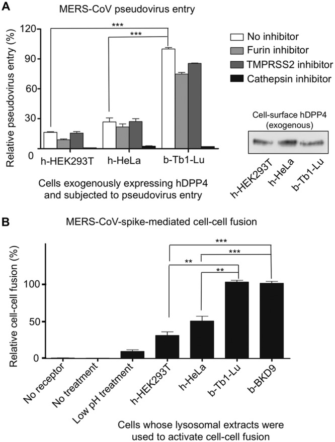
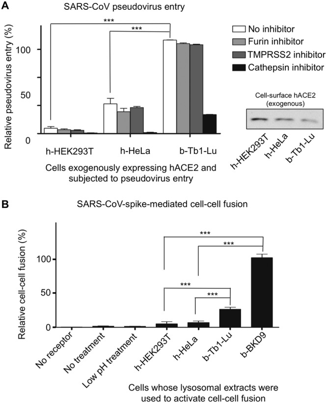
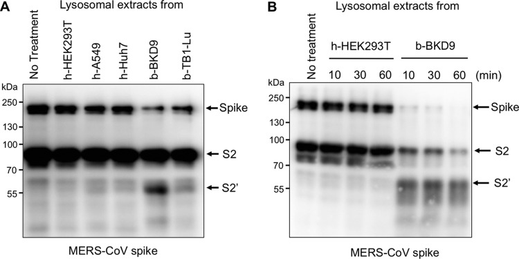
Cell entry by coronaviruses involves two principal steps, receptor binding and membrane fusion; the latter requires activation by host proteases, particularly lysosomal proteases. Despite the importance of lysosomal proteases in both coronavirus entry and cell metabolism, the correlation between lysosomal proteases and cell tropism of coronaviruses has not been established. Here, we examined the roles of lysosomal proteases in activating coronavirus surface spike proteins for membrane fusion, using the spike proteins from severe acute respiratory syndrome coronavirus (SARS-CoV) and Middle East respiratory syndrome coronavirus (MERS-CoV) as the model system. To this end, we controlled the contributions from receptor binding and other host proteases, thereby attributing coronavirus entry solely or mainly to the efficiency of lysosomal proteases in activating coronavirus spike-mediated membrane fusion. Our results showed that lysosomal proteases from bat cells support coronavirus spike-mediated pseudovirus entry and cell-cell fusion more effectively than their counterparts from human cells. Moreover, purified lysosomal extracts from bat cells cleave cell surface-expressed coronavirus spikes more efficiently than their counterparts from human cells. Overall, our study suggests that different lysosomal protease activities from different host species and tissue cells are an important determinant of the species and tissue tropism of coronaviruses.IMPORTANCE Coronaviruses are capable of colonizing new species, as evidenced by the recent emergence of SARS and MERS coronaviruses; they can also infect multiple tissues in the same species. Lysosomal proteases play critical roles in coronavirus entry by cleaving coronavirus surface spike proteins and activating the fusion of host and viral membranes; they also play critical roles in cell physiology by processing cellular products. How do different lysosomal protease activities from different cells impact coronavirus entry? Here, we controlled the contributions from known factors that function in coronavirus entry so that lysosomal protease activities became the only or the main determinant of coronavirus entry. Using pseudovirus entry, cell-cell fusion, and biochemical assays, we showed that lysosomal proteases from bat cells activate coronavirus spike-mediated membrane fusion more efficiently than their counterparts from human cells. Our study provides the first direct evidence supporting lysosomal proteases as a determinant of the species and tissue tropisms of coronaviruses.

Keywords: coronavirus spike protein; lysosomal proteases; species tropism; tissue tropism.


Figures

FIG 1
FIG 1
Screening for cell lines that are suitable for studying lysosomal protease-activated coronavirus entry. To screen for cell lines that endogenously express no or low levels of receptor protein for the coronavirus of interest, MERS-CoV pseudoviruses (A) or SARS-CoV pseudoviruses (B) were used to enter a number of cells from different tissues of different host species (human, monkey, and bat). Entry efficiency was characterized using luciferase activity accompanying entry and calibrated against the highest entry efficiency (MERS-CoV entry into MRC5 cells was taken as 100% in panel A, whereas SARS-CoV entry into Vero cells was taken as 100% in panel B). Mock, no pseudoviruses were added. The error bars indicate standard errors of the mean (SEM) (n = 5). (C) To screen for cell lines that could be easily transfected and hence controlled to exogenously express receptor protein for the coronavirus of interest, different cells were transfected with a plasmid encoding human DPP4 (hDPP4); subsequently, the expression level of human DPP4 in each of the cell lines was detected through Western blotting using an antibody recognizing its C-terminal C9 tag. The expression level of β-actin in each of the cell lines was used as a positive control. (D) MTT cell viability assay, showing that the viabilities of three types of cells were not affected by the presence of different protease inhibitors. The error bars indicate SEM (n = 5). OD570, optical density at 570 nm. There was no statistical significance under different conditions within each cell group (P > 0.05, based on a two-tailed t test).
FIG 2
FIG 2
Roles of lysosomal proteases in MERS-CoV spike-mediated membrane fusion. (A) Roles of lysosomal proteases in MERS-CoV pseudovirus entry. Three types of cells, h-HEK293T, h-HeLa, and b-Tb1-Lu, were controlled to exogenously express human DPP4 as shown in Fig. 1C and then subjected to MERS-CoV pseudovirus entry as shown in Fig. 1A. The furin inhibitor chloromethyl ketone, the cell surface protease (i.e., TMPRSS2) inhibitor camostat, and the lysosomal protease (i.e., cathepsin) inhibitor E64d were used in parallel experiments to investigate the relative contributions of the different proteases to MERS-CoV pseudovirus entry. The expression levels of cell surface-associated C9-tagged human DPP4 were measured through Western blot analysis using an anti-C9 tag monoclonal antibody and were further calibrated across the three types of cells. (B) MERS-CoV spike-mediated cell-cell fusion in the presence of lysosomal extracts. h-HEK293T cells exogenously expressing MERS-CoV spike and h-HEK293T cells exogenously expressing human DPP4 were mixed at pH 5.6 in the presence of lysosomal extracts from h-HEK293T cells, h-HeLa cells, b-Tb1-Lu cells, or b-BKD9 cells. Cell-cell fusion efficiency was characterized using luciferase activity accompanying fusion and calibrated against the highest fusion efficiency (i.e., in the presence of lysosomal extracts from b-Tb1-Lu cells). Three negative controls were used: (i) cells not expressing human DPP4 were used (No receptor); (ii) no lysosomal proteases were added to the medium, and the medium was at neutral pH (No treatment); (iii) no lysosomal proteases were added, but the medium was at pH 5.6 (Low pH treatment). Statistical analyses were performed using a two-tailed t test. The error bars indicate SEM (n = 4). ***, P < 0.001; **, P < 0.01.
FIG 3
FIG 3
Characterization of the purity of lysosomal extracts from different cell lines. Because ALP and CPR are enzymatic markers of plasma and the ER, respectively, the purified lysosomal extracts and whole-cell lysates from different cell lines (for each cell line, lysosomal extracts and whole-cell lysates were at equal concentrations) were assayed for their ALP activities (A) and CPR activities (B) to evaluate potential contaminants from other cell organelles. The error bars indicate SEM (n = 3; some of the error bars may be too small to be seen).
FIG 4
FIG 4
Roles of lysosomal proteases in SARS-CoV spike-mediated membrane fusion. The experiments were performed in the same way as for Fig. 2, except that SARS-CoV spike and its receptor, human ACE2 (hACE2), replaced MERS-CoV spike and human DPP4, respectively.
FIG 5
FIG 5
Cleavage of cell surface-expressed MERS-CoV spike using purified lysosomal extracts. (A) Cleavage of cell surface-expressed MERS-CoV spike using lysosomal extracts from a number of cell lines. MERS-CoV spike was exogenously expressed on the surfaces of h-HEK293T cells and then treated with 50 µg/ml lysosomal extracts (from different types of cells) at pH 5.6 for 30 min. The cleavage state of MERS-CoV spike was detected through Western blotting using an antibody recognizing its C-terminal C9 tag. (B) Cleavage of cell surface-expressed MERS-CoV spike using 100 µg/ml lysosomal extracts (from two types of cells) at pH 5.6 in a time-dependent manner (10, 30, and 60 min). The experiments were repeated five times, and representative results are shown.
FIG 6
FIG 6
Cleavage of cell surface-expressed HKU4 spike using purified lysosomal extracts. The experiments were performed in the same way as for Fig. 5A, except that HKU4 spike (either wild type [WT] or containing an N762 mutation that removed a glycosylation site from the lysosomal protease motif) replaced MERS-CoV spike. The experiments were repeated five times, and representative results are shown.

References

    1. Nomaguchi M, Fujita M, Miyazaki Y, Adachi A. 2012. Viral tropism. Front Microbiol 3:281. doi:10.3389/fmicb.2012.00281. - DOI - PMC - PubMed
    1. Dimitrov DS. 2004. Virus entry: molecular mechanisms and biomedical applications. Nat Rev Microbiol 2:109–122. doi:10.1038/nrmicro817. - DOI - PMC - PubMed
    1. Skehel JJ, Wiley DC. 2000. Receptor binding and membrane fusion in virus entry: the influenza hemagglutinin. Annu Rev Biochem 69:531–569. doi:10.1146/annurev.biochem.69.1.531. - DOI - PubMed
    1. Li WH, Wong SK, Li F, Kuhn JH, Huang IC, Choe H, Farzan M. 2006. Animal origins of the severe acute respiratory syndrome coronavirus: insight from ACE2-S-protein interactions. J Virol 80:4211–4219. doi:10.1128/JVI.80.9.4211-4219.2006. - DOI - PMC - PubMed
    1. Luzio JP, Pryor PR, Bright NA. 2007. Lysosomes: fusion and function. Nat Rev Mol Cell Biol 8:622–632. doi:10.1038/nrm2217. - DOI - PubMed

Publication types

MeSH terms

Substances

LinkOut - more resources