Virulent Pseudorabies Virus Infection Induces a Specific and Lethal Systemic Inflammatory Response in Mice
- PMID: 30258005
- PMCID: PMC6258956
- DOI: 10.1128/JVI.01614-18
Virulent Pseudorabies Virus Infection Induces a Specific and Lethal Systemic Inflammatory Response in Mice
Abstract
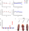
Pseudorabies virus (PRV) is an alphaherpesvirus that infects the peripheral nervous system (PNS). The natural host of PRV is the swine, but it can infect most mammals, including cattle, rodents, and dogs. In these nonnatural hosts, PRV always causes a severe acute and lethal neuropathy called the "mad itch," which is uncommon in swine. Thus far, the pathophysiological and immunological processes leading to the development of the neuropathic itch and the death of the animal are unclear. Using a footpad inoculation model, we established that mice inoculated with PRV-Becker (virulent strain) develop a severe pruritus in the foot and become moribund at 82 h postinoculation (hpi). We found necrosis and inflammation with a massive neutrophil infiltration only in the footpad and dorsal root ganglia (DRGs) by hematoxylin and eosin staining. PRV load was detected in the foot, PNS, and central nervous system tissues by quantitative reverse transcription-PCR. Infected mice had elevated plasma levels of proinflammatory cytokines (interleukin-6 [IL-6] and granulocyte colony-stimulating factor [G-CSF]) and chemokines (Gro-1 and monocyte chemoattractant protein 1). Significant IL-6 and G-CSF levels were detected in several tissues at 82 hpi. High plasma levels of C-reactive protein confirmed the acute inflammatory response to PRV-Becker infection. Moreover, mice inoculated with PRV-Bartha (attenuated, live vaccine strain) did not develop pruritus at 82 hpi. PRV-Bartha also replicated in the PNS, and the infection spread further in the brain than PRV-Becker. PRV-Bartha infection did not induce the specific and lethal systemic inflammatory response seen with PRV-Becker. Overall, we demonstrated the importance of inflammation in the clinical outcome of PRV infection in mice and provide new insights into the process of PRV-induced neuroinflammation.IMPORTANCE Pseudorabies virus (PRV) is an alphaherpesvirus related to human pathogens such as herpes simplex virus 1 and varicella-zoster virus (VZV). The natural host of PRV is the swine, but it can infect most mammals. In susceptible animals other than pigs, PRV infection always causes a characteristic lethal pruritus known as the "mad itch." The role of the immune response in the clinical outcome of PRV infection is still poorly understood. Here, we show that a systemic host inflammatory response is responsible for the severe pruritus and acute death of mice infected with virulent PRV-Becker but not mice infected with attenuated strain PRV-Bartha. In addition, we identified IL-6 and G-CSF as two main cytokines that play crucial roles in the regulation of this process. Our findings give new insights into neuroinflammatory diseases and strengthen further the similarities between VZV and PRV infections at the level of innate immunity.
Keywords: cytokines; immunopathogenesis; inflammation; mice; nervous system; pseudorabies virus.
Copyright © 2018 American Society for Microbiology.
Figures


Similar articles
-
The Neuropathic Itch Caused by Pseudorabies Virus.Pathogens. 2020 Mar 31;9(4):254. doi: 10.3390/pathogens9040254. Pathogens. 2020. PMID: 32244386 Free PMC article. Review.
-
Alphaherpesvirus infection of mice primes PNS neurons to an inflammatory state regulated by TLR2 and type I IFN signaling.PLoS Pathog. 2019 Nov 1;15(11):e1008087. doi: 10.1371/journal.ppat.1008087. eCollection 2019 Nov. PLoS Pathog. 2019. PMID: 31675371 Free PMC article.
-
Retrograde, transneuronal spread of pseudorabies virus in defined neuronal circuitry of the rat brain is facilitated by gE mutations that reduce virulence.J Virol. 1999 May;73(5):4350-9. doi: 10.1128/JVI.73.5.4350-4359.1999. J Virol. 1999. PMID: 10196333 Free PMC article.
-
Histamine Is Responsible for the Neuropathic Itch Induced by the Pseudorabies Virus Variant in a Mouse Model.Viruses. 2022 May 17;14(5):1067. doi: 10.3390/v14051067. Viruses. 2022. PMID: 35632808 Free PMC article.
-
A Tug of War: Pseudorabies Virus and Host Antiviral Innate Immunity.Viruses. 2022 Mar 6;14(3):547. doi: 10.3390/v14030547. Viruses. 2022. PMID: 35336954 Free PMC article. Review.
Cited by
-
An improved animal model for herpesvirus encephalitis in humans.PLoS Pathog. 2020 Mar 30;16(3):e1008445. doi: 10.1371/journal.ppat.1008445. eCollection 2020 Mar. PLoS Pathog. 2020. PMID: 32226043 Free PMC article.
-
Pseudorabies Virus UL4 protein promotes the ASC-dependent inflammasome activation and pyroptosis to exacerbate inflammation.PLoS Pathog. 2024 Sep 24;20(9):e1012546. doi: 10.1371/journal.ppat.1012546. eCollection 2024 Sep. PLoS Pathog. 2024. PMID: 39316625 Free PMC article.
-
Epizootic situation of Aujeszky's disease within the territory of the Republic of Kazakhstan.Open Vet J. 2021 Jan-Mar;11(1):135-143. doi: 10.4314/ovj.v11i1.20. Epub 2021 Feb 19. Open Vet J. 2021. PMID: 33898295 Free PMC article.
-
Pseudorabies virus as a zoonosis: scientific and public health implications.Virus Genes. 2025 Feb;61(1):9-25. doi: 10.1007/s11262-024-02122-2. Epub 2024 Dec 18. Virus Genes. 2025. PMID: 39692808 Review.
-
Characteristics of the pseudorabies virus strain GDWS2 with severe neurological signs and high viral shedding capacity in pigs.Front Vet Sci. 2025 Apr 14;12:1530765. doi: 10.3389/fvets.2025.1530765. eCollection 2025. Front Vet Sci. 2025. PMID: 40297827 Free PMC article.
References
-
- Gustafson DP. 1986. Pseudorabies, p 209–223. In Leman AD, Glock RD, Mengeling WL, Penny RHC, Scholl E, Straw B (ed), Diseases of swine, 6th ed Iowa State University Press, Ames, IA.
-
- Wittmann G, Rziha H-J. 1989. Aujeszky’s disease (pseudorabies) in pigs, p 230–325. In Knipe DM, Howley PM (ed), Herpesvirus diseases of cattle, horses, and pigs, vol 9 Kluwer Academic Publishers, Boston, MA.
Publication types
MeSH terms
Substances
Grants and funding
LinkOut - more resources
Full Text Sources
Other Literature Sources
Research Materials

