. 2018 Sep 26;9(10):1007.
doi: 10.1038/s41419-018-1041-8.

Neuroprotection of retinal ganglion cells by a novel gene therapy construct that achieves sustained enhancement of brain-derived neurotrophic factor/tropomyosin-related kinase receptor-B signaling

Affiliations

Neuroprotection of retinal ganglion cells by a novel gene therapy construct that achieves sustained enhancement of brain-derived neurotrophic factor/tropomyosin-related kinase receptor-B signaling

Andrew Osborne et al. Cell Death Dis. .

Abstract

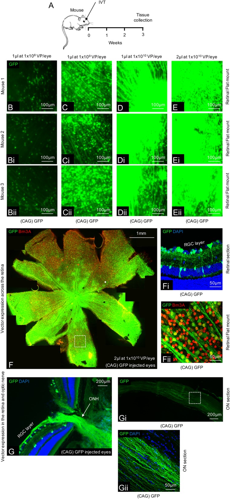
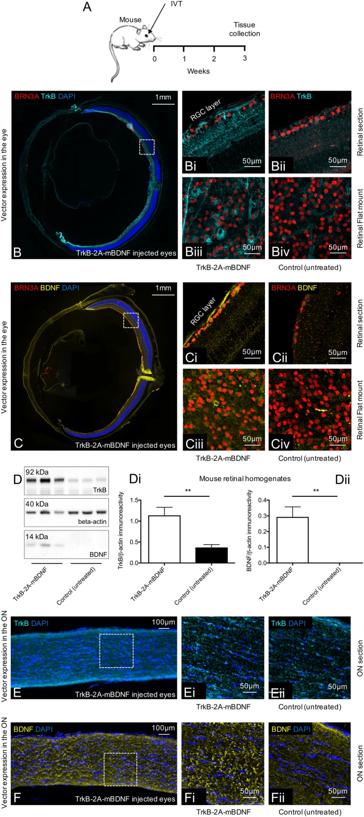
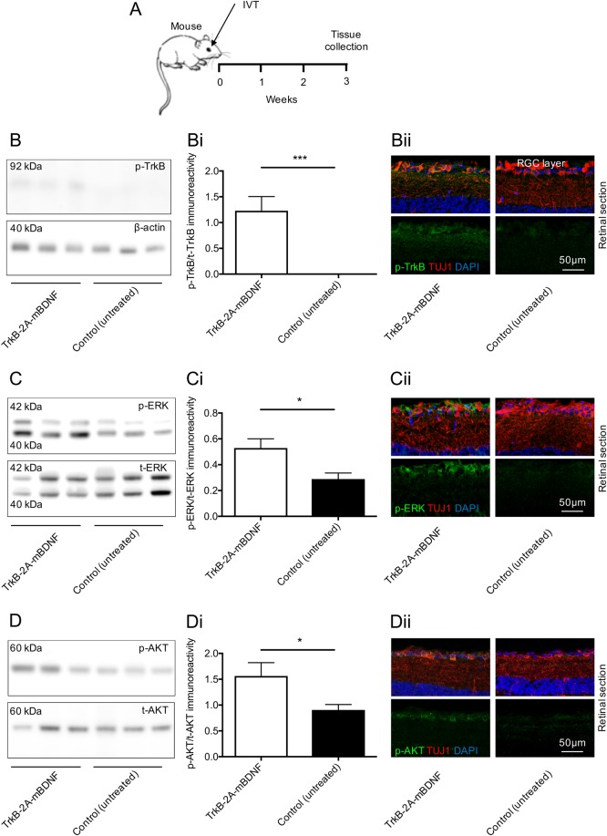
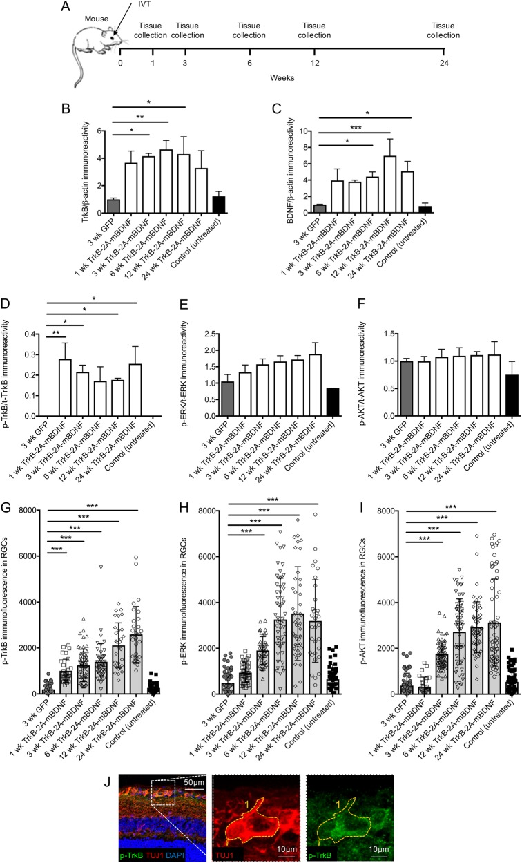
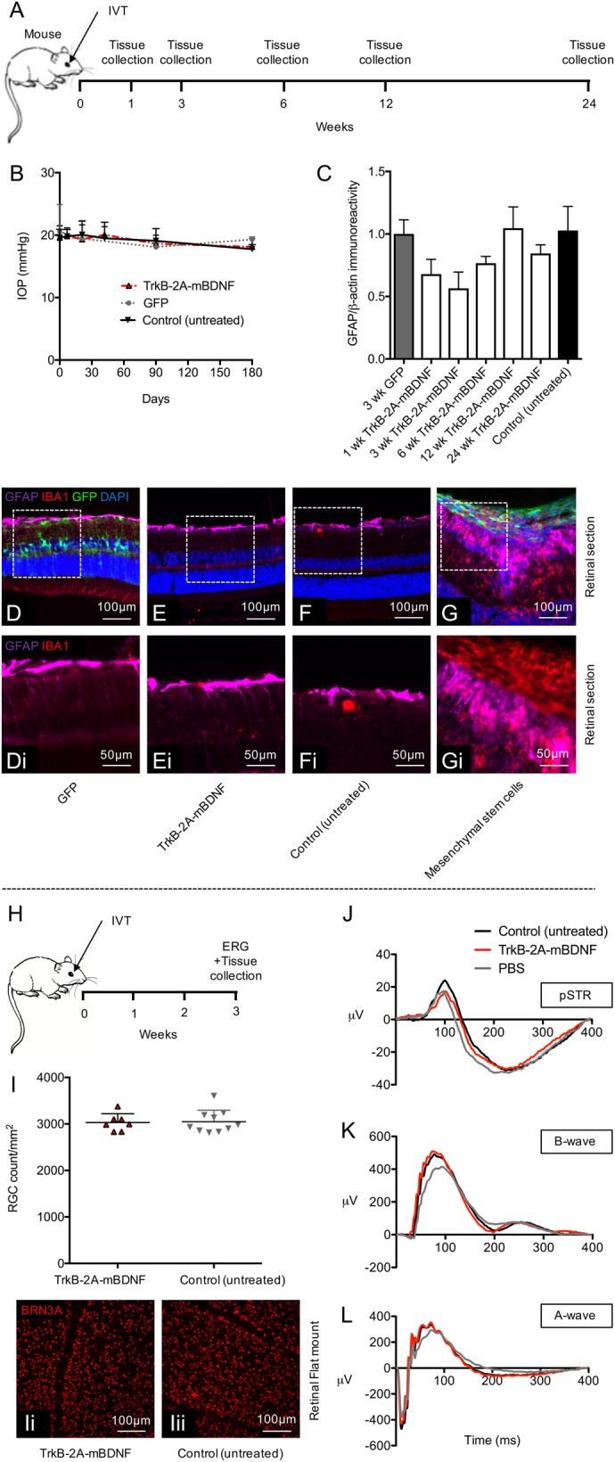
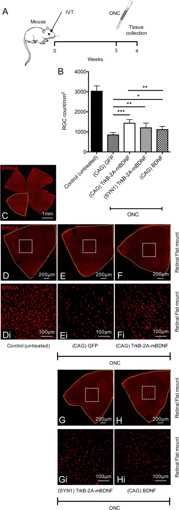
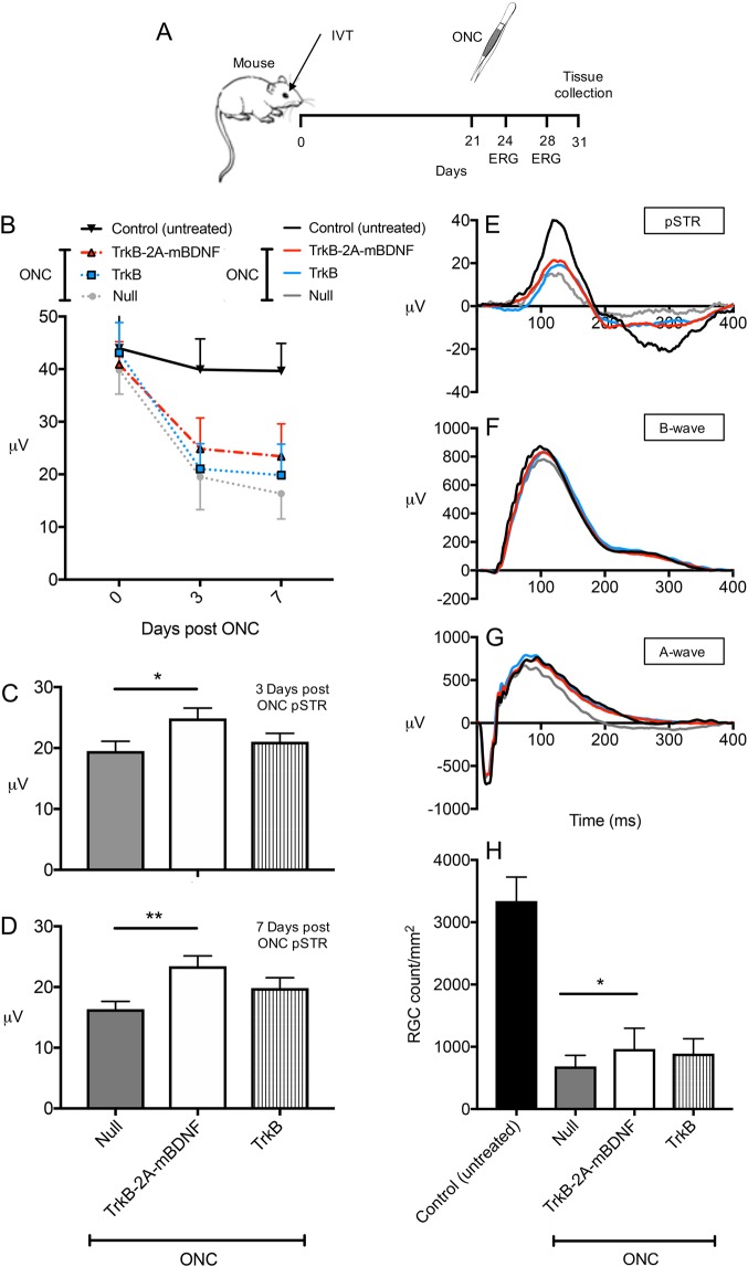
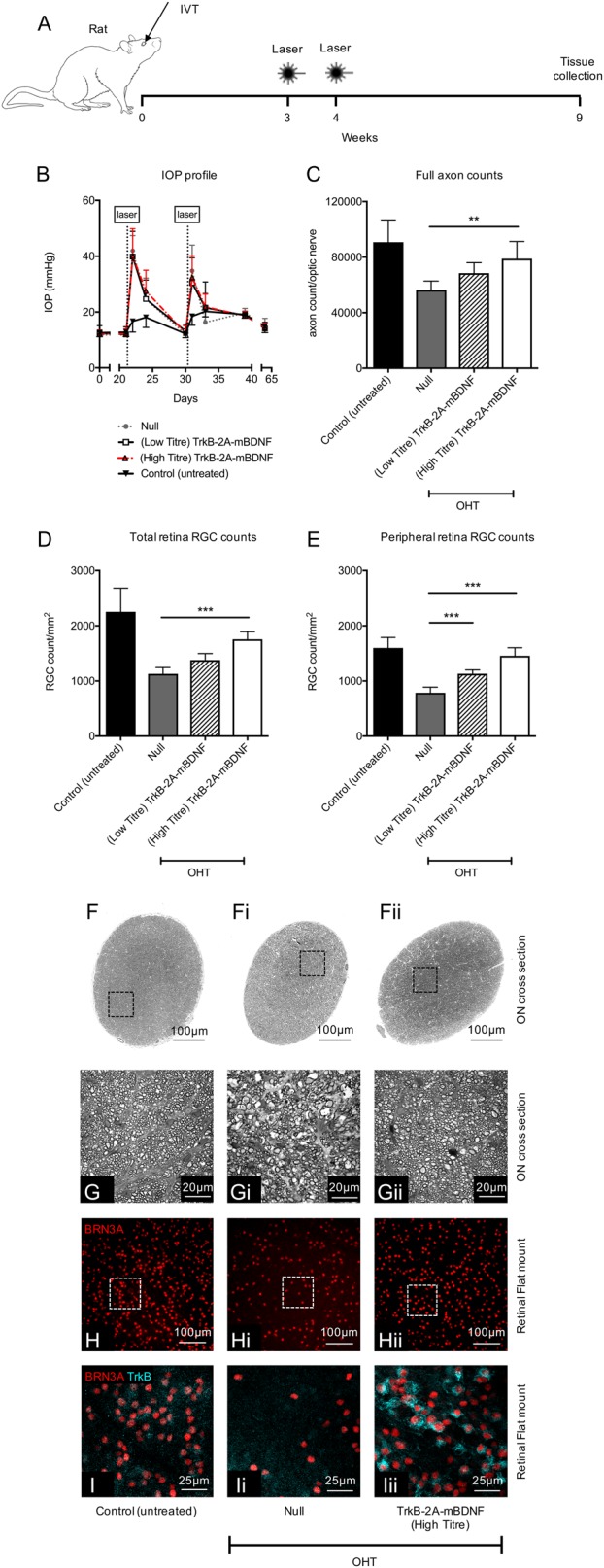
Previous studies have demonstrated that intravitreal delivery of brain-derived neurotrophic factor (BDNF) by injection of recombinant protein or by gene therapy can alleviate retinal ganglion cell (RGC) loss after optic nerve injury. BDNF gene therapy can improve RGC survival in experimental models of glaucoma, the leading cause of irreversible blindness worldwide. However, the therapeutic efficacy of BDNF supplementation alone is time limited at least in part due to BDNF receptor downregulation. Tropomyosin-related receptor kinase-B (TrkB) downregulation has been reported in many neurological diseases including glaucoma, potentially limiting the effect of sustained or repeated BDNF delivery.Here, we characterize a novel adeno-associated virus (AAV) gene therapy (AAV2 TrkB-2A-mBDNF) that not only increases BDNF production but also improves long-term neuroprotective signaling by increasing expression of the BDNF receptor (TrkB) within the inner retina. This approach leads to significant and sustained elevation of survival signaling pathways ERK and AKT within RGCs over 6 months and avoids the receptor downregulation which we observe with treatment with AAV2 BDNF alone. We validate the neuroprotective efficacy of AAV2 TrkB-2A-mBDNF in a mouse model of optic nerve injury, where it outperforms conventional AAV2 BDNF or AAV2 TrkB therapy, before showing powerful proof of concept neuroprotection of RGCs and axons in a rat model of chronic intraocular pressure (IOP) elevation. We also show that there are no adverse effects of the vector on retinal structure or function as assessed by histology and electroretinography in young or aged animals. Further studies are underway to explore the potential of this vector as a candidate for progression into clinical studies to protect RGCs in patients with glaucoma and progressive visual loss despite conventional IOP-lowering treatment.


Conflict of interest statement

P.S.W. and K.R.M. have a financial interest in Quethera Ltd, a company working to develop gene therapy approaches to neurodegenerative diseases. A.O., K.H., and K.R.M. have received grant support from Quethera Ltd. T.Z.K., L.S., A.C.B., and G.Y.X.K. have no disclosures.

Figures

Fig. 1
Fig. 1. Ensuring adequate transfection in the mouse retina using an AAV2 CAG vector system.
a Schematic of the procedure performed. be Expression of GFP throughout the retina using various titres and volumes of AAV2 GFP (n = 4/group). f A tile scan of a representative retinal wholemount 3 weeks after injection of 2 µl AAV2 GFP at 1 × 1010 vector particles/eye. fi GFP expression is localized to the inner retina with transduction of predominantly RGCs and some amacrine/Müller glia cells. fii Magnification of the wholemount shows co-localization between GFP and Brn3A+ RGCs. ggii GFP expression can also be seen transported anterogradely down the optic nerve in both proximal (g) and distal (gi, ii) locations
Fig. 2
Fig. 2. Expression of TrkB and BDNF in mouse eyes 3 weeks after intravitreal delivery of AAV2 TrkB-2A-mBDNF (2 µl, 9.14 × 109 vector particles/eye).
a Schematic of the procedure performed. bbiv Increased TrkB receptor expression on the cell surface of transduced RGCs (Brn3A+ cells) throughout the mouse retina after treatment with AAV2 TrkB-2A-mBDNF compared to untreated eyes. cciv Increased BDNF expression within transduced RGCs compared to untreated eyes (n = 3 for retinal sections, n = 3 for retinal flat mount). ddii Increased TrkB and BDNF expression was detected in retinal lysates following AAV2 TrkB-2A-mBDNF delivery compared to contralateral, untreated eyes (n = 6), **P < 0.01. eeii TrkB receptor expression did not appear elevated within the optic nerve 3 weeks after AAV2 TrkB-2A-mBDNF delivery (n = 4). ffii Increased BDNF anterograde transport was observed in the optic nerve proximal and distal to the globe after intravitreal injection of AAV2 TrkB-2A-mBDNF (n = 4). Graphs show mean ± SEM with P values obtained via Student’s two-sample t-tests
Fig. 3
Fig. 3. Elevated BDNF/TrkB signaling was measurable in the mouse retina 3 weeks after intravitreal injection of AAV2 TrkB-2A-mBDNF (2 µl, 9.14 × 109 vector particles/eye).
a Schematic of the procedure performed. b, bi Increased phosphorylated TrkB(Y515) was detected in retinal lysates following AAV2 TrkB-2A-mBDNF delivery compared to contralateral, untreated eyes (n = 6). bii p-TrkB immunoreactivity was upregulated specifically in RGCs (TUJ1+ cells) after treatment with AAV2 TrkB-2A-mBDNF (n = 3). c, ci Increased phosphorylated ERK was detected in retinal lysates following AAV2 TrkB-2A-mBDNF delivery compared to contralateral, untreated eyes (n = 6). cii p-ERK immunoreactivity was upregulated in RGCs (TUJ1+ cells) after treatment with TrkB-2A-mBDNF compared to untreated eyes (n = 3). d, di Phosphorylated AKT was upregulated in retinas transduced with AAV2 TrkB-2A-mBDNF compared to untreated eyes (n = 5). dii p-AKT immunoreactivity was upregulated in RGCs (TUJ1+ cells) after treatment with AAV2 TrkB-2A-mBDNF compared to untreated eyes (n = 3). *P < 0.05 and ***P < 0.001. Graphs show mean ± SEM with P values obtained via Student’s two-sample t-tests
Fig. 4
Fig. 4. Long-term vector expression and signaling in the mouse retina after intravitreal injection of AAV2 GFP or AAV2 TrkB-2A-mBDNF (2 µl, 1 × 1010 vector particles/eye).
a Schematic of the procedure performed and the time points in which tissues were collected. b, c Increased TrkB and BDNF expression was detected in retinal lysates at multiple time points post injection of AAV2 TrkB-2A-mBDNF compared to AAV2 GFP (n = 3/time point). d Increased p-TrkB was measured in retinal lysates expressing AAV2 TrkB-2A-mBDNF compared to AAV2 GFP or untreated controls (n = 3/time point). e, f Activated ERK (p-ERK) and AKT (p-AKT) showed a trend for increased expression following transduction with AAV2 TrkB-2A-mBDNF when assessed in whole retinal lysates (n = 3/time point). gi p-TrkB, p-ERK, and p-AKT immunofluorescence within individual RGCs (TUJ1+ cells) was significantly increased in eyes injected with AAV2 TrkB-2A-mBDNF compared to controls (n = 2000 RGCs from 14 retinas). j Representative image showing how immunofluorescence was measured within individual RGCs (TUJ1+ cells). *P < 0.05, **P < 0.01 and ***P < 0.001 compared to 3-week GFP. Graphs show mean ± SEM with P values obtained via a one-way ANOVA followed by Bonferroni-modified t-tests for multiple comparisons
Fig. 5
Fig. 5. Delivery of AAV2 TrkB-2A-mBDNF (2 µl, 1 × 1010 vector particles/eye) to the mouse retina had no adverse effects on retinal health or function.
a Schematic of the procedure performed and the time points in which tissues were collected. b Intraocular pressure (IOP) did not change pre- and post-injection with AAV2 GFP or AAV2 TrkB-2A-mBDNF compared to control, non-injected eyes (n = 5/time point). c Gliosis, measured by GFAP expression over time from AAV2 treated retinal lysates compared to control, non-injected eyes (n = 3/time point) also showed no increase in reactivity. df Representative images of GFAP immunoreactivity and inflammatory cell marker IBA1 in retinal sections 3 weeks post-treatment. g Gliosis and substantial inflammation was observed 3 weeks after intravitreal injection of 20,000 GFP+ mesenchymal stem cells (2 µl) into the eye, serving as a positive control. iiii RGC (Brn3A+ cell) counts 3 weeks after intravitreal injection of AAV2 TrkB-2A-mBDNF compared to untreated control eyes showed no detrimental effect on cell number (n = 7–10). jl Representative pSTRs, characteristic of RGC function, A-wave (bipolar and Müller activity), and mixed A-wave/B-wave (photoreceptor function) profiles between untreated, vector treated, or PBS injected eyes were also not altered when transduced with AAV2 TrkB-2A-mBDNF (n = 7–8) h Schematic of the procedure performed and the time points in which tissues were collected and ERGs recorded
Fig. 6
Fig. 6. Neuroprotection using AAV2 TrkB-2A-mBDNF vs AAV2 BDNF in a mouse optic nerve crush (ONC) injury model.
a Schematic of the procedure performed. Vectors were administered 3 weeks prior to ONC (2 µl, 9.14 × 109 vector particles/eye). b RGC (Brn3A+ cells) were counted 1 week after ONC, 8 images quantified/retina. c Representative image of an entire mouse retinal flat mount. dhi Representative image of a retinal quadrant showing the size of a single zoomed in image (white box) which was quantified and used to estimate RGC number. Control (untreated) n = 10, (CAG) GFP n = 9, (CAG) TrkB-2A-mBDNF n = 7, (SYN1) TrkB-2A-mBDNF n = 6, (CAG) BDNF n = 7. *P < 0.05, **P < 0.01 and ***P < 0.001 compared to AAV2 GFP transduced eyes which had ONC. The graph shows mean ± SD with P values obtained via a one-way ANOVA followed by Bonferroni-modified t-tests for multiple comparisons. CAG = cytomegalovirus/chicken beta-actin hybrid promoter, SYN1 = synapsin-1 promoter
Fig. 7
Fig. 7. Neuroprotection and functional improvement using AAV2 TrkB-2A-mBDNF vs AAV2 TrkB in a mouse optic nerve crush (ONC) injury model.
a Schematic of the procedure performed and the time points in which ERG recordings were taken. Vectors were administered 3 weeks prior to ONC (2 µl, 1 × 1010 vector particles/eye). b Loss of inner retinal function over time post ONC with evidence of partial improvement in pSTR responses in the AAV2 TrkB-2A-mBDNF group. c, d Preserved pSTR response compared to AAV2 Null vector injected eyes at 3 and 7 days post ONC. eg Representative pSTRs, characteristic of RGC function, A-wave (bipolar and Müller activity), and mixed A-wave/B-wave (photoreceptor function) profiles of untreated, AAV2 TrkB-2A-mBDNF, AAV2 TrkB and AAV2 Null transduced retinas 7 days after ONC. h RGC (Brn3A+ cells) counts 10 days after ONC, 8 images quantified/retina. *P < 0.05, **P < 0.01 compared to AAV2 Null transduced eyes which had ONC. Control (untreated) n = 16, Null n = 11, TrkB-2A-mBDNF n = 14, TrkB n = 12. Graphs show mean ± SEM for functional readouts and mean ± SD for survival readouts with P values obtained via a one-way ANOVA followed by Bonferroni-modified t-tests for multiple comparisons
Fig. 8
Fig. 8. Neuroprotection using AAV2 TrkB-2A-mBDNF in rats with laser-induced ocular hypertension (OHT).
a Schematic of the procedure performed. Vectors were administered 3 weeks prior to the first laser treatment (5 µl, 1 × 1010 vector particles/eye unless stated otherwise). b Intraocular pressure post intravitreal injection (0–21 days) and then post laser (21–65 days) across the four groups. c Axonal counts from within the optic nerve 6 weeks after the onset of OHT. d RGC (Brn3A+ cells) counts from the entire rat retinal flat mount (24 images/retina). e Peripheral RGC (Brn3A+ cells) counts from 16 images around the edge of the retina. f-fii Representative images through the optic nerve used to calculate cross-sectional area of each nerve. Black boxes (ggii) highlight a single region used for axon quantification. hhii Representative images of retinal flatmounts 6 weeks after OHT onset for each of the treatment groups. iiii TrkB expression on RGCs transduced with AAV2 TrkB-2A-mBDNF compared to untreated and AAV2 Null vector transduced retinas. Control (untreated) n = 19, Null n = 7, (low titre) TrkB-2A-mBDNF n = 7, (high titre) TrkB-2A-mBDNF n = 8. **P < 0.01 and ***P < 0.001 compared to AAV2 Null transduced eyes which had OHT. Low titre = 1 × 109 vector particles/eye, high titre = 1 × 1010 vector particles/eye. Graphs show mean ± SD with P values obtained via a one-way ANOVA followed by Bonferroni-modified t-tests for multiple comparisons

Similar articles

Cited by

References

    1. Jonas J, et al. Glaucoma. Lancet. 2017;390:2183–2193. doi: 10.1016/S0140-6736(17)31469-1. - DOI - PubMed
    1. Miglior S, et al. Results of the European Glaucoma Prevention Study. Ophthalmology. 2005;112:366–375. doi: 10.1016/j.ophtha.2005.06.020. - DOI - PubMed
    1. Garway-Heath D, et al. Latanoprost for open-angle glaucoma (UKGTS): a randomised, multicentre, placebo-controlled trial. Lancet. 2015;385:1295–1304. doi: 10.1016/S0140-6736(14)62111-5. - DOI - PubMed
    1. Caprioli J, et al. Trabeculectomy can improve long-term visual function in glaucoma. Ophthalmology. 2016;123:117–128. doi: 10.1016/j.ophtha.2015.09.027. - DOI - PubMed
    1. Heijl A, Buchholz P, Norrgren G, Bengtsson B. Rates of visual field progression in clinical glaucoma care. Acta Ophthalmol. 2013;91:406–412. doi: 10.1111/j.1755-3768.2012.02492.x. - DOI - PMC - PubMed

Publication types

MeSH terms

Substances