. 2018 Sep 26;9(10):1004.
doi: 10.1038/s41419-018-1019-6.

HMGB1 promotes ERK-mediated mitochondrial Drp1 phosphorylation for chemoresistance through RAGE in colorectal cancer

Affiliations

HMGB1 promotes ERK-mediated mitochondrial Drp1 phosphorylation for chemoresistance through RAGE in colorectal cancer

Chih-Yang Huang et al. Cell Death Dis. .

Abstract

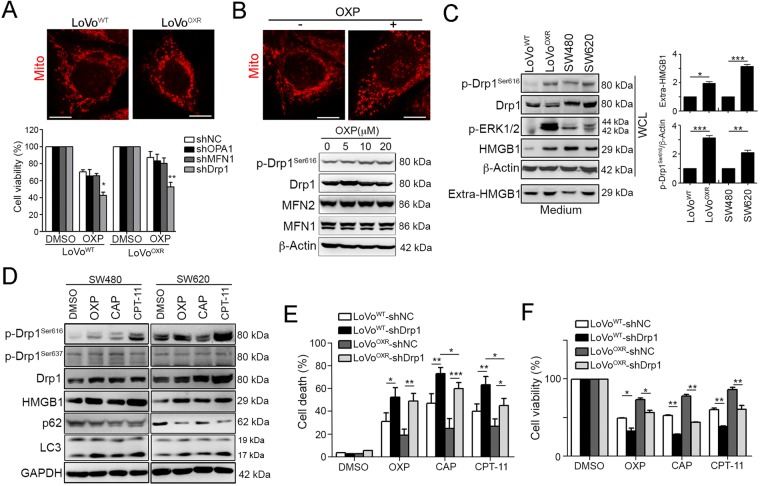
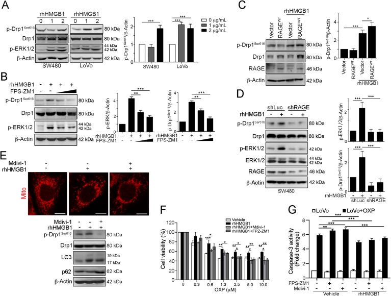
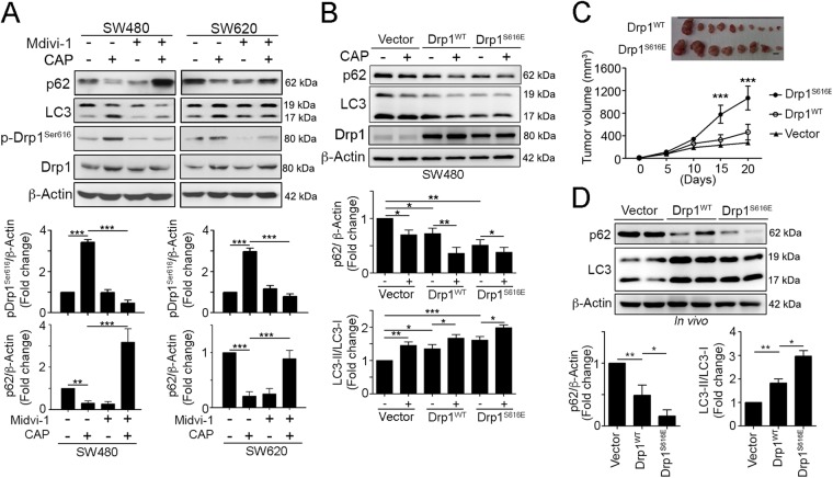
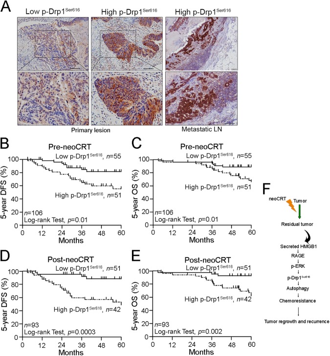
Dysfunctional mitochondria have been shown to enhance cancer cell proliferation, reduce apoptosis, and increase chemoresistance. Chemoresistance develops in nearly all patients with colorectal cancer, leading to a decrease in the therapeutic efficacies of anticancer agents. However, the effect of dynamin-related protein 1 (Drp1)-mediated mitochondrial fission on chemoresistance in colorectal cancer is unclear. Here, we found that the release of high-mobility group box 1 protein (HMGB1) in conditioned medium from dying cells by chemotherapeutic drugs and resistant cells, which triggered Drp1 phosphorylation via its receptor for advanced glycation end product (RAGE). RAGE signals ERK1/2 activation to phosphorylate Drp1 at residue S616 triggerring autophagy for chemoresistance and regrowth in the surviving cancer cells. Abolishment of Drp1 phosphorylation by HMGB1 inhibitor and RAGE blocker significantly enhance sensitivity to the chemotherapeutic treatment by suppressing autophagy. Furthermore, patients with high phospho-Drp1Ser616 are associated with high risk on developing tumor relapse, poor 5-year disease-free survival (DFS) and 5-year overall survival (OS) after neoadjuvant chemoradiotherapy (neoCRT) treatment in locally advanced rectal cancer (LARC). Moreover, patients with RAGE-G82S polymorphism (rs2070600) are associated with high phospho-Drp1Ser616 within tumor microenvironment. These findings suggest that the release of HMGB1 from dying cancer cells enhances chemoresistance and regrowth via RAGE-mediated ERK/Drp1 phosphorylation.


Conflict of interest statement

The authors declare that they have no conflict of interest.

Figures

Fig. 1
Fig. 1. Drp1 is highly phosphorylated upon cytotoxic insults to promote chemoresistance.
a LoVoWT and LoVoOXR were infected by lentivirus carrying shRNA against OPA1, Drp1, and MFN1. These cells were treated with oxaliplatin (OXP, 10 μM) for 48 h. The cell viability was analyzed using an MTT assay (n = 3). *p < 0.01 and **p < 0.01. b Mitochondria morphology was observed by MitoTracker Red staining. Scale bar = 10 μm. c LoVoWT cells were treated with diverse concentrations of OXP for 24 h. Cell lysates were analyzed by immunoblotting. Mitochondria morphology was observed by MitoTracker Red staining. Scale bar = 10 μm. d Cell lysate and medium were analyzed by immunoblotting. Quantification of these results is shown (n = 3). *p < 0.05, **p < 0.01 and ***p < 0.001. e SW480 and SW620 cells were treated with OXP (10 μM), CAP (10 μM), or CPT-11 (5 μM) for 24 h. Cell lysates were analyzed by immunoblotting. f LoVo and LoVoOxR cells with shNC and shDrp1 were treated with OXP (10 μM), CAP (10 μM), or CPT-11 (10 μM) for 48 h. Cell death was analyzed using an AnnexinV/PI assay (n = 3). *p < 0.05, **p < 0.01 and, ***p < 0.001. g LoVo and LoVoOxR cells carrying shNC and shDrp1 were treated with OXP (10 μM Oxaliplatin), CAP (10 μM), or CPT-11 (5 μM) for 48 h. The cell viability was analyzed using an MTT assay (n = 3). *p < 0.05 and **p < 0.01
Fig. 2
Fig. 2. Extracellular HMGB1 and phosphorylated Drp1 were highly upregulated in the chemoresistant CRC in vivo.
a Athymic nude BALB/c mice were subcutaneously inoculated with LoVoWT and LoVoOXR cells into the flank of each mouse. (1 × 106 cells/ mice, n = 6). The tumor volume was measured at various time intervals throughout the study. Quantification of these results is shown (n = 6). *p < 0.05. b Xenografted tumors from representative mice were stained for phospho-Drp1Ser616 by immunohistochemistry. Bar: 10 μm. Xenografted tumors from representative mice were homogenized and isolated for the cytoplasmic and mitochondrial fraction analysis. Then, these fractions were analyzed by immunoblotting. Quantification of these results is shown (n = 3). *p < 0.05 and **p < 0.01. c Xenografted tumors from LoVoWT- and LoVoOXR-injected mice were analyzed by immunohistochemistry. Bar: 10 μm. Serum from LoVo-injected and LoVoOxR-injected mice was analyzed by ELISA. Quantification of these results is shown (n = 6). *p < 0.05. d Xenografted tumors from representative mice were collected for lysis and analyzed by immunoblotting. Quantification of these results is shown (n = 3). *p < 0.05 and **p < 0.01. These data were obtained from three independent experiments, and the values represent the means ± S.D
Fig. 3
Fig. 3. HMGB1 triggered ERK-mediated Drp1 phosphorylation via RAGE in CRC.
a SW480 and LoVo cells were treated with the recombinant human HMGB1 protein (0, 1, and 2 μg/mL) for 24 h. Cell lysate was analyzed by immunoblotting. Quantification of these results is shown (n = 3). ***p < 0.001. b SW480 cells were treated with the recombinant human HMGB1 protein (1 μg/mL) and RAGE inhibitor (10 μM FPS-ZM1) for 24 h. Cell lysate was analyzed by immunoblotting. Quantification of these results is shown (n = 3). **p < 0.01 and ***p < 0.001. c SW480 cells were transfected with the pcDNA3.1-vector, pcDNA3.1-wtRAGE for 24 h and then treated with the recombinant human HMGB1 protein (1 μg/mL) for 24 h. Cell lysate was analyzed by immunoblotting. Quantification of these results is shown (n = 3). **p < 0.01. d SW480 cells were silenced with lentivirus carrying shRNA again RAGE, and then treated with the recombinant human HMGB1 protein (1 μg/mL) for 24 h. Cell lysate was analyzed by immunoblotting. e LoVo cells were treated with recombinant human HMGB1 (1 μg/mL) for 24 h. Cell lysate was analyzed by immunoblotting. Quantification of these results is shown (n = 3). ***p < 0.001. Mitochondria morphology was observed by MitoTracker Red staining. Scale bar = 10 μm. f LoVo cells were treated with RAGE inhibitor (10 μM FPS-ZM1), Drp1 inhibitor (50 μM Mdivi-1) and recombinant human HMGB1 (1 μg/mL) for 1 h prior to addition of different dosages of OXP for 48 h. The cell viability was analyzed using an MTT assay (n = 3). *p < 0.05. g LoVo cells were treated with RAGE inhibitor (10 μM FPS-ZM1), Drp1 inhibitor (50 μM Mdivi-1) and recombinant human HMGB1 (1 μg/mL) for 1 h prior to addition of OXP (10 μM) for 48 h. The caspase-3 activity was analyzed (n = 3). **p < 0.01. These data were obtained from three independent experiments, and the values represent the means ± S.D
Fig. 4
Fig. 4. Extracellular HMGB1 by chemotherapy triggered ERK-mediated Drp1 phosphorylation for autophagy and chemoresistance via RAGE in CRC.
a SW480 and SW620 cells were treated with OXP (10 μM), CAP (10 μM), or CPT-11 (5 μM) for 24 h. Conditioned medium were analyzed by immunoblotting. Quantification of these results is shown (n = 3). *p < 0.05, **p < 0.01 and ***p < 0.001. b SW480 cells carrying shNC, shHMGB1#1 and shHMGB1#2 were treated with various concentration of oxaliplatin for 48 h. The cell viability was analyzed using an MTT assay (n = 3). **p < 0.01. c SW480 cells were treated with CAP (10 μM) and HMGB1 inhibitor (Quercetin, 50 μM) for 24 h. Cell lysate was analyzed by immunoblotting. Quantification of these results is shown (n = 3). **p < 0.01 and ***p < 0.001. d SW480 cells were treated with OXP (10 μM) and PD98059 (10 μM) for 24 h. Cell lysate was analyzed by immunoblotting. e SW480 cells were transfected with the pcDNA3.1-vector, pcDNA3.1-wtRAGE and pcDNA3.1-RAGEΔCyto for 24 h and then treated with OXP (10 μM), CAP (10 μM), or CPT-11 (5 μM) for 48 h. The cell viability was analyzed using an MTT assay (n = 3). *p < 0.05. f SW480 cells carrying shNC and shRAGE were treated with OXP (10 μM Oxaliplatin), CAP (10 μM), or CPT-11 (5 μM) for 48 h. The caspase-3 activity was analyzed (n = 3). **p < 0.01. g SW480 cells carrying shNC and shRAGE were treated with OXP (10 μM), CAP (10 μM), or CPT-11 (5 μM) for 48 h. Cell death was analyzed using an AnnexinV/PI assay (n = 3). *p < 0.05 and ***p < 0.001. h SW480 cells were treated with CAP (10 μM) and RAGE inhibitor (FPS-ZM1, 10 μM) for 24 h. Cell lysate was analyzed by immunoblotting. Quantification of these results is shown (n = 3). **p < 0.01 and ***p < 0.001. i LoVo cells were pre-incubated with anti-RAGE or anti-HMGB1 neutralizing antibodies (2.5 μg/mL) for 30 min prior to addition of recombinant HMGB1 and OXP for 48 h. The cell viability was analyzed using an MTT assay (n = 3). *p < 0.05. These data were obtained from three independent experiments, and the values represent the means ± S.D
Fig. 5
Fig. 5. Hyperactivated Drp1 is required for autophagy turnover and tumor growth of CRC in vivo.
a SW480 and SW620 cells were treated with CAP (10 μM) and a Drp1 inhibitor (50 μM Mdivi-1) for 24 h. Cell lysate were analyzed by immunoblotting. b SW480 cells were transfected with a GFP vector, GFP-wtDrp1 or GFP-Drp1S616E for 24 h and then treated with CAP (10 μM Capecitabine) for 24 h. Cell lysate was analyzed by immunoblotting. Quantification of these results is shown (n = 3). *p < 0.05, **p < 0.01, and ***p < 0.001. c 1 × 106 cells were injected subcutaneously into the flank of each mouse. Tumor volume, body weight and survival time were measured at various time intervals throughout the study. Quantification of these results is shown (n = 5). **p < 0.01. d Xenografted tumors from representative mice were collected for lysis and analyzed by immunoblotting. Quantification of these results is shown (n = 3). *p < 0.05 and **p < 0.01. These data were obtained from three independent experiments, and the values represent the means ± S.D
Fig. 6
Fig. 6. Hyperactivated Drp1 is associated with poor 5-year DFS and 5-year OS tumor growth for advanced metastasis in CRC.
a The expression of phospho-Drp1S616 in the pre-neoCRT biopsies and metastatic lymph nodes. b High phospho-Drp1S616 within the tumor microenvironment of pre-neoCRT biopsies was associated with a poor 5-year DFS. c High phospho-Drp1S616 within the tumor microenvironment of pre-neoCRT biopsies was associated with a poor 5-year OS. d High phospho-Drp1S616 within the tumor microenvironment of post-neoCRT surgical tissues was associated with a poor 5-year DFS and 5-year OS. e High phospho-Drp1S616 within the tumor microenvironment of post-neoCRT surgical tissues was associated with a poor 5-year OS. f The proposed mechanism of HMGB1-mediated Drp1 phosphorylation via RAGE-ERK1/2 signaling to promote chemoresistance in colorectal cancer

References

    1. Siegel RL, et al. Colorectal cancer statistics, 2017. Cancer J. Clin. 2017;67:177–193. doi: 10.3322/caac.21395. - DOI - PubMed
    1. Adlard JW, Richman SD, Seymour MT, Quirke P. Prediction of the response of colorectal cancer to systemic therapy. Lancet Oncol. 2002;3:75–82. doi: 10.1016/S1470-2045(02)00648-4. - DOI - PubMed
    1. Goldberg RM, et al. A randomized controlled trial of fluorouracil plus leucovorin, irinotecan, and oxaliplatin combinations in patients with previously untreated metastatic colorectal cancer. J. Clin. Oncol. 2004;22:23–30. doi: 10.1200/JCO.2004.09.046. - DOI - PubMed
    1. Wang KC, Brooks DA, Botting KJ, Morrison JL. IGF-2R-mediated signaling results in hypertrophy of cultured cardiomyocytes from fetal sheep. Biol. Reprod. 2012;86:183. - PubMed
    1. Zhao J, et al. Mitochondrial dynamics regulates migration and invasion of breast cancer cells. Oncogene. 2013;32:4814–4824. doi: 10.1038/onc.2012.494. - DOI - PMC - PubMed

Publication types

MeSH terms