. 2018 Sep 26;8(1):14398.
doi: 10.1038/s41598-018-32851-y.

ERK activating peptide, AES16-2M promotes wound healing through accelerating migration of keratinocytes

Affiliations

ERK activating peptide, AES16-2M promotes wound healing through accelerating migration of keratinocytes

Sora Lee et al. Sci Rep. .

Abstract

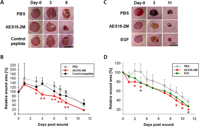
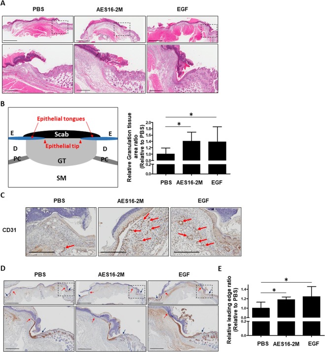
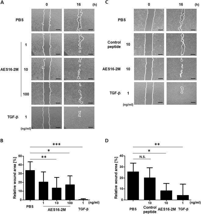
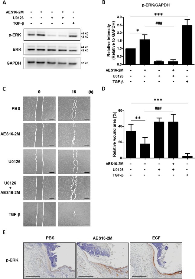
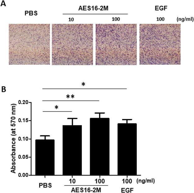
Wound healing is an important issue that influences quality of life, and the need for products associated with wound healing is growing annually. New materials and therapies for skin wounds are being continuously researched and developed in order to increase treatment efficacy. Here, we show that the peptide AES16-2M comprised of five short amino acid sequences (REGRT) demonstrates efficacy in wound healing. AES16-2M exerted more effective healing than the control in an acute wound model, and tissue regeneration was similar to that of normal tissue in AES16-2M-treated skin. We found that the increase in re-epithelialization by AES16-2M early in wound development was due to migration of keratinocytes; a scratch assay using a human keratinocyte cell line (HaCaT) also demonstrated effective wound closure by AES16-2M. The migration of keratinocytes effected by AES16-2M was promoted through ERK phosphorylation and blocked with U0126, an ERK inhibitor. Moreover, AES16-2M treatment stimulated human dermal fibroblast (HDF) migration as well as keratinocyte. Taken together, these results suggest that AES16-2M can be an effective therapeutic agent for wound healing by promoting migration of keratinocytes and fibroblasts via ERK phosphorylation.


Conflict of interest statement

The authors declare no competing interests.

Figures

Figure 1
Figure 1
AES16-2M treatment accelerated wound healing in an in vivo model. Full-thickness wounds were created on the back of BALB/c wild-type (A,B) and BALB/c-nude (C,D) mice. Each group was treated with 50 μl Pluronic® F-127 gel containing either PBS, AES16-2M, control peptide, or EGF. (A) A photographic record of wound healing was processed on days 0, 3, and 8 post-injury. Scale bar: 1 cm. (B) The residual wound area was calculated as the ratio of the relative wound area to the original wound area on day 0 post-injury (n = 5). Error bars, mean ± SD. *p < 0.05, **p < 0.01 (AES16-2M treatment group versus PBS treatment group), two-tailed student t test. (C) A photographic record of wound healing was processed on days 0, 3, and 11 post-injury. Scale bar: 1 cm. (D) The residual wound area was calculated as the ratio of the relative wound area to the original wound area on day 0 post-injury (n = 8). Error bars, mean ± SD. *p < 0.05 (AES16-2M or EGF treatment group versus PBS treatment group), two-tailed student t test.
Figure 2
Figure 2
AES16-2M promoted re-epithelialization via keratinocyte migration. On post-wound day 4, serial sections of skin tissue were prepared and examined. (A) H&E staining was performed and photographed. (B) Schematic representation of wound histology and comparison of granulation tissue area in the AES16-2M treatment group. The area of granulation tissue was measured and graphed based on an H&E-stained section of panel A (n = 8). Error bars, mean ± SD. *p < 0.05 (AES16-2M or EGF treatment group versus PBS treatment group), two-tailed student t test. (C) Immunohistochemistry was performed with anti-CD31 antibody and photographed. Scale bar: 300 μm. (D) Slides were stained with anti-keratin 14 antibody and visualized. The blue arrows indicate the wound margin and the red arrows indicate the end of the epithelial tips. (E) Measurements of leading-edge ratio in panel D. The leading-edge ratio was calculated as the ratio of the length of the wound to the length of the epithelial tongues (n = 6). Error bars, mean ± SD. *p < 0.05 (AES16-2M or EGF treatment group versus PBS treatment group), two-tailed student t test. (A,D) The dashed line in the upper panel is magnified and shown in the lower panel. Upper panel scale bar: 2 mm. Lower panel scale bar: 500 μm.
Figure 3
Figure 3
AES16-2M enhanced HaCaT cell migration. HaCaT cells were cultured in 12-well plates. Cells pre-treated with mitomycin C (10 μg/ml) were scratched and incubated with the indicated dose of AES16-2M, control peptide (10 ng/ml), or TGF-β (1 ng/ml). HaCaT cell migration was visualized and recorded using a microscope at 0 and 16 hr post-scratch. The line indicates the border of wound. Scale bar: 500 μm. The residual area of the wound was measured by Image J. Relative wound area was calculated as the ratio of the remaining area at the 16-hr time point to the 0-hr starting point. The six pairs of time points were separately measured and averaged. (A) The migration effect according to AES16-2M concentration. (B) Graph of measurements in panel A. (C) Comparison of the migration effects of the control peptide and AES16-2M. (D) Graph of measurements in panel C. Error bars, mean ± SD. *p < 0.05, **p < 0.01, ***p < 0.001 (AES16-2M or TGF-β treatment group versus PBS treatment group). N.S. = No significance (control peptide versus PBS treatment group), one-way ANOVA followed by Tukey post-hoc test.
Figure 4
Figure 4
AES16-2M promoted ERK phosphorylation for HaCaT cell migration. (A) HaCaT cells were treated with AES16-2M (10 ng/ml), TGF-β (1 ng/ml), and PBS for 30 min under serum-free conditions. The ERK inhibitor (U0126 at 0.1 μM) was added to cells 1 hr prior to treatment, as indicated. Western blotting was performed for phospho-ERK, total-ERK, and GAPDH. The image presented here is a cropped image; full-length blots are presented in Figure S3. (B) The band intensity of panel A was measured with Image J and relative density was analysed as the ratio of phospho-ERK to GAPDH (n = 3). Error bars, mean ± SD. *p < 0.05, ***p < 0.001 (AES16-2M or TGF-β treatment group versus PBS group), ###p < 0.001 (AES16-2M versus AES16-2M + U0126 treatment group), one-way ANOVA followed by Tukey post-hoc test. (C) For the scratch assay, HaCaT cells were treated with ERK inhibitor (U0126 at 0.1 μM) 1 hr before AES16-2M treatment. Scale bar: 500 μm. (D) The residual area of the wound was measured by Image J and the relative wound area was calculated as the ratio of the remaining area to that at 0 hr (n = 6). Error bars, mean ± SD. **p < 0.01, ***p < 0.001 (AES16-2M or TGF-β treatment group versus PBS group), ###p < 0.001 (AES16-2M versus AES16-2M + U0126 treatment group), one-way ANOVA followed by Tukey post-hoc test. (E) Immunohistochemistry was performed with anti-phosphorylated (p-)ERK antibody and photographed. Scale bar: 300 μm.
Figure 5
Figure 5
AES16-2M induced HDF cell migration. A transwell migration assay was performed with HDF cells. HDF cells were treated with AES16-2M at the indicated doses or EGF (100 ng/ml) and incubated overnight. HDF cells (1 × 104 cells) were seeded onto a transwell plate and incubated for 24 hr. (A) Transwells were stained and recorded by photomicrograph. (B) Bar graph showing the eluted density of migrated cells in each group (n = 3). The eluted cells were measured by ELISA. Error bars, mean ± SD. *p < 0.05, **p < 0.01 (AES16-2M or TGF-β treatment group versus control group), one-way ANOVA followed by Tukey post-hoc test.

References

    1. Li J, Chen J, Kirsner R. Pathophysiology of acute wound healing. Clinics in dermatology. 2007;25:9–18. doi: 10.1016/j.clindermatol.2006.09.007. - DOI - PubMed
    1. Gurtner GC, Werner S, Barrandon Y, Longaker MT. Wound repair and regeneration. Nature. 2008;453:314–321. doi: 10.1038/nature07039. - DOI - PubMed
    1. Greaves NS, Ashcroft KJ, Baguneid M, Bayat A. Current understanding of molecular and cellular mechanisms in fibroplasia and angiogenesis during acute wound healing. Journal of dermatological science. 2013;72:206–217. doi: 10.1016/j.jdermsci.2013.07.008. - DOI - PubMed
    1. Santoro MM, Gaudino G. Cellular and molecular facets of keratinocyte reepithelization during wound healing. Experimental cell research. 2005;304:274–286. doi: 10.1016/j.yexcr.2004.10.033. - DOI - PubMed
    1. Demidova-Rice TN, Hamblin MR, Herman IM. Acute and impaired wound healing: pathophysiology and current methods for drug delivery, part 1: normal and chronic wounds: biology, causes, and approaches to care. Advances in skin & wound care. 2012;25:304–314. doi: 10.1097/01.ASW.0000416006.55218.d0. - DOI - PMC - PubMed

Publication types

LinkOut - more resources