

Impact of Age and Diastolic Function on Novel, 4D flow CMR Biomarkers of Left Ventricular Blood Flow Kinetic Energy
- PMID: 30258186
- PMCID: PMC6158175
- DOI: 10.1038/s41598-018-32707-5
Impact of Age and Diastolic Function on Novel, 4D flow CMR Biomarkers of Left Ventricular Blood Flow Kinetic Energy
Abstract
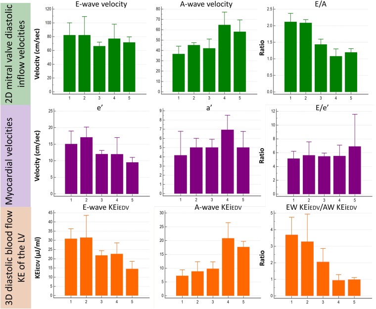
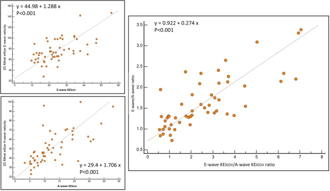
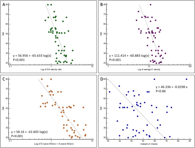
Two-dimensional (2D) methods of assessing mitral inflow velocities are pre-load dependent, limiting their reliability for evaluating diastolic function. Left ventricular (LV) blood flow kinetic energy (KE) derived from four-dimensional flow cardiovascular magnetic resonance imaging (4D flow CMR) may offer improvements. It remains unclear whether 4D LV blood flow KE parameters are associated with physiological factors, such as age when compared to 2D mitral inflow velocities. Fifty-three healthy volunteers underwent standard CMR, plus 4D flow acquisition. LV blood flow KE parameters demonstrated good reproducibility with mean coefficient of variation of 6 ± 2% and an accuracy of 99% with a precision of 97%. The LV blood flow KEiEDV E/A ratio demonstrated good association to the 2D mitral inflow E/A ratio (r = 0.77, P < 0.01), with both decreasing progressively with advancing age (P < 0.01). Furthermore, peak E-wave KEiEDV and A-wave KEiEDV displayed a stronger association to age than the corresponding 2D metrics, peak E-wave and A-wave velocity (r = -0.51 vs -0.17 and r = 0.65 vs 0.46). Peak E-wave KEiEDV decreases whilst peak A-wave KEiEDV increases with advancing age. This study presents values for various LV blood flow KE parameters in health, as well as demonstrating that they show stronger and independent correlations to age than standard diastolic metrics.
Conflict of interest statement
The authors declare no competing interests.
Figures

References
-
- Nagueh SF, et al. Recommendations for the Evaluation of Left Ventricular Diastolic Function by Echocardiography: An Update from the American Society of Echocardiography and the European Association of Cardiovascular Imaging. J. Am. Soc. Echocardiogr. 2016;29:277–314. doi: 10.1016/j.echo.2016.01.011. - DOI - PubMed
-
- Erhayiem B, et al. 85 Newly Diagnosed, Treatment-Naive Patients with Rheumatoid Arthritis have early Abnormalities of Vascular and Myocardial Function. Heart. 2015;101:A46–A47.
Publication types
MeSH terms
Grants and funding
LinkOut - more resources
Full Text Sources
Other Literature Sources
Medical
