. 2018 Sep 26;8(1):14385.
doi: 10.1038/s41598-018-32773-9.

Neprilysin inhibition promotes corneal wound healing

Affiliations

Neprilysin inhibition promotes corneal wound healing

Rachel M Genova et al. Sci Rep. .

Abstract

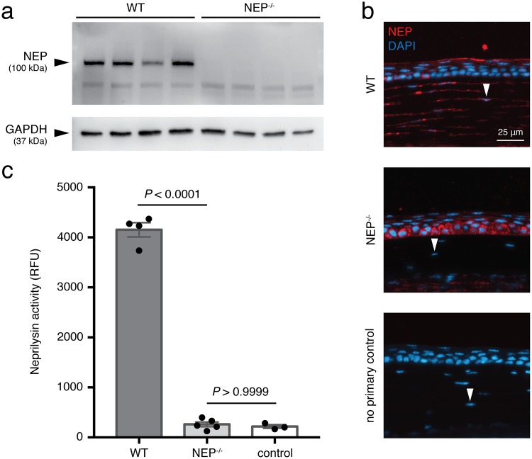
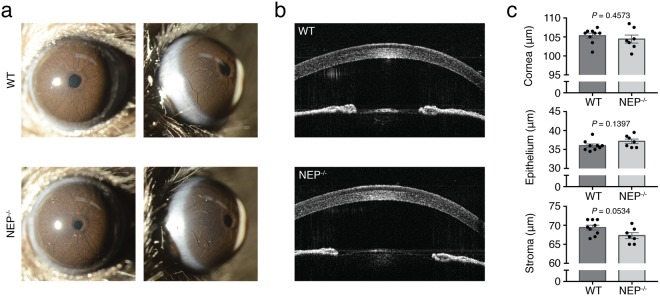
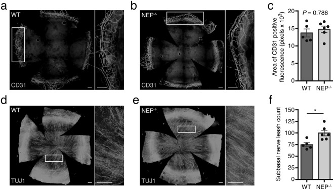
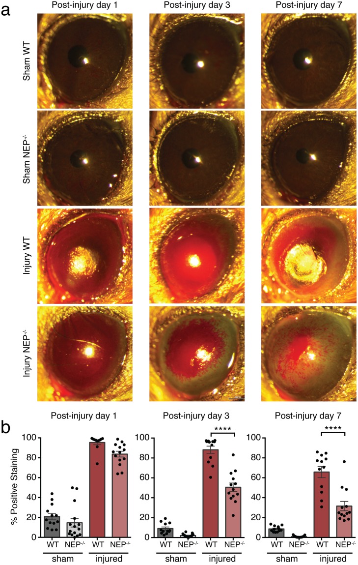
Neprilysin (NEP), an ectoenzyme that modulates inflammation by degrading neuropeptides, was recently identified in the human corneal epithelium. The cornea expresses many NEP substrates, but the function of NEP in homeostatic maintenance and wound healing of the cornea is unknown. We therefore investigated the role of this enzyme under naive and injured conditions using NEP-deficient (NEP-/-) and wild type (WT) control mice. In vivo ocular surface imaging and histological analysis of corneal tissue showed no differences in limbal vasculature or corneal anatomy between naive NEP-/- and WT mice. Histological examination revealed increased corneal innervation in NEP-/- mice. In an alkali burn model of corneal injury, corneal wound healing was significantly accelerated in NEP-/- mice compared to WT controls 3 days after injury. Daily intraperitoneal administration of the NEP inhibitor thiorphan also accelerated corneal wound healing after alkali injury in WT mice. Collectively, our data identify a previously unknown role of NEP in the cornea, in which pharmacologic inhibition of its activity may provide a novel therapeutic option for patients with corneal injury.


Conflict of interest statement

The authors declare no competing interests.

Figures

Figure 1
Figure 1
Functional expression of NEP in the mouse cornea. (a) Western blot showing expression of NEP in whole cornea lysates from WT and NEP−/− mice with glyceraldehyde 3-phosphate dehydrogenase (GAPDH) run as a loading control on the same gel. Membrane was cut at 50 kDa. (b) Representative immunofluorescence images showing immunostaining for NEP (red) and DAPI (blue) in WT corneal sections (n = 3) compared to NEP−/− (n = 3) and no primary control (n = 2) sections. NEP localizes to superficial epithelial cells and stromal keratocytes (arrowheads) in WT sections. Original magnification, 40×; scale bar, 25 μm. (c) NEP activity in whole cornea lysates, determined with a FRET-based assay (mean ± SEM; ****P < 0.0001; two-tailed t-test).
Figure 2
Figure 2
Gross corneal phenotype of NEP−/− mice. (a) Broad beam slit lamp images of WT and NEP−/− eyes showing corneal surfaces and limbal regions (n = 3 mice/group). (b) Representative images of WT and NEP−/− corneas obtained with anterior segment optical coherence tomography. (c) Quantification of central corneal thickness and thickness of epithelial and stromal layers (n = 7–9 mice/group; mean ± SEM; two-tailed t-test).
Figure 3
Figure 3
Limbal vasculature and corneal innervation in WT and NEP−/− mice. Representative corneal flat mounts from a WT (a) and NEP−/− (b) mouse immunostained with the vascular marker CD31. Higher magnification images of boxed areas in (a) and (b) are shown to the right of each image, respectively. (c) Area of positive CD31 immunofluorescence (n = 5–6 mice/group, mean ± SEM; two-tailed t-test). Representative corneal flat mounts from a WT (d) and NEP−/− (e) mouse immunostained with the pan-neuronal marker βIII tubulin (TUJ1). Higher magnification images of boxed areas in (d) and (e) are shown to the right of each image, respectively. (f) Subbasal nerve leashes (n = 5–6 mice/group, mean ± SEM; *P = 0.0103; two-tailed t-test). Original magnification, 10×; scale bar, 250 μm for all images.
Figure 4
Figure 4
Corneal wound healing after alkali burn in WT and NEP−/− mice. (a) Representative broad beam slit lamp images obtained at 1, 3, and 7 days post-injury with rose bengal instillation to visualize corneal defects. (b) Quantification of positive rose bengal staining reported as a percentage of total corneal area at 1, 3, and 7 days post-injury (n = 12–18 mice/group at each timepoint; mean ± SEM; ****P < 0.0001; one-way ANOVA with Bonferroni correction).
Figure 5
Figure 5
Corneal wound healing after alkali burn with thiorphan administration in WT mice. (a) Representative broad beam slit lamp images obtained at 1, 3, and 7 days post-injury with rose bengal instillation to visualize corneal defects. (b) Quantification of positive rose bengal staining reported as a percentage of total corneal area at 1, 3, and 7 days post-injury (n = 5–10 mice/group at each timepoint; mean ± SEM; ***P = 0.0004; one-way ANOVA with Bonferroni correction). V, vehicle; 5, 5 mg/kg thiorphan; 15, 15 mg/kg thiorphan.

References

    1. Eghrari AO, Riazuddin SA, Gottsch JD. Overview of the cornea: Structure, function, and development. Prog. Mol. Biol. Transl. Sci. 2015;134:7–23. doi: 10.1016/bs.pmbts.2015.04.001. - DOI - PubMed
    1. Négrel AD, Thylefors B. The global impact of eye injuries. Ophthalmic Epidemiol. 1998;5:143–169. doi: 10.1076/opep.5.3.143.8364. - DOI - PubMed
    1. Haring RS, Canner JK, Haider AH, Schneider EB. Ocular injury in the United States: Emergency department visits from 2006–2011. Injury. 2016;47:104–108. doi: 10.1016/j.injury.2015.07.020. - DOI - PubMed
    1. Haring RS, Sheffield ID, Channa R, Canner JK, Schneider EB. Epidemiologic trends of chemical ocular burns in the United States. JAMA Ophthalmol. 2016;134:1119–1124. doi: 10.1001/jamaophthalmol.2016.2645. - DOI - PubMed
    1. Rozenbaum D, Baruchin AM, Dafna Z. Chemical burns of the eye with special reference to alkali burns. Burns. 1991;17:136–140. doi: 10.1016/0305-4179(91)90137-6. - DOI - PubMed

Publication types