. 2018 Sep 26;8(1):14413.
doi: 10.1038/s41598-018-32840-1.

Cell type-specific activation of mitogen-activated protein kinase in D1 receptor-expressing neurons of the nucleus accumbens potentiates stimulus-reward learning in mice

Affiliations

Cell type-specific activation of mitogen-activated protein kinase in D1 receptor-expressing neurons of the nucleus accumbens potentiates stimulus-reward learning in mice

Md Ali Bin Saifullah et al. Sci Rep. .

Abstract

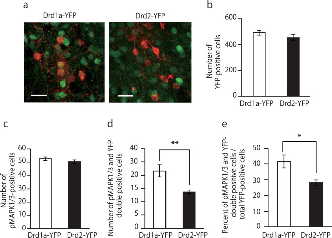
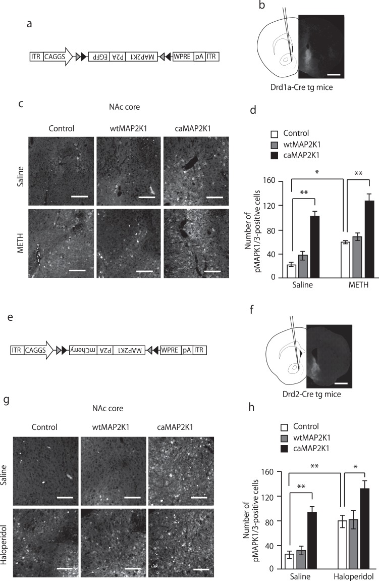
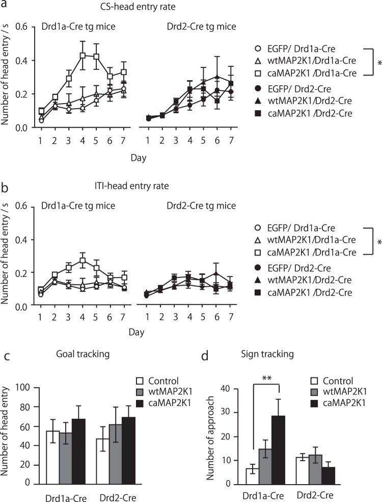
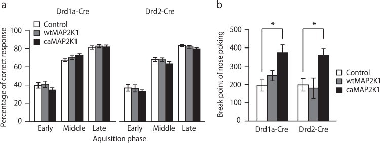
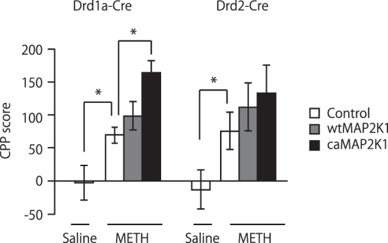
Medium spiny neurons (MSN) in the nucleus accumbens (NAc) are a fundamental component of various aspects of motivated behavior. Although mitogen-activated protein kinase (MAPK) signaling plays a crucial role in several types of learning, the cell type-specific role of MAPK pathway in stimulus-reward learning and motivation remains unclear. We herein investigated the role of MAPK in accumbal MSNs in reward-associated learning and memory. During the acquisition of Pavlovian conditioning, the number of phosphorylated MAPK1/3-positive cells was increased significantly and exclusively in the NAc core by 7-days of extensive training. MAPK signaling in the respective D1R- and D2R-MSNs was manipulated by transfecting an adeno-associated virus (AAV) plasmid into the NAc of Drd1a-Cre and Drd2-Cre transgenic mice. Potentiation of MAPK signaling shifted the learning curve of Pavlovian conditioning to the left only in Drd1a-Cre mice, whereas such manipulation in D2R-MSNs had negligible effects. In contrast, MAPK manipulation in D2R-MSNs of the NAc core significantly increased motivation for food rewards as found in Drd1a-Cre mice. These results suggest that MAPK signaling in the D1R-MSNs of NAc core plays an important role in stimulus-reward learning, while MAPK signaling in both D1R- and D2R-MSNs is involved in motivation for natural rewards.


Conflict of interest statement

The authors declare no competing interests.

Figures

Figure 1
Figure 1
Phosphorylation of MAPK1/3 in the NAc during acquisition of the Pavlovian conditional approach. (a) Schematic representation of the Pavlovian conditioning procedure. (b) CS-head entry rate in paired CS-US and unpaired CS-US control mice. In unpaired CS-US control group, the timing of food pellet delivery changed randomly within a 60-s trial throughout the session, so that there was no contingency between CS and food reward delivery. (Unpaired CS-US: n = 7; paired CS-US: n = 12). (c) Head entry rate during ITI period in C57BL/6 mice (unpaired CS-US: n = 7; paired CS-US: n = 12). (d) Confocal images of pMAPK1/3-positive cells in the NAc core after training session in the unpaired CS-US and paired CS-US group. (e) Number of pMAPK1/3 in unpaired CS-US and paired CS-US group after training session on day 1 and day 7. (f) Confocal images of total MAPK1/3-positive cells in the NAc core. (g) Number of total MAPK1/3-positive cells in the NAc core of C57BL/6 mice after the training session of Pavlovian conditioning on days 1 and 7 (Day 1: unpaired CS-US, n = 4; paired CS-US, n = 4; Day 7: unpaired CS-US, n = 4; paired CS-US, n = 5). Data are presented as the mean ± SEM. *p < 0.05. Scale bars represent 100 μm.
Figure 2
Figure 2
Proportion of phosphorylated MAPK1/3-positive cells in the NAc core of Pavlovian-conditioned mice. (a) Representative confocal images of pMAPK1/3-positive cells in the NAc core of Drd1a-YFP and Drd2-YFP mice after the training session of Pavlovian conditioning on day 7. (bd) Quantification of YFP-positive (b), pMAPK1/3-positive (c) and double-positive (d) cells in each mouse line. (e) Percentage of pMAPK1/3 and YFP double-positive cells in all pMAPK1/3-positive cells in each mouse line (n = 4 for Drd1a-YFP and n = 5 for Drd2-YFP). Data are presented as the mean ± SEM. *p < 0.01, **p < 0.01. Scale bars represent 25 μm.
Figure 3
Figure 3
Manipulation of MAPK signaling in accumbal D1R and D2R-MSNs. (a) Schematic diagram shows the AAV construct. (b) Stereotaxic injection of AAVs into the NAc of Drd1a-Cre transgenic mice and representative coronal brain slices showing the expression of EGFP (shown in grey) 3 weeks after the AAV injection. The scale bar represents 1 mm. (c,d) pMAPK1/3-positive cells in the NAc of AAV-mutant MAP2K1-injected Drd1a-Cre transgenic mice. Drd1a-Cre transgenic mice were microinjected with AAV-mutant MAP2K1 into the NAc. Three weeks after the treatment, mice were administered saline or METH (10 mg/kg, i.p.) 30 minutes before perfusion. (c) Representative confocal images of pMAPK1/3. The upper panel represents saline-treated mice. The lower panel represents METH-treated mice. Scale bars represent 100 μm. (d) Quantification of pMAPK1/3-positive cells in the NAc core (n = 5 for the saline-treated control, n = 3 for saline-treated wtMAP2K1, n = 3 for saline-treated caMAP2K1, n = 4 for the METH-treated control, n = 3 for METH-treated wtMAP2K1, n = 4 for METH-treated caMAP2K1). (e) Schematic diagram shows the AAV construct. (f) Stereotaxic injection of AAVs into the NAc of Drd2-Cre transgenic mice and representative coronal brain slices showing the expression of mCherry (shown in grey) 3 weeks after the AAV injection. The scale bar represents 1 mm. (g,h) pMAPK1/3-positive cells in the NAc of AAV-mutant MAP2K1-injected Drd2-Cre transgenic mice. Drd2-Cre transgenic mice were microinjected with AAV-mutant MAP2K1 into the NAc. Three weeks after the treatment, mice were administered saline or haloperidol (0.5 mg/kg, i.p.) 15 minutes before perfusion. (g) Representative confocal images of pMAPK1/3. The upper panel represents saline-treated mice. The lower panel represents haloperidol-treated mice. Scale bars represent 100 μm. (h) Quantification of pMAPK1/3-positive cells in the NAc core (n = 5 for the saline-treated control, n = 3 for saline-treated wtMAP2K1, n = 3 for saline-treated caMAP2K1, n = 4 for the haloperidol-treated control, n = 3 for haloperidol-treated wtMAP2K1, n = 3 for haloperidol-treated caMAP2K1). Data are presented as the mean ± SEM. *p < 0.01, **p < 0.01.
Figure 4
Figure 4
Hyperactivity of MAPK signaling in accumbal D1R-MSNs, but not in D2R-MSNs potentiates learning in Pavlovian conditioning. (a,b) Head entry rate during CS presentation (a) and ITI (b) period in mutant MAP2K1-transfected Drd1a-Cre and Drd2-Cre mice. (c,d) Number of conditional approaches for goal-tracking (c) and sign-tracking (d) behaviors in mutant MAP2K1-transfected Drd1a-Cre and Drd2-Cre mice on day 7 (n = 9 for the control/Drd1a-Cre, n = 13 for wtMAP2K1/Drd1a-Cre, n = 8 for caMAP2K1/Drd1a-Cre, n = 11 for the control/Drd2-Cre, n = 9 for wtMAP2K1/Drd2-Cre, n = 12 for caMAP2K1/Drd2-Cre). Data are presented as the mean ± SEM. *p < 0.01, **p < 0.01.
Figure 5
Figure 5
Hyperactivity of MAPK signaling in accumbal D1R-MSNs and D2R-MSNs increases motivation for food rewards but not instrumental learning. (a) Percentage of correct responses during the acquisition phase of the FR2 random nose-poking task in AAV-mutant MAP2K1-microinjected Drd1a-Cre and Drd2-Cre mice. (b) Break point of nose poking during the progressive ratio task in AAV-mutant MAP2K1-microinjected Drd1a-Cre and Drd2-Cre mice (n = 9 for the control/Drd1a-Cre, n = 13 for wtMAP2K1//Drd1a-Cre, n = 8 for caMAP2K1/Drd1a-Cre, n = 11 for the control/Drd2-Cre, n = 9 for wtMAP2K1/Drd2-Cre, n = 12 for caMAP2K1/Drd2-Cre). Data are presented as the mean ± SEM. *p < 0.05.
Figure 6
Figure 6
Manipulation of MAPK signaling in accumbal D1R-MSNs controls METH-induced CPP. Effects of the AAV-mediated expression of mutant MAP2K1 on methamphetamine-induced CPP in Drd1a-Cre and Drd2-Cre mice (n = 16 for the saline-treated control/ Drd1a-Cre, n = 18 for the METH-treated control/Drd1a-Cre, n = 19 for METH-treated wtMAP2K1/Drd1a-Cre, n = 17 for METH-treated caMAP2K1/Drd1a-Cre, n = 13 for the saline-treated control/Drd2-Cre, n = 10 for the METH-treated control/Drd2-Cre, n = 11 for METH-treated wtMAP2K1/Drd2-Cre, n = 11 for METH-treated caMAP2K1/Drd2-Cre). Data are presented as the mean ± SEM. *p < 0.05.

References

    1. Eyny YS, Horvitz JC. Opposing roles of D1 and D2 receptors in appetitive conditioning. J. Neurosci. 2003;23:1584–1587. doi: 10.1523/JNEUROSCI.23-05-01584.2003. - DOI - PMC - PubMed
    1. Gore BB, Zweifel LS. Genetic reconstruction of dopamine D1 receptor signaling in the nucleus accumbens facilitates natural and drug reward responses. J. Neurosci. 2013;33:8640–8649. doi: 10.1523/jneurosci.5532-12.2013. - DOI - PMC - PubMed
    1. Brown PL, Jenkins HM. Auto-shaping of the pigeon’s key-peck. J. Exp. Anal. Behav. 1968;11:1–8. doi: 10.1901/jeab.1968.11-1. - DOI - PMC - PubMed
    1. Boakes RA, Poli M, Lockwood MJ, Goodall G. A study of misbehavior: token reinforcement in the rat. J. Exp. Anal. Behav. 1978;29:115–134. doi: 10.1901/jeab.1978.29-115. - DOI - PMC - PubMed
    1. Wise RA. Dopamine, learning and motivation. Nat. Rev. Neurosci. 2004;5:483–494. doi: 10.1038/nrn1406. - DOI - PubMed

Publication types

Substances