

Biomimetic matrices for rapidly forming mineralized bone tissue based on stem cell-mediated osteogenesis
- PMID: 30258220
- PMCID: PMC6158243
- DOI: 10.1038/s41598-018-32794-4
Biomimetic matrices for rapidly forming mineralized bone tissue based on stem cell-mediated osteogenesis
Abstract
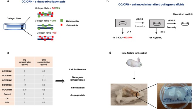
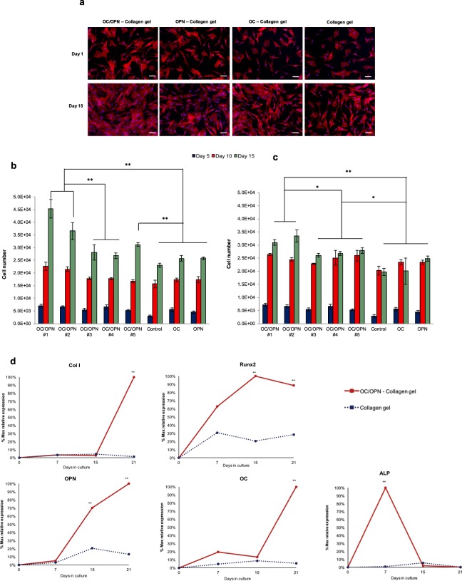
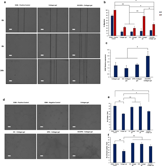
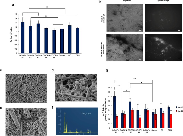
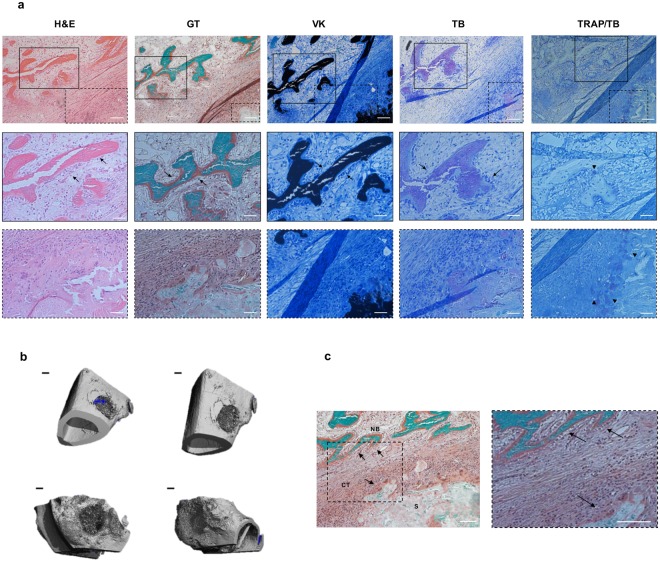
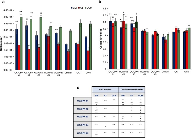
Bone regeneration, following fracture, relies on autologous and allogenic bone grafts. However, majority of fracture population consists of older individuals with poor quality bone associated with loss and/or modification of matrix proteins critical for bone formation and mineralization. Allografts suffer from same limitations and carry the risk of delayed healing, infection, immune rejection and eventual fracture. In this work, we apply a synergistic biomimetic strategy to develop matrices that rapidly form bone tissue - a critical aspect of fracture healing of weight bearing bones. Collagen matrices, enhanced with two selected key matrix proteins, osteocalcin (OC) and/or osteopontin (OPN), increased the rate and quantity of synthesized bone matrix by increasing mesenchymal stem/stromal cell (MSC) proliferation, accelerating osteogenic differentiation, enhancing angiogenesis and showing a sustained bone formation response from MSC obtained from a variety of human tissue sources (marrow, fat and umbilical cord). In vivo assessment of OC/OPN mineralized scaffolds in a critical sized-defect rabbit long-bone model did not reveal any foreign body reaction while bone tissue was being formed. We demonstrate a new biomimetic strategy to rapidly form mineralized bone tissue and secure a sustained bone formation response by MSC from multiple sources, thus facilitating faster patient recovery and treatment of non-union fractures in aging and diseased population. Acellular biomimetic matrices elicit bone regeneration response from MSC, obtained from multiple tissue sources, and can be used in variety of scaffolds and made widely available.
Conflict of interest statement
Patent application (US serial No. 15/570,942) was filed under 35 U.S.C. § 371 of International application number PCT/US2016/030410 by Rensselaer Polytechnic Institute with A. Poundarik, M. Carvalho and D. Vashishth as inventors. Composition of OC/OPN presented in this paper were included in the application to show bone regeneration. This research was supported by NSF grant #1462613. A. Poundarik and D. Vashishth have an equity interest in Orthograft LLC, a company that may potentially benefit from the research results. The terms of this arrangement have been reviewed and approved by Rensselaer Polytechnic Institute in accordance with its conflict of interest policies.
Figures
Similar articles
-
Synergistic effect of extracellularly supplemented osteopontin and osteocalcin on stem cell proliferation, osteogenic differentiation, and angiogenic properties.J Cell Biochem. 2019 Apr;120(4):6555-6569. doi: 10.1002/jcb.27948. Epub 2018 Oct 25. J Cell Biochem. 2019. PMID: 30362184
-
Guided bone regeneration in pig calvarial bone defects using autologous mesenchymal stem/progenitor cells - a comparison of different tissue sources.J Craniomaxillofac Surg. 2012 Jun;40(4):310-20. doi: 10.1016/j.jcms.2011.05.004. Epub 2011 Jun 30. J Craniomaxillofac Surg. 2012. PMID: 21723141
-
Rapid biomimetic mineralization of collagen fibrils and combining with human umbilical cord mesenchymal stem cells for bone defects healing.Mater Sci Eng C Mater Biol Appl. 2016 Nov 1;68:43-51. doi: 10.1016/j.msec.2016.05.104. Epub 2016 May 25. Mater Sci Eng C Mater Biol Appl. 2016. PMID: 27523994
-
Recapitulating endochondral ossification: a promising route to in vivo bone regeneration.J Tissue Eng Regen Med. 2015 Aug;9(8):889-902. doi: 10.1002/term.1918. Epub 2014 Jun 11. J Tissue Eng Regen Med. 2015. PMID: 24916192 Review.
-
Genetically Engineered-MSC Therapies for Non-unions, Delayed Unions and Critical-size Bone Defects.Int J Mol Sci. 2019 Jul 12;20(14):3430. doi: 10.3390/ijms20143430. Int J Mol Sci. 2019. PMID: 31336890 Free PMC article. Review.
Cited by
-
Role of offset and gradient architectures of 3-D melt electrowritten scaffold on differentiation and mineralization of osteoblasts.Biomater Res. 2020 Jan 3;24:2. doi: 10.1186/s40824-019-0180-z. eCollection 2020. Biomater Res. 2020. PMID: 31911842 Free PMC article.
-
Biodegradable Hydrogel Beads Combined with Calcium Phosphate Bone Cement for Bone Repair: In Vitro and In Vivo Characterization.Polymers (Basel). 2022 Jan 27;14(3):505. doi: 10.3390/polym14030505. Polymers (Basel). 2022. PMID: 35160495 Free PMC article.
-
Efficacy of Three-Dimensional Bioactive Composites in Long Bone Repair with Photobiomodulation.Materials (Basel). 2025 Apr 9;18(8):1704. doi: 10.3390/ma18081704. Materials (Basel). 2025. PMID: 40333272 Free PMC article.
-
Application of shear stress for enhanced osteogenic differentiation of mouse induced pluripotent stem cells.Sci Rep. 2022 Nov 8;12(1):19021. doi: 10.1038/s41598-022-21479-8. Sci Rep. 2022. PMID: 36347883 Free PMC article.
-
Osteopontin - The stirring multifunctional regulatory factor in multisystem aging.Front Endocrinol (Lausanne). 2022 Dec 22;13:1014853. doi: 10.3389/fendo.2022.1014853. eCollection 2022. Front Endocrinol (Lausanne). 2022. PMID: 36619570 Free PMC article. Review.
References
-
- MDI, U.S. Markets for Biomaterials; Medical Data International, Inc.: Santa Ana, CA, USA, 2000.
Publication types
MeSH terms
Substances
Grants and funding
LinkOut - more resources
Full Text Sources
Other Literature Sources
Research Materials
