Review
. 2018 Oct;21(10):1341-1349.
doi: 10.1038/s41593-018-0238-6. Epub 2018 Sep 26.

Propagation and spread of pathogenic protein assemblies in neurodegenerative diseases

Affiliations
Review

Propagation and spread of pathogenic protein assemblies in neurodegenerative diseases

Mathias Jucker et al. Nat Neurosci. 2018 Oct.

Abstract

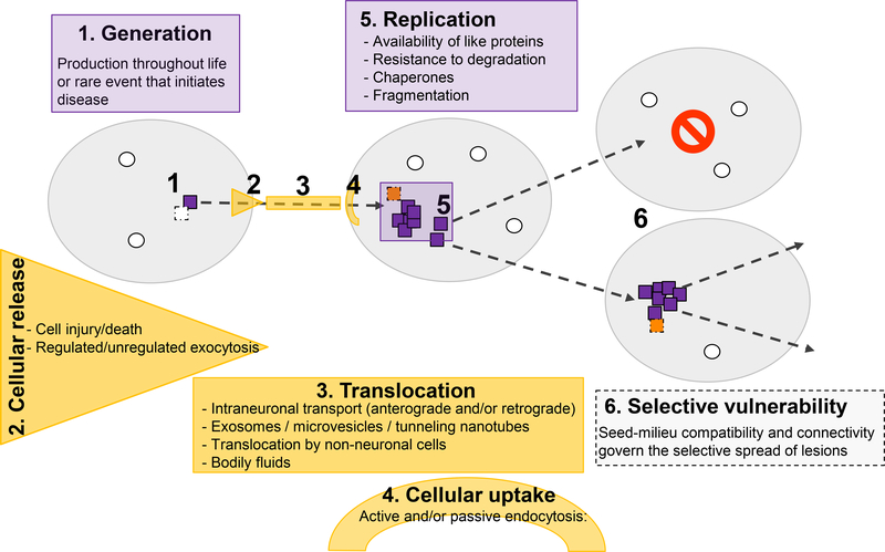
Many neurodegenerative diseases, such as Alzheimer's disease, Parkinson's disease, and amyotrophic lateral sclerosis, are characterized by the progressive appearance of abnormal proteinaceous assemblies in the nervous system. Studies in experimental systems indicate that the assemblies originate from the prion-like seeded aggregation of specific misfolded proteins that proliferate and amass to form the intracellular and/or extracellular lesions typical of each disorder. The host in which the proteopathic seeds arise provides the biochemical and physiological environment that either supports or restricts their emergence, proliferation, self-assembly, and spread. Multiple mechanisms influence the spatiotemporal spread of seeds and the nature of the resulting lesions, one of which is the cellular uptake, release, and transport of seeds along neural pathways and networks. The characteristics of cells and regions in the affected network govern their vulnerability and thereby influence the neuropathological and clinical attributes of the disease. The propagation of pathogenic protein assemblies within the nervous system is thus determined by the interaction of the proteopathic agent and the host milieu.


Conflict of interest statement

Competing interests: The authors declare no competing interests.

Figures

Figure 1.
Figure 1.
Factors governing the genesis, replication, and spread of proteopathic seeds. Disease-specific seeds (orange) are generated when certain normally produced proteins (green) misfold, in which state they structurally corrupt like proteins and self-assemble into multimers. Whether seeds are produced throughout life and usually are actively removed or whether this is a rare event that inevitably marks the beginning of disease remains to be determined. The seeds move from one location to another by any of several potential mechanisms; in some instances, the affected site can be extracellular. All of these phenomena may contribute to selective local vulnerability; they can vary in different cell types and regions of the nervous system, where such factors as the presence of auxiliary agents for replication, transport and uptake mechanisms differ (dark blue, a cell in which different agents restrict further propagation). In addition, the expression level or isoform of the cognate proteins may differ among cells and compartments, thereby further supporting or restricting the spread of the seeds and/or defining the strain of seed that is propagated (see Figs. 2 and 3).
Figure 2.
Figure 2.
Compatibility of seed and cognate host protein regulates the propagation of proteopathic seeds at the organismic level. A,B, Proteopathic seeds isolated from a human brain are conformationally heterogeneous (colored dots). In a murine host brain milieu that is permissive for amplification of the dominant conformation (orange), lesions with a molecular structure similar to that in the donor brain will be preferentially propagated following introduction of the exogenous seeds. A, To facilitate seeding, the host mice are often transgenic (Tg), and are engineered to express the human protein that forms specific lesions in the human brain. Seeding efficiency is augmented by high (transgenic) expression of the cognate protein in the host (orange mouse). B, Wild-type (WT) mice usually are more restrictive in propagating the human conformation (for instance, owing to different amino acid sequences in the proteins), but in some cases they may permit the propagation of a subconformation (gray dots). The more abundant the exogenous seeds and the closer their structural characteristics to the host protein, the more likely and efficient their propagation in the host. Hence, transmission of proteopathic lesions from a human donor to a WT mouse (gray) typically requires longer incubation times and sometimes may never occur during the lifetime of the mouse (see also Table 1).
Figure 3.
Figure 3.
Compatibility of seed and cognate cellular protein governs propagation at the cellular or compartmental level. For seeded propagation and spread, proteopathic seeds must translocate from cell-to-cell or compartment-to-compartment, and they must be replicated at each successive location (see also Fig. 1). Both steps are dependent on the host, and can vary in different cell types such as neurons and glia (upper and lower cells, respectively, in panels A–C), in which such factors as protein expression, isoforms, and auxiliary molecules influence cell tropism. As a result, some cells resist seeding, and others may select for particular proteopathic conformations (different colored dots). A, Neurons and glia both select for the same strains. B, Neurons and glia select for different strains; here the glia generate secondary seeds that differ from the initial seed. C, The glia are incapable of replicating any pathogenic form of the protein.

References

    1. Paget S The distribution of secondary growths in cancer of the breast. The Lancet 133, 571–573 (1889). - PubMed
    1. Prusiner SB Novel proteinaceous infectious particles cause scrapie. Science 216, 136–144 (1982). - PubMed
    1. Prusiner SB Biology and genetics of prions causing neurodegeneration. Annu. Rev. Genet 47, 601–623 (2013). - PMC - PubMed
    1. Mead S & Reilly MM A new prion disease: relationship with central and peripheral amyloidoses. Nat. Rev. Neurol 11, 90–97 (2015). - PubMed
    1. Imran M & Mahmood S An overview of animal prion diseases. Virol J. 8, 493 (2011). - PMC - PubMed

Publication types