

Clinical Presentation, Neuroimaging Findings, and Predictors of Brain Parenchymal Lesions in Cerebral Vein and Dural Sinus Thrombosis: A Retrospective Study
- PMID: 30258263
- PMCID: PMC6137629
- DOI: 10.4103/aian.AIAN_470_17
Clinical Presentation, Neuroimaging Findings, and Predictors of Brain Parenchymal Lesions in Cerebral Vein and Dural Sinus Thrombosis: A Retrospective Study
Abstract
Introduction: Cerebral venous sinus thrombosis (CVST) is an unusual cause of stroke with potentially serious consequences. This study was designed to investigate the clinical and neuroimaging features in patients with CVST and to analyze the predictors of brain parenchymal lesions.
Materials and methods: A retrospective study of 181 patients with CVST was conducted in a tertiary care hospital.
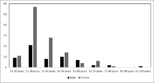
Results: Of 181 patients (age range 14-96 years, mean age: 34.64 ± 14.66 years), 121 were female (66.9%). Most of the patients were in their third decade of life. Headache (47.51%) was the most common clinical presentation followed by seizures (24.31%). Transverse sinus (TS) (77.9%) was the most common site of venous sinus thrombosis. Brain parenchymal lesions were present in 63%, and each patient had subarachnoid and intraventricular hemorrhage. Hemorrhagic venous infarct was the most common brain parenchymal lesion (37.57%). Frontal region (25.4%) was the most common site of brain parenchymal lesions followed by frontoparietal region (21.9%). Women were more likely to have brain parenchymal lesions (72.4%, P = 0.034). Headache was the most common clinical presentation in patients without brain parenchymal lesions while seizures with brain parenchymal lesions. Straight sinus thrombosis was more likely to be associated with brain parenchymal lesions (P = 0.009).
Conclusion: CVST presents in young and more commonly in females. TS was the most common site of venous sinus thrombosis. Female gender, seizures, altered sensorium and focal neurological deficit at presentation, and straight sinus thrombosis were more likely associated with the presence of brain parenchymal lesions.
Keywords: Magnetic resonance venography; straight sinus; superior sagittal sinus; transverse sinus.
Conflict of interest statement
There are no conflicts of interest.
Figures

References
-
- Stam J. Thrombosis of the cerebral veins and sinuses. N Engl J Med. 2005;352:1791–8. - PubMed
-
- ISCVT Investigators. Ferro JM, Canhão P, Stam J, Bousser MG, Barinagarrementeria F, et al. Prognosis of cerebral vein and dural sinus thrombosis: Results of the international study on cerebral vein and dural sinus thrombosis (ISCVT) Stroke. 2004;35:664–70. - PubMed
-
- Wasay M, Bakshi R, Bobustuc G, Kojan S, Sheikh Z, Dai A, et al. Cerebral venous thrombosis: Analysis of a multicenter cohort from the United States. J Stroke Cerebrovasc Dis. 2008;17:49–54. - PubMed
-
- Dormont D, Anxionnat R, Evrard S, Louaille C, Chiras J, Marsault C, et al. MRI in cerebral venous thrombosis. J Neuroradiol. 1994;21:81–99. - PubMed
LinkOut - more resources
Full Text Sources
Other Literature Sources
