

Vibroacoustographic System for Tumor Identification
- PMID: 30258308
- PMCID: PMC6153624
Vibroacoustographic System for Tumor Identification
Abstract
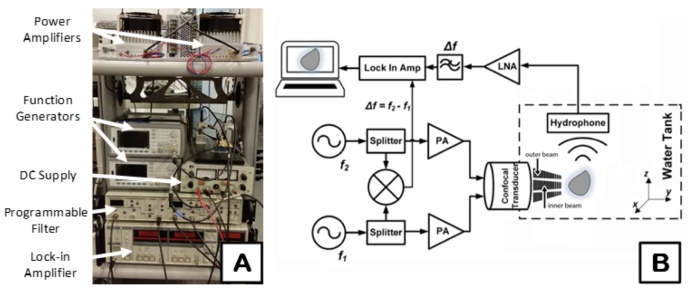
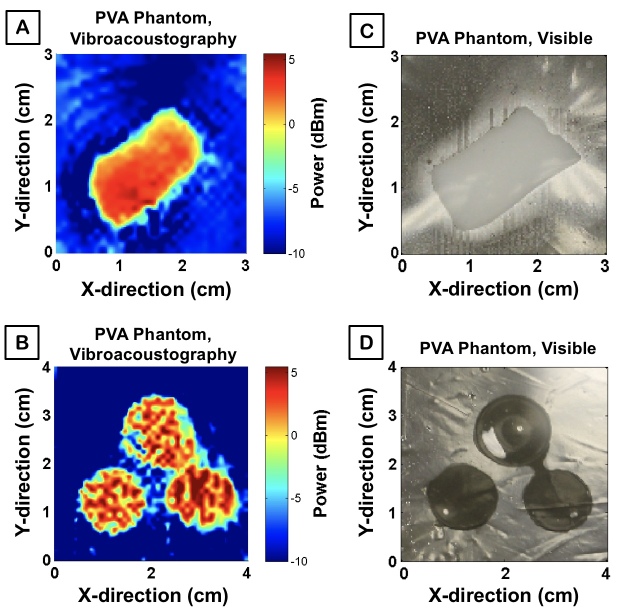
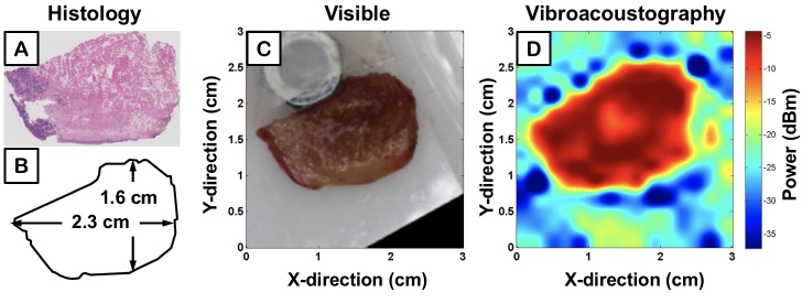
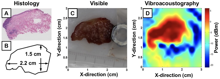
Oral and head and neck squamous cell carcinoma (OSCC) is the sixth most common cancer worldwide. The primary management of OSCC relies on complete surgical resection of the tumor. Margin-free resection, however, is difficult given the devastating effects of aggressive surgery. Currently, surgeons determine where cuts are made by palpating edges of the tumor. Accuracy varies based on the surgeon's experience, the location and type of tumor, and the risk of damage to adjacent structures limiting resection margins. To fulfill this surgical need, we contrast tissue regions by identifying disparities in viscoelasticity by mixing two ultrasonic beams to produce a beat frequency, a technique termed vibroacoustography (VA). In our system, an extended focal length of the acoustic stress field yields surgeons' high resolution to detect focal lesions in deep tissue. VA offers 3D imaging by focusing its imaging plane at multiple axial cross-sections within tissue. Our efforts culminate in production of a mobile VA system generating image contrast between normal and abnormal tissue in minutes. We model the spatial direction of the generated acoustic field and generate images from tissue-mimicking phantoms and ex vivo specimens with squamous cell carcinoma of the tongue to qualitatively demonstrate the functionality of our system. These preliminary results warrant additional validation as we continue clinical trials of ex vivo tissue. This tool may prove especially useful for finding tumors that are deep within tissue and often missed by surgeons. The complete primary resection of tumors may reduce recurrence and ultimately improve patient outcomes.
Keywords: head and neck cancer; intraoperative imaging; tumor imaging.
Figures

Similar articles
-
Dynamic optical contrast imaging as a novel modality for rapidly distinguishing head and neck squamous cell carcinoma from surrounding normal tissue.Cancer. 2017 Mar 1;123(5):879-886. doi: 10.1002/cncr.30338. Epub 2016 Oct 20. Cancer. 2017. PMID: 27763689
-
Evaluation of tongue squamous cell carcinoma resection margins using ex-vivo MR.Int J Comput Assist Radiol Surg. 2017 May;12(5):821-828. doi: 10.1007/s11548-017-1524-6. Epub 2017 Jan 27. Int J Comput Assist Radiol Surg. 2017. PMID: 28130702 Free PMC article.
-
Optical coherence tomography in the assessment of oral squamous cell carcinoma resection margins.Photodiagnosis Photodyn Ther. 2016 Mar;13:211-217. doi: 10.1016/j.pdpdt.2015.07.170. Epub 2015 Jul 22. Photodiagnosis Photodyn Ther. 2016. PMID: 26210067
-
Imaging in head and neck oncology.Surg Oncol Clin N Am. 2004 Jan;13(1):13-35. doi: 10.1016/S1055-3207(03)00124-8. Surg Oncol Clin N Am. 2004. PMID: 15062359 Review.
-
Determining Adequate Margins in Head and Neck Cancers: Practice and Continued Challenges.Curr Oncol Rep. 2016 Sep;18(9):54. doi: 10.1007/s11912-016-0540-y. Curr Oncol Rep. 2016. PMID: 27469263 Review.
References
-
- Mignogna MD, Fedele S, Russo LL. The World Cancer Report and the burden of oral cancer. Eur J Cancer Prev. 2004;13(2):139–42. - PubMed
-
- Mignogna MD, Fedele S, Russo LL, Ruoppo E, Muzio LL. Oral and pharyngeal cancer: lack of prevention and early detection by health care providers. Eur J Cancer Prev. 2001;10(4):381–3. - PubMed
Publication types
MeSH terms
Grants and funding
LinkOut - more resources
Full Text Sources
Medical
Molecular Biology Databases