

Combining Three-Dimensional Quantitative Phase Imaging and Fluorescence Microscopy for the Study of Cell Pathophysiology
- PMID: 30258314
- PMCID: PMC6153632
Combining Three-Dimensional Quantitative Phase Imaging and Fluorescence Microscopy for the Study of Cell Pathophysiology
Abstract
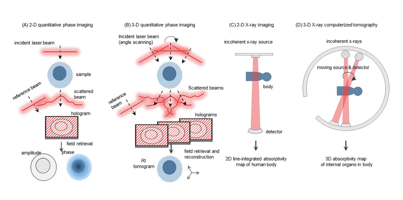
Quantitative phase imaging (QPI) has emerged as one of the powerful imaging tools for the study of live cells in a non-invasive manner. In particular, multimodal approaches combining QPI and fluorescence microscopic techniques have been recently developed for superior spatiotemporal resolution as well as high molecular specificity. In this review, we briefly summarize recent advances in three-dimensional QPI combined with fluorescence techniques for the correlative study of cell pathophysiology. Through this review, biologists and clinicians can be provided with insights on this rapidly growing field of research and may find broader applications to investigate unrevealed nature in cell physiology and related diseases.
Keywords: correlative imaging; fluorescence imaging; holotomography; label-free imaging; microscopy; quantitative phase imaging.
Figures
References
-
- Popescu G. Quantitative phase imaging of cells and tissues. New York: McGraw Hill Professional; 2011.
-
- Popescu G, Ikeda T, Dasari RR, Feld MS. Diffraction phase microscopy for quantifying cell structure and dynamics. Opt Lett. 2006;31(6):775–7. - PubMed
Publication types
MeSH terms
LinkOut - more resources
Full Text Sources
Medical