Review
. 2018 Sep 12:11:317.
doi: 10.3389/fnmol.2018.00317. eCollection 2018.

Structure-Function Relationships of Glycine and GABAA Receptors and Their Interplay With the Scaffolding Protein Gephyrin

Affiliations
Review

Structure-Function Relationships of Glycine and GABAA Receptors and Their Interplay With the Scaffolding Protein Gephyrin

Vikram B Kasaragod et al. Front Mol Neurosci. .

Abstract

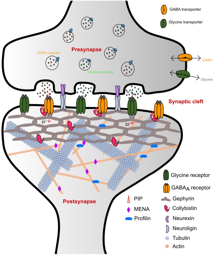
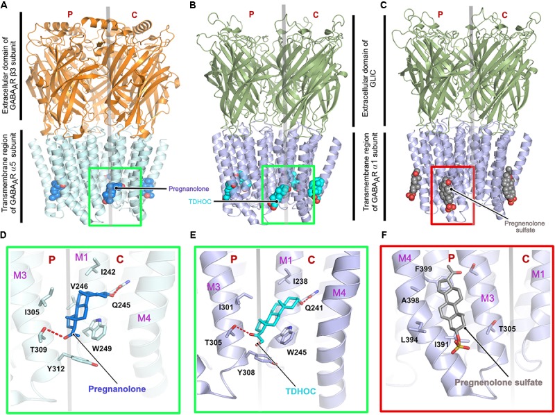
Glycine and γ-aminobutyric acid (GABA) are the major determinants of inhibition in the central nervous system (CNS). These neurotransmitters target glycine and GABAA receptors, respectively, which both belong to the Cys-loop superfamily of pentameric ligand-gated ion channels (pLGICs). Interactions of the neurotransmitters with the cognate receptors result in receptor opening and a subsequent influx of chloride ions, which, in turn, leads to hyperpolarization of the membrane potential, thus counteracting excitatory stimuli. The majority of glycine receptors and a significant fraction of GABAA receptors (GABAARs) are recruited and anchored to the post-synaptic membrane by the central scaffolding protein gephyrin. This ∼93 kDa moonlighting protein is structurally organized into an N-terminal G-domain (GephG) connected to a C-terminal E-domain (GephE) via a long unstructured linker. Both inhibitory neurotransmitter receptors interact via a short peptide motif located in the large cytoplasmic loop located in between transmembrane helices 3 and 4 (TM3-TM4) of the receptors with a universal receptor-binding epitope residing in GephE. Gephyrin engages in nearly identical interactions with the receptors at the N-terminal end of the peptide motif, and receptor-specific interaction toward the C-terminal region of the peptide. In addition to its receptor-anchoring function, gephyrin also interacts with a rather large collection of macromolecules including different cytoskeletal elements, thus acting as central scaffold at inhibitory post-synaptic specializations. Dysfunctions in receptor-mediated or gephyrin-mediated neurotransmission have been identified in various severe neurodevelopmental disorders. Although biochemical, cellular and electrophysiological studies have helped to understand the physiological and pharmacological roles of the receptors, recent high resolution structures of the receptors have strengthened our understanding of the receptors and their gating mechanisms. Besides that, multiple crystal structures of GephE in complex with receptor-derived peptides have shed light into receptor clustering by gephyrin at inhibitory post-synapses. This review will highlight recent biochemical and structural insights into gephyrin and the GlyRs as well as GABAA receptors, which provide a deeper understanding of the molecular machinery mediating inhibitory neurotransmission.

Keywords: GABAA receptors; cytoskeletal proteins; gephyrin; glycine receptors; inhibitory post-synaptic specialization; moonlighting protein.


Figures

FIGURE 1
FIGURE 1
Schematic representation of inhibitory synaptic specializations. While the simultaneous presence of both GlyRs and GABAARs was chosen for illustrative purposes, such a mixed receptor population exists in spinal cord neurons (Dumoulin et al., 2000). Please note that many interaction partners of gephyrin including DLC and Pin1 have been omitted from the schematic to improve clarity.
FIGURE 2
FIGURE 2
Architecture of glycine receptors. (A–C) Cartoon representation of the overall architecture of the homopentameric α3 GlyR in complex with glycine elucidated by X-ray crystallography (A, PDB: 5TIN) as well as the α1 GlyR in complex with strychnine (B, PDB: 3JAD) and ivermectin (C, PBD: 3JAF) by cryo-EM. The ECD is colored in light orange and the TMD in blue, bound ligands are shown in space filling representation. Enlarged views of the ligand binding pocket of the agonist glycine (black, D), the antagonist strychnine (green, E) and the positive allosteric modulator ivermectin (magenta, F). Bound ligands and critical residues which mediate the binding of the molecules are displayed in stick representation.
FIGURE 3
FIGURE 3
Architecture of the homopentameric GABAAR. (A) Crystal structure of the GABAAR β3 homopentamer in the desensitized state displayed in cartoon with the bound benzamidine in space filling representation (PDB: 4COF). (B) Top view of the receptor displaying the ion channel lining contributed by TM2. (C) Enlarged view of the agonist benzamidine (green) binding pocket where the bound ligand and crucial residues are shown in stick representation. (D) Enlarged view of the intracellular loops connecting the TM helices displaying the artificially shortened TM3-TM4 loop which aided in structure determination. (E) Cartoon representation of a GABAAR β3 monomer showing its relationship to a left arm.
FIGURE 4
FIGURE 4
Architecture of the chimeric GABAARs. (A) Crystal structure of a chimeric GABAAR, resulting from the fusion of the transmembrane domain of the α5 subunit and the extracellular domain (ECD) of the β3 subunit, in complex with pregnanolone (PDB: 5O8F). Crystal structure of a chimeric GABAAR, resulting from the fusion of the transmembrane domain of the α1 subunit and the ECD of the prokaryotic GLIC, in complex with TDHOC (B, PDB: 5OSB) and pregnenolone sulfate (C, PDB: 5OSC). In all cases, the bound ligand is shown in space filling representation and the receptor as cartoon. Enlarged views of the ligand binding pocket of the potentiators, pregnanolone (D) and TDHOC (E) as well as the inhibitor pregnenolone sulfate (F). Residues contributed by either the principal or complementary subunit are separated by a transparent gray line. The binding of the potentiators are mediated by the M3 helix from the principal and the M1 helix from the complementary subunit (D,E), in contrast, binding of the inhibitory pregnenolone sulfate is mediated by helices M3 and M4 from the principal subunit (F).
FIGURE 5
FIGURE 5
Structure and organization of a heteropentameric GABAAR. (A) Side view of the cryo-EM structure of the α1β2γ2 heteropentamer in complex with GABA and flumazenil shown in space-filling representation and with their C-atoms colored in green and blue, respectively. (B) View of the receptor from the ECD with the N-linked glycans (stick representation) of the α1-subunits pointing into the extracellular vestibule. (C) View of the receptor from the intracellular side clearly demonstrating how the γ2 subunit breaks the fivefold symmetry and obstructs the pore of the receptor. (D) Close-up view of the neurotransmitter binding pocket displaying the interaction of GABA and the β2-α1 receptor subunits. (E) Zoom into the benzodiazepam binding pocket displaying how flumazenil is bound at the α1-γ2 interface.
FIGURE 6
FIGURE 6
Structure of gephyrin. (A) Crystal structure of the N-terminal GephG trimer where one monomer is shown in cartoon representation (yellow) and the other two in surface representation in gray (PDB: 1JLJ). (B) Crystal structure of the C-terminal GephE dimer with one monomer colored according to its four subdomains (border lining residues of each subdomain are shown in the schematic diagram below the crystal structure) and the other monomer in surface representation in gray (PDB: 5ERQ). (C) Surface view of the ensemble of models of the full-length gephyrin derived from SAXS studies, where the compact state of the protein is represented in orange, the moderately extended state in gray and the fully extended state in green. (D) Schematic representation of the domain architecture of gephyrin.
FIGURE 7
FIGURE 7
Structures of the gephyrin-receptor interactions. (A) Schematic representation of a single subunit of an inhibitory GABAAR or GlyR. The core binding motifs mediating the interaction of the receptor with gephyrin are underlined in red. (B) Overall architecture of the superimposed crystal structure of GephE in complex with the GlyR-β49 (PDB:2FTS) and GephE-GABAAR α311 peptide (PDB: 4TK1), where the GephE subdomains are colored according to the scheme shown below the superimposed structure and are labeled with Roman numerals. Close-up view of the interaction of GlyR β49 (C, shown as light brown sticks) and GABAAR α3 (D, light pink sticks) with GephE. The receptor counterpart and residues of GephE, which are crucial for binding, are shown in stick representation and the others as a cartoon model. Please note that Asp327 of the E domain effectively interacts with the GlyR peptide forming both side chain-side chain and side chain-main chain interactions in contrast to the GABAAR α3 peptide and also the favorable interaction of Leu404 of the GlyR peptide in contrast to the partially polar Thr374 of GABAAR α3. These interaction primarily determine the different receptor affinities for GephE.
FIGURE 8
FIGURE 8
Domain swapped crystal structures of GephE-receptor derived peptide complexes. (A) Crystal structure of the GephE dimer in the apo form. Crystal structures of domain swapped GephE-peptide complexes determined in the presence of high affinity dimeric peptide derived from either GlyR β (B, PDB: 4U91) or GABAAR α3 (C, PDB: 4U90) subunits. The peptide takes a straight trajectory in case of the GlyR-β peptide in contrast to the GABAAR α3 peptide which adopts a cross trajectory to connect two GephE monomers. (D,E) Enlarged views of the N-terminal region of the GABAAR α3 and GlyR-β core binding motifs. Please note that in the case of the GABAAR α3 peptide (D) Asn369 (underlined label) is forced to exhibit dual conformations of its side chain to match the hydrogen bonding potentials of its partners, in contrast, a single conformation of Ser399 in GlyR-β effectively stabilizes the GephE-GlyR interaction.
FIGURE 9
FIGURE 9
Post-translational modifications (PTMs) of gephyrin. Schematic representation of well-characterized PTMs of gephyrin where the different PTMs are colored according to the accompanying scheme. Location of the PTM as indicated by corresponding residue number and type.
FIGURE 10
FIGURE 10
Alternative splicing of gephyrin. Schematic representation of alternative splicing of gephyrin with GephG specific cassettes colored in blue, linker cassettes in red and E domain cassettes in green along with the length of the cassettes and also the insertion site is shown for each cassette. The full-length protein is numbered according to the P1 splice variant nomenclature.

References

    1. Agarwal S., Tannenberg R. K., Dodd P. R. (2008). Reduced expression of the inhibitory synapse scaffolding protein gephyrin in Alzheimer’s disease. J. Alzheimers Dis. 14 313–321. 10.3233/JAD-2008-14305 - DOI - PubMed
    1. Althoff T., Hibbs R. E., Banerjee S., Gouaux E. (2014). X-ray structures of GluCl in apo states reveal a gating mechanism of Cys-loop receptors. Nature 512 333–337. 10.1038/nature13669 - DOI - PMC - PubMed
    1. Antonelli R., Pizzarelli R., Pedroni A., Fritschy J. M., Del Sal G., Cherubini E., et al. (2014). Pin1-dependent signalling negatively affects GABAergic transmission by modulating neuroligin2/gephyrin interaction. Nat. Commun. 5:5066. 10.1038/ncomms6066 - DOI - PMC - PubMed
    1. Belelli D., Lambert J. J. (2005). Neurosteroids: endogenous regulators of the GABA(A) receptor. Nat. Rev. Neurosci. 6 565–575. 10.1038/nrn1703 - DOI - PubMed
    1. Betz H., Langosch D., Hoch W., Prior P., Pribilla I., Kuhse J., et al. (1991). Structure and expression of inhibitory glycine receptors. Adv. Exp. Med. Biol. 287 421–429. 10.1007/978-1-4684-5907-4_37 - DOI - PubMed