Review
. 2018 Sep 12:9:538.
doi: 10.3389/fendo.2018.00538. eCollection 2018.

The Molecular Basis for Specificity at the Level of the Protein Kinase a Catalytic Subunit

Affiliations
Review

The Molecular Basis for Specificity at the Level of the Protein Kinase a Catalytic Subunit

Kristoffer Søberg et al. Front Endocrinol (Lausanne). .

Abstract

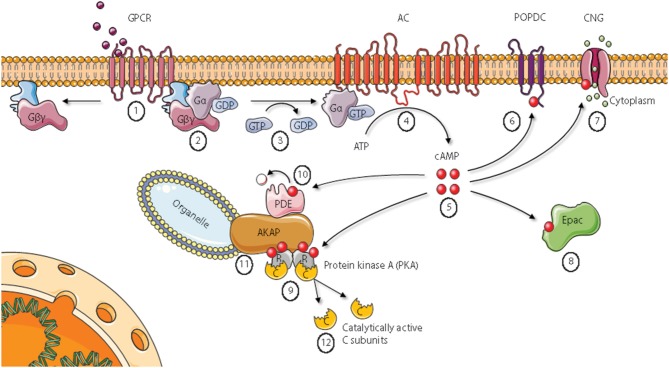
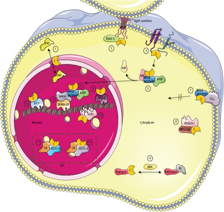
Assembly of multi enzyme complexes at subcellular localizations by anchoring- and scaffolding proteins represents a pivotal mechanism for achieving spatiotemporal regulation of cellular signaling after hormone receptor targeting [for review, see (1)]. In the 3' 5'-cyclic adenosine monophosphate (cAMP) dependent protein kinase (PKA) signaling pathway it is generally accepted that specificity is secured at several levels. This includes at the first level stimulation of receptors coupled to heterotrimeric G proteins which through stimulation of adenylyl cyclase (AC) forms the second messenger cAMP. Cyclic AMP has several receptors including PKA. PKA is a tetrameric holoenzyme consisting of a regulatory (R) subunit dimer and two catalytic (C) subunits. The R subunit is the receptor for cAMP and compartmentalizes cAMP signals through binding to cell and tissue-specifically expressed A kinase anchoring proteins (AKAPs). The current dogma tells that in the presence of cAMP, PKA dissociates into an R subunit dimer and two C subunits which are free to phosphorylate relevant substrates in the cytosol and nucleus. The release of the C subunit has raised the question how specificity of the cAMP and PKA signaling pathway is maintained when the C subunit no longer is attached to the R subunit-AKAP complex. An increasing body of evidence points toward a regulatory role of the cAMP and PKA signaling pathway by targeting the C subunits to various C subunit binding proteins in the cytosol and nucleus. Moreover, recent identification of isoform specific amino acid sequences, motifs and three dimensional structures have together provided new insight into how PKA at the level of the C subunit may act in a highly isoform-specific fashion. Here we discuss recent understanding of specificity of the cAMP and PKA signaling pathway based on C subunit subcellular targeting as well as evolution of the C subunit structure that may contribute to the dynamic regulation of C subunit activity.

Keywords: PKA; anchoring; catalytic subunit; molecular determinants; specificity.


Figures

Figure 1
Figure 1
Cyclic AMP signaling pathways. Epac, Exchange protein directly activated by cAMP; AKAP, A Kinase Anchoring Protein; PDE, phosphodiesterase. See main text for details. Figure based on Wong and Scott (2). Figure created using the Servier Medical Art resource (http://www.servier.com).
Figure 2
Figure 2
Subcellular localization of C kinase anchoring proteins—C-KAPs. The PKA C subunit associates and regulates the activity of proteins located to multiple cellular compartments and molecules. These compartments include the outer cell membrane, the cell cytoplasm and the cell nucleus. In the nucleus, C-KAPs co-locate the PKA C subunit with DNA and components of the splicing factor compartment (SFC). In the cytoplasm the PKA C subunit in addition to associate with the PKA R subunit interacts with the PKA inhibitor PKI (1), the small G protein Rab13 (2), PDE7A1 (8) the Rsk1 kinase through regulation by ERK (9), and finally IkB (5) which is a component of the cytoplasmic NFkB/AKIP complex. In the outer membrane compartment PKA C subunits associate with caveolin-1, p75NTR (3), and the heterotrimeric G protein Gα0 (7). In the nucleus, the PKA C subunit regulates DNA activity through interaction with SAF-1 (4), HSF-1 (10), HA95 (12), and p73 (11). Finally, the PKA C subunit is also involved in regulating mRNA splicing in SFC by direct interaction with serine and arginine (SR) proteins such as SFSR17A (13), SRSF1 and SRSF7 (14).
Figure 3
Figure 3
Three dimensional structure of the PKA C subunit. (A) The C subunit is composed of a small lobe, large lobe, and an active site cleft with a binding site for an ATP molecule (yellow sticks) and two Mg2+ ions (yellow spheres). The figure is based on the experimental structure with Protein Data Bank (PDB) identifier 3FJQ (169). (B) Schematic representation of the active site cleft of PKA Cα1. Motifs and residues described in the text are indicated. Dashed lines indicate the chain of interactions leading from pThr197 to Phe185 in the DFG motif when the enzyme is in the active conformation. The structure is solved with Mn2+ as the divalent cations, although Mg2+ is thought to be the most relevant biological chelating agent (170). ATP and the Mn2+ ions are shown in yellow, and the DFG motif (teal), Gly-rich loop (salmon), catalytic loop (yellow), and activation loop (cyan) are also highlighted. PDB identifier 3FJQ (169).
Figure 4
Figure 4
Core and tail structures of the PKA C subunit. (A) C- and R-spines in Cα1. In the active conformation of kinases, the C- and R-spines are assembled. In the case of PKA Cα1, the residues constituting the C-spine (yellow) are Ala70, Val57, Leu173, Ile174, Leu172, Met128, Met231, and Leu227. The adenine nucleobase of ATP (slate stick presentation) is also part of the C-spine. The R-spine (red) consists of Cα1 residues Leu106, Leu95, Phe185, and Tyr164. One-letter aa abbreviations are used in the figure. Figure is based upon (175). PDB identifier 3FJQ (169). (B) Presentation of the conserved kinase core (rendered as a surface in slate) of Cα1, including the N-tail (salmon) and C-tail (green) in cartoon presentations. Myristic acid (gray) is shown bound to the hydrophobic pocket. Selected structures and residues in the N- and C-tails are highlighted and described in the text. PDB identifier 1CMK (73).
Figure 5
Figure 5
Different configurations of N terminal parts of C subunit isoforms. For all figures, the C subunit is represented in cyan with the hydrophobic pocket highlighted in purple. Alternative exon 1 encoded parts of the C subunit are in orange cartoon presentation, in addition to the mainly exon 2 encoded A helix. Hypothesized (i.e., not supported by published crystal structure data) structures of N-terminal residues are colored red. (A) Representation of human myristoylated Cα1. The structure of unphosphorylated (Ser10), unmodified (i.e., not deamidated) Asn2, and Gly1-myristoylated Cα1 shows a fully ordered N-terminus. The modifiable residues in the 5′ encoded exon are highlighted in stick presentations, and myristic acid (yellow) occupies the hydrophobic pocket. Based on the structure with PDB identifier 1CMK (73). (B) Proposed model of N-terminal structure of CαL/Cβ2 homologs. Our study identified a conserved Trp59 (human Cβ2 numbering) (stick presentation, slate) residue which potentially occupies the hydrophobic pocket. The most conserved part of the N-terminus was predicted to encode a helix structure, which we hypothesize may be ordered upon binding to interaction partners. The figure is modeled from the experimental structure of Cα1, with the N-terminal residues encoded by exon 1 modeled. PDB identifier 1CMK (73). (C) Proposed model of N-terminal structure of CαShort variants. Short N-terminal transcripts in Cα were identified in most vertebrate species investigated. The short N-terminal end displays the open hydrophobic pocket as earlier proposed for Cα2. The figure is based upon the experimental structure of human Cα2 with PDB identifier 4AE9 (63).
Figure 6
Figure 6
Hypothesis of localized pools of isoform-specific PKA signaling. (A) Most of the variations in the Core16−350 residues in Cα and Cβ proteins are located to 11 solvent exposed residues in the small lobe. This opens for the possibility of Cα- and Cβ-specific interaction partners interacting with the small lobe [described in (196)], possibly locating the two subunits into separate intracellular signaling pools. (B) Evolution of alternative N-termini in Cα and Cβ provides another mechanism for acquiring localized pools of isoform-specific PKA signaling [described in (188)]. The Cα1 and Cβ1 pool (left) shares the myristic acid with a regulatory mechanism, evolved in mammals, through phosphorylation/dephosphorylation of Ser10 for switching myristic acid in and out of the hydrophobic pocket (phosphate group presented as a red dot, and myristic acid presented as a yellow chain). This represents the main source of PKA C activity in most human cells. The conserved, putative inducible α-helix opens for the possibility of a CαL/Cβ2-specific pool (middle), docking the C subunits via a flexible linker to a CαL/Cβ2-specific subcellular assembly of proteins (purple). The Cα2 protein (CaShort pool, right) has a conserved sperm-specific expression in all mammals, and possibly interacts with Cα2-specific proteins (bright yellow) binding to the hydrophobic pocket. Similar CαShort-specific proteins may exist in other tissues in non-mammals. Figure created using the Servier Medical Art resource (http://www.servier.com/Powerpoint-image-bank).
Figure 7
Figure 7
Model of evolution of PKA C subunits. The catalytic core is a conserved feature of the eukaryotic-like kinases (ELKs). The ePKs differ from ELKs through the attainment of the activation loop, typically involving a phosphorylatable Thr which can regulate the catalytic core into active/inactive conformations, and the G, H, and I helices (“GHI domain”), serving as docking motifs for substrates (197). The C-tail is a conserved feature of the AGC group of ePKs, and is highly regulated and essential for catalytic activity (171). The N-tail of PKA Cα and Cβ includes the A helix, which interacts with AKIP in Cα1 residues 15–29 (128). This segment is shared among all C subunit isoforms, whereas the alternative N-termini are located N-terminal to the AKIP-docking site. These alterations give rise to possible functional effects in different C subunit isoforms (“myristic acid,” “inducible helix,” “…”). Figure inspired by Taylor et al. (171, 197). PDB, 3FJQ.

References

    1. Scott JD, Pawson T. Cell signaling in space and time: where proteins come together and when they're apart. Science (2009) 326:1220–4. 10.1126/science.1175668 - DOI - PMC - PubMed
    1. Wong W, Scott JD. AKAP signalling complexes: focal points in space and time. Nat Rev Mol Cell Biol. (2004) 5:959–70. 10.1038/nrm1527 - DOI - PubMed
    1. Skålhegg BS, Taskén K. Specificity in the cAMP/PKA signaling pathway. Differential expression, regulation, and subcellular localization of subunits of PKA. Front Biosci. (2000) 5:678–93. - PubMed
    1. Zhang F, Zhang L, Qi Y, Xu H. Mitochondrial cAMP signaling. Cell Mol Life Sci. (2016) 73:4577–90. 10.1007/s00018-016-2282-2 - DOI - PMC - PubMed
    1. Hur EM, Kim KT. G protein-coupled receptor signalling and cross-talk: Achieving rapidity and specificity. Cell Signal. (2002) 14:397–405. 10.1016/S0898-6568(01)00258-3 - DOI - PubMed

LinkOut - more resources