. 2018 Sep 12:9:2192.
doi: 10.3389/fmicb.2018.02192. eCollection 2018.

The Cellular DExD/H-Box RNA Helicase UAP56 Co-localizes With the Influenza A Virus NS1 Protein

Affiliations

The Cellular DExD/H-Box RNA Helicase UAP56 Co-localizes With the Influenza A Virus NS1 Protein

Shiho Chiba et al. Front Microbiol. .

Abstract

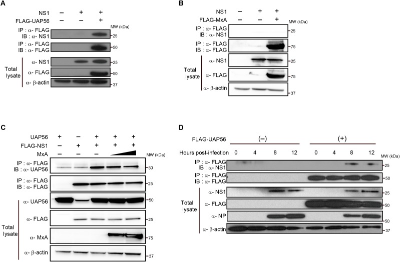
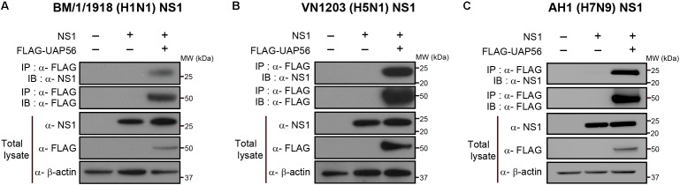
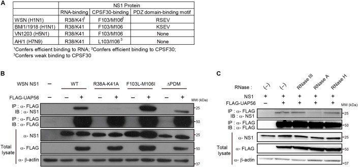
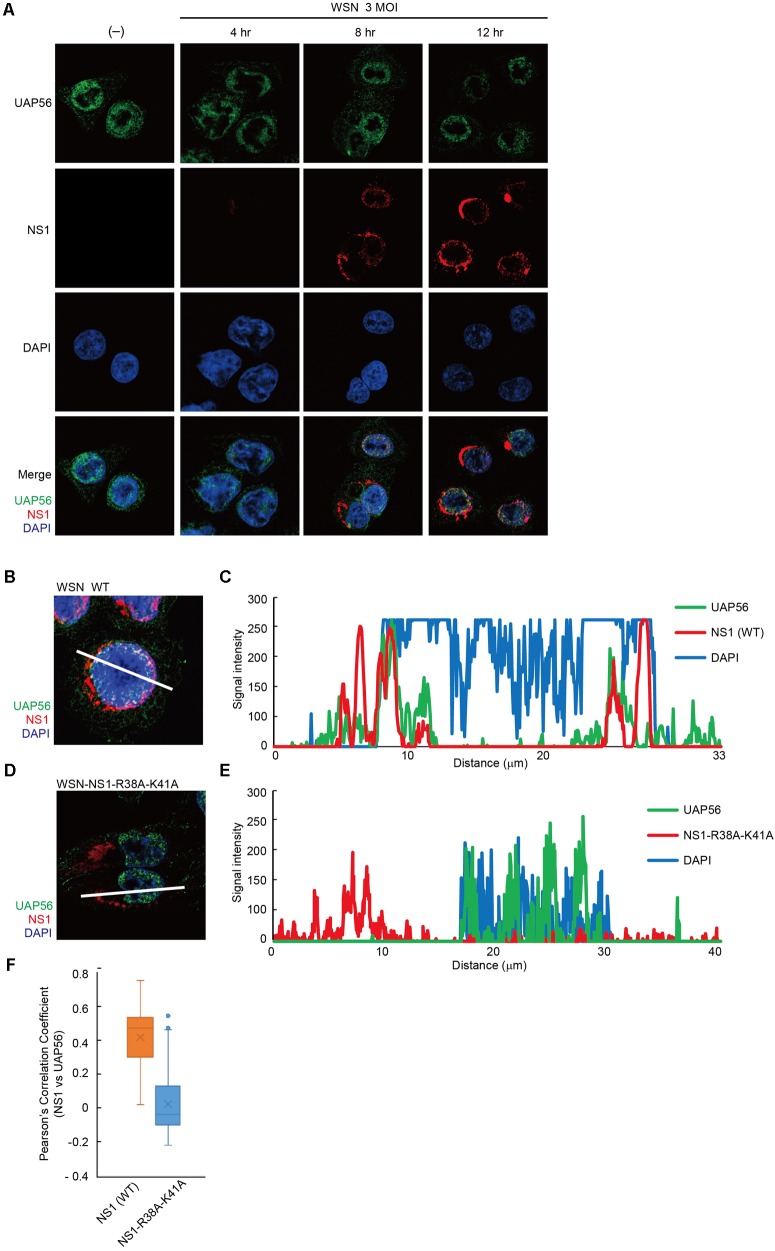
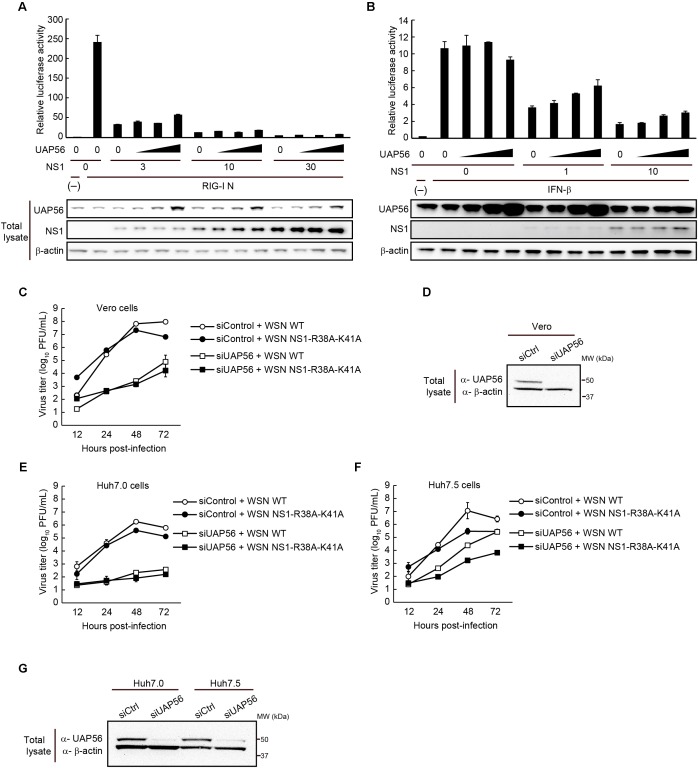
UAP56, a member of the DExD/H-box RNA helicase family, is essential for pre-mRNA splicing and mRNA export in eukaryotic cells. In influenza A virus-infected cells, UAP56 mediates viral mRNA nuclear export, facilitates viral ribonucleoprotein complex formation through direct interaction with the viral nucleoprotein, and may indirectly affect antiviral host responses by binding to and/or facilitating the activation of the antiviral host factors MxA and PKR. Here, we demonstrate that UAP56 also co-localizes with the influenza A viral NS1 protein, which counteracts host cell innate immune responses stimulated by virus infection. The UAP56-NS1 association relies on the RNA-binding residues R38 and K41 in NS1 and may be mediated by single-stranded RNA. UAP56 association with NS1 does not affect the NS1-mediated downregulation of cellular innate immune pathways in reporter gene assays, leaving in question the exact biological role and relevance of the UAP56-NS1 association.

Keywords: UAP56; host factors; influenza A NS1; influenza A virus; nuclear localization.


Figures

FIGURE 1
FIGURE 1
In vitro coprecipitation of NS1 with UAP56 or MxA. (A) In vitro interaction of NS1 with UAP56. HEK293T cells were transfected with protein expression plasmids for WSN-NS1 and/or FLAG-tagged UAP56. At 48 h post-transfection, cells were lyzed and immunoprecipitated (IP) with anti-FLAG antibody. Co-precipitated proteins were analyzed by immunoblotting (IB) with anti-NS1 antibody. (B) In vitro interaction of NS1 with MxA. HEK293T cells were transfected with protein expression plasmids for WSN-NS1 and/or FLAG-tagged MxA. Co-immunoprecipitation and IB were carried out as described in A. (C) Overexpression of MxA does not affect the UAP56–NS1 interaction. HEK293T cells were transfected with plasmids expressing UAP56 and/or FLAG-tagged NS1, and increasing amounts of MxA. Forty-eight hours later, the cells were lyzed and IP with anti-FLAG antibody. Co-immunoprecipitation and IB were carried out as described in A. (D) UAP56–NS1 interaction in influenza A virus-infected cells. HEK293T cells were transfected with pCAGGS-FLAG-UAP56 or a control vector, and infected with WSN virus at a multiplicity of infection (MOI) of one. The cells were harvested and lyzed at the indicated time points post-infection and IP with anti-FLAG antibody. Co-precipitated proteins were analyzed by IB with anti-NS1 antibody; the NP expression levels in the total cell lysate were analyzed as an infection control.
FIGURE 2
FIGURE 2
UAP56 interacts with NS1 proteins derived from different influenza A viruses. HEK293T cells were transfected with plasmids expressing FLAG-tagged UAP56 proteins and/or the NS1 proteins of A/Brevig Mission/1/1918 (H1N1; BM/1/1918) (A), A/Vietnam/1203/2004 (H5N1; VN1203) (B), or A/Anhui/1/2013 (H7N9; AH1) (C). At 48 h post-transfection, the cells were lyzed and immunoprecipitated with anti-FLAG M2 antibody-conjugated magnetic beads. Co-precipitated proteins were analyzed by immunoblotting with anti-NS1 antibody.
FIGURE 3
FIGURE 3
Identification of NS1 residues critical for the UAP56–NS1 interaction. (A) Comparison of RNA-binding residues, CPSF30-interacting residues, and PDZ domain binding motifs among the NS1 proteins of WSN, BM/1/1918, VN1203, and AH1. (B) Interaction of UAP with mutant NS1 proteins. HEK293T cells were transfected with a protein expression vector for wild-type (WT) or mutant WSN-NS1 protein and FLAG-tagged UAP56 or a control vector. At 48 h post-transfection, the cells were lyzed and immunoprecipitated with anti-FLAG M2 antibody-conjugated magnetic beads. Co-precipitated proteins were analyzed by immunoblotting with anti-NS1 antibody. ΔPDM: deletion of the “PDZ domain-binding motif.” (C) Effect of RNase treatment on the UAP56–NS1 interaction. HEK293T cells were transfected with plasmids for the expression of WSN-NS1 and FLAG-tagged UAP56. At 48 h post-transfection, the cells were lyzed and the collected cell lysate was mock-treated or treated with the indicated RNases at 37°C for 20 min. The lysates were incubated with anti-FLAG antibody-conjugated magnetic beads, and co-precipitated proteins were analyzed by immunoblotting.
FIGURE 4
FIGURE 4
Localization of UAP56 and NS1 in WSN-infected A549 cells. (A) A549 cells were infected with WSN virus at an MOI of three. At 4, 8, or 12 h post-infection, the cells were fixed with 4% paraformaldehyde in PBS. The cells were then analyzed with specific antibodies against UAP56 and NS1. (B–E) Representative immunofluorescence images of A549 cells at 12 h post-infection with wild-type (WT) WSN virus (B) or WSN-NS1-R38A-K41A mutant virus (D) at an MOI of 3. The cells were analyzed with anti-UAP56 (green) antibody, anti-NS1 (red) antibody, and DAPI (blue). (C,E) Signal intensities across the line shown in B,D, respectively, were plotted and analyzed by using LSM510 META and ZEN2009 software. (F) A549 cells infected with WSN WT virus (n = 102 cells) or WSN-NS1-R38A-K41A mutant virus (n = 100 cells) at an MOI of three for 12 h were imaged, and Pearson’s correlation coefficients for UAP56 and NS1 co-localization were determined for individual cells.
FIGURE 5
FIGURE 5
Effect of UAP56 overexpression on the IFN-antagonist activity of NS1. (A) UAP56 overexpression does not affect NS1’s ability to suppress IFN-β promoter activity. HEK293T cells were transfected with plasmids expressing firefly luciferase under the control of an IFN-β promoter, pRL-TK luciferase (as an internal control), a constitutively active form of RIG-I (pCAGGS-RIG-I N), increasing amounts of pCAGGS-NS1 (0, 3, 10, or 30 ng), and pCAGGS-UAP56 (0, 3, 10, or 30 ng). Twenty-four hours later, the cells were lyzed and firefly and Renilla luciferase activities were measured. The firefly luciferase activity was normalized by the internal control value. The total amount of transfected vector was adjusted in all wells using the pCAGGS control vector. Data are shown as the mean ± SD (n = 3; biological replicates). UAP56 and NS1 expression levels in cells were examined by immunoblotting. (B) UAP56 overexpression does not affect NS1’s ability to suppress ISRE-driven gene expression. HEK293T cells were transfected with plasmids expressing firefly luciferase under the control of an ISRE promoter element, pRL-TK luciferase (as an internal control), and increasing amounts of pCAGGS-NS1 (0, 1, or 10 ng) and pCAGGS-UAP56 (0, 3, 10, or 30 ng). Twenty-four hours later, the cells were mock-treated or treated with IFN-β (10,000 U/mL), and cultured for 24 h. Luciferase activities were measured and data were analyzed as described in A. Data are shown as the mean ± SD (n = 3; biological replicates). UAP56 and NS1 expression levels in cells were examined by immunoblotting. (C–G) WSN virus or WSN-NS1-R38A-K41A mutant virus replication in cells transfected with siRNA targeting UAP56 or with a control siRNA. Vero (C), Huh7.0 (E), or Huh7.5 (F) cells were transfected with siRNA targeting UAP56, or a control siRNA. At 24 (Vero cells) or 48 h (Huh7.0 and Huh7.5 cells) post-transfection, the cells were infected with WSN virus or WSN-NS1-R38A-K41A mutant virus at an MOI of 0.01 and incubated at 37°C. Culture supernatants were collected at the indicated times post-infection and viral titers were analyzed by performing plaque assays in MDCK cells. The data are shown as the mean ± SD (n = 3; biological replicates). Levels of UAP56 expression in Vero (D) or Huh7.0 and Huh7.5 (G) cells transfected with siRNA targeting UAP56, or with a control siRNA. UAP56 expression levels were analyzed by immunoblotting at 48 (Vero cells) or 72 h (Huh7.0 and Huh7.5 cells) after siRNA transfection.

Similar articles

Cited by

References

    1. Anastasina M., Le May N., Bugai A., Fu Y., Soderholm S., Gaelings L., et al. (2016). Influenza virus NS1 protein binds cellular DNA to block transcription of antiviral genes. Biochim. Biophys. Acta 1859 1440–1448. 10.1016/j.bbagrm.2016.09.005 - DOI - PubMed
    1. Egorov A., Brandt S., Sereinig S., Romanova J., Ferko B., Katinger D., et al. (1998). Transfectant influenza A viruses with long deletions in the NS1 protein grow efficiently in Vero cells. J. Virol. 72 6437–6441. - PMC - PubMed
    1. Ehrhardt C., Marjuki H., Wolff T., Nurnberg B., Planz O., Pleschka S., et al. (2006). Bivalent role of the phosphatidylinositol-3-kinase (PI3K) during influenza virus infection and host cell defence. Cell. Microbiol. 8 1336–1348. 10.1111/j.1462-5822.2006.00713.x - DOI - PubMed
    1. Ehrhardt C., Wolff T., Pleschka S., Planz O., Beermann W., Bode J. G., et al. (2007). Influenza A virus NS1 protein activates the PI3K/Akt pathway to mediate antiapoptotic signaling responses. J. Virol. 81 3058–3067. 10.1128/JVI.02082-06 - DOI - PMC - PubMed
    1. Fan S., Macken C. A., Li C., Ozawa M., Goto H., Iswahyudi N. F., et al. (2013). Synergistic effect of the PDZ and p85beta-binding domains of the NS1 protein on virulence of an avian H5N1 influenza A virus. J. Virol. 87 4861–4871. 10.1128/JVI.02608-12 - DOI - PMC - PubMed

LinkOut - more resources