

Zinner syndrome presenting with intermittent scrotal pain in a young man
- PMID: 30258511
- PMCID: PMC6148829
- DOI: 10.1016/j.radcr.2018.08.012
Zinner syndrome presenting with intermittent scrotal pain in a young man
Abstract
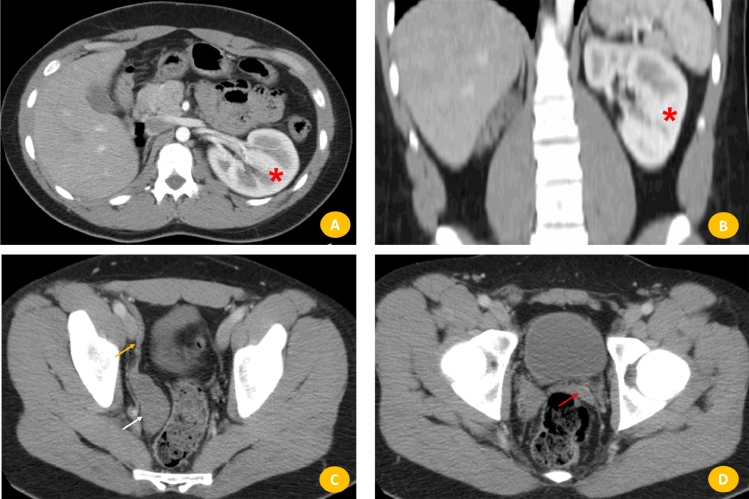
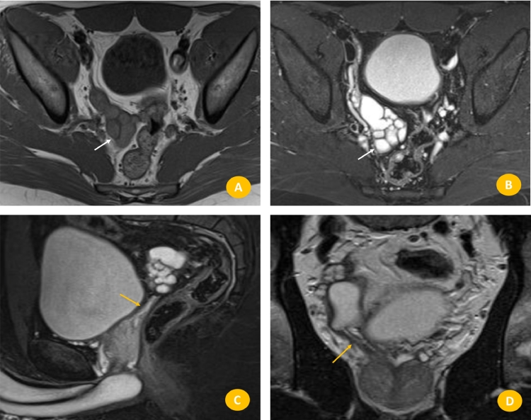
Congenital malformations of the seminal vesicle are uncommon, and most of them are cystic malformations. If an insult occurs during the first trimester of gestation, the embryogenesis of the kidney, ureter, seminal vesicle, and vas deferens could be altered. The mutual embryological origins of the seminal vesicle and ureteral bud from the mesonephric (Wolffian) duct result in association between ipsilateral renal agenesis and seminal vesical cysts. Zinner syndrome is a rare condition comprising a triad of unilateral renal agenesis, ipsilateral seminal vesicle obstruction and ipsilateral ejaculatory duct obstruction. This syndrome were first described by Zinner in 1914, and 200 cases have been reported in the literature. Most patients with this anomaly are asymptomatic until the second or third decade of life. Some cases have nonspecific symptoms such as prostatism, urinary urgency, dysuria, painful ejaculation, and perineal discomfort. In this paper, we present a uncommon case of a 21-year-old patient which the initial presentation of this condition was intermittent scrotal pain. A brief review of the literature is undertaken, regarding the main clinical, imaging implications, and the developmental anomalies that are involved in this congenital anomaly.
Keywords: Ejaculation; Genital diseases; Hemospermia; Infertility; Mesonephric duct abnormality; Zinner syndrome.
Figures
References
-
- Zinner A. Ein fall von intravesikaler samenblasenzyste. Weien Med Wschr. 1914;64:604–610.
-
- Pereira BJ, Sousa L, Azinhais P, Conceição P, Borges R, Leão R. Zinner's syndrome: an up-to-date review of the literature based on a clinical case. Andrologia. 2009;41(5):322–330. - PubMed
-
- Beeby DI. Seminal vesicle cyst associated with ipsilateral renal agenesis: case report and review of literature. J Urol. 1974;112:120–122. - PubMed
-
- Levisay GL, Holder J, Weigel JW. Ureteral ectopia associated with seminal vesicle cyst and ipsilateral renal agenesis. Radiology. 1975;114(3):575–576. - PubMed
-
- King BF, Hattery RR, Lieber MM, Berquist TH, Williamson B, Jr., Hartman GW. Congenital cystic disease of the seminal vesicle. Radiology. 1991;178(1):207–211. - PubMed
Publication types
LinkOut - more resources
Full Text Sources
Other Literature Sources
Molecular Biology Databases
