

Balint syndrome (chronic visual-spatial disorder) presenting without known cause
- PMID: 30258515
- PMCID: PMC6148828
- DOI: 10.1016/j.radcr.2018.08.026
Balint syndrome (chronic visual-spatial disorder) presenting without known cause
Abstract
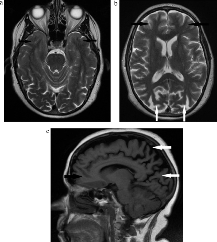
Balint's syndrome is a rare disorder characterized by a triad of simultanagnosia, optic apraxia, and ocular apraxia. The syndrome manifests when there is an injury to the posterior parietal and occipital lobes and is often bilateral. Several causes of this syndrome were published in the literature, such as trauma, infarctions, infections, tumors, and pre-eclampsia. It can also be the presenting feature of several neurodegenerative disorders, such as atypical Alzheimer's disease. We report a case of a 62-year-old lady who presented with simultanagnosia, optic apraxia, and ocular ataxia which are the typical signs and symptoms of this syndrome. Neuropsychological evaluation revealed severe affection of the visual-spatial function with intact memory, language, and cognition. Brain imaging confirmed atrophy and decreased perfusion in the posterior parietal and occipital lobes. No underlying cause could be identified to explain the brain parenchymal atrophy. The follow-up neuropsychological assessment and brain imaging did not show any progression confirming the static course of the disease.
Keywords: Balint's syndrome; Optic apraxia; Parieto-occipital atrophy; Simultanagnosia; Visual-spatial disorder.
Figures

References
-
- Moscote-Salazar LR, Calderon-Miranda WG, Carmona-Meza ZA, Alvis-Miranda HR, Churio NZ, Alcalá-Cerra G. Post-traumatic Balint's syndrome: a case report and review of the literature. Bull Emerg Trauma [Internet] 2016 Apr 4;4(2):113–115. http://www.ncbi.nlm.nih.gov/pmc/articles/PMC4897994 Available from. - PMC - PubMed
-
- Chechlacz M, Humphreys GW. The enigma of Bálint's syndrome: neural substrates and cognitive deficits. Front Hum Neurosci [Internet]. 2014/03/07. 2014;8:123. https://www.ncbi.nlm.nih.gov/pubmed/24639641 Available from. - PMC - PubMed
-
- Udesen H, Madsen AL. [Balint's syndrome–visual disorientation]. [Review] [Danish] Ugeskr Laeger [Internet] 1992 May 18;154(21):1492–1494. http://www.ncbi.nlm.nih.gov/pubmed/1598720 cited 2018 Jul 7Available from. - PubMed
-
- Ribai P, Vokaer M, De Tiege X, Massat I, Slama H, Bier JC. Acute Balint's syndrome is not always caused by a stroke. Eur J Neurol [Internet] 2006;13(3):310–312. http://doi.wiley.com/10.1111/j.1468-1331.2006.01144.x cited 2018 Jul 7 Available from. - DOI - PubMed
-
- Amalnath Sd, Kumar S, Deepanjali S, Dutta T. Balint syndrome. Ann Indian Acad Neurol [Internet] 2014;17(1):10. http://www.ncbi.nlm.nih.gov/pubmed/24753652 cited 2018 Jul 7Available from. - PMC - PubMed
Publication types
LinkOut - more resources
Full Text Sources
Other Literature Sources
Molecular Biology Databases
