

Acute portal vein thrombosis in a 59-year-old male with JAK2 V617F mutation
- PMID: 30258517
- PMCID: PMC6148830
- DOI: 10.1016/j.radcr.2018.08.023
Acute portal vein thrombosis in a 59-year-old male with JAK2 V617F mutation
Abstract
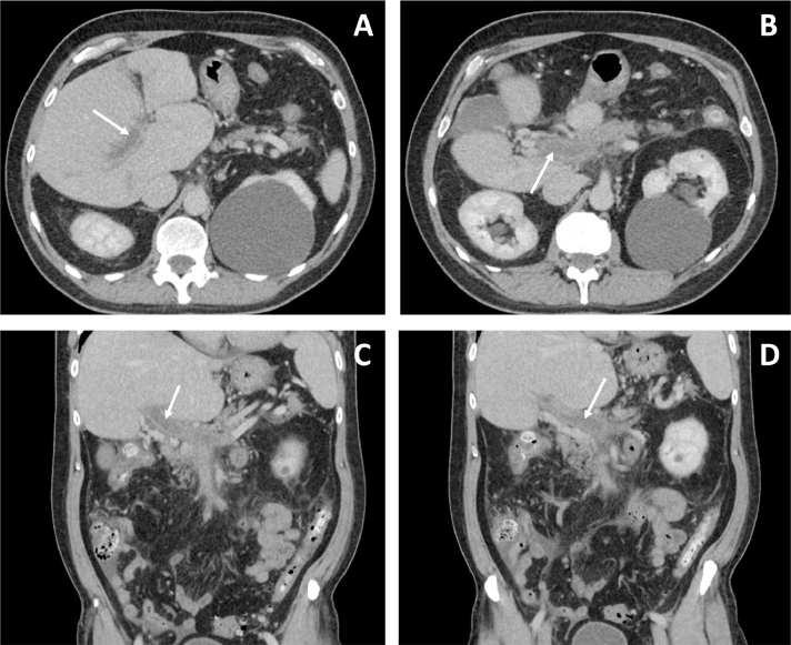
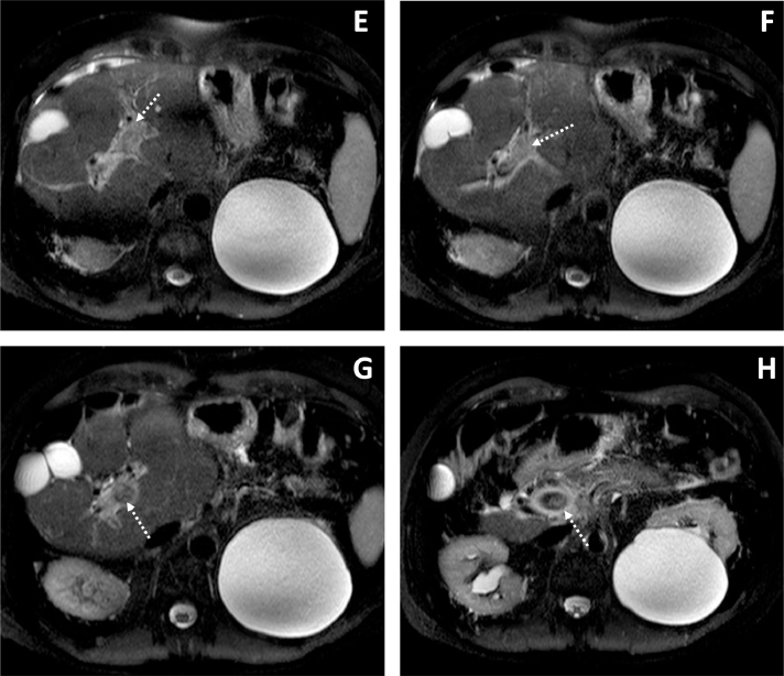
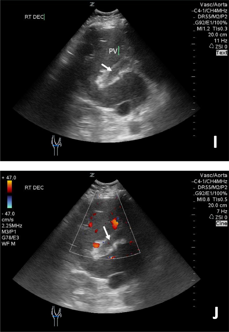
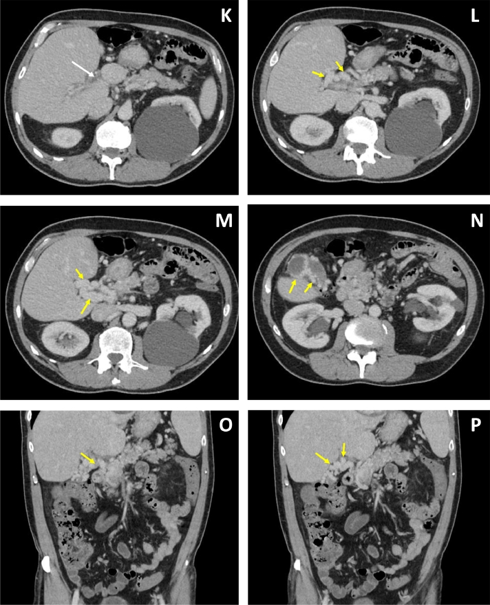
Portal vein thrombosis is an uncommon finding that typically arises in the context of cirrhosis. In the acute setting, it may present with abdominal pain, portal hypertension, ascites, gastrointestinal bleeding, or mesenteric ischemia. Local risk factors that predispose its formation include: cirrhosis, hepatocellular carcinoma, pancreatitis, and intraabdominal infection. Systemic factors, including hypercoagulable states and sepsis, also pose an increased risk. JAK2 V617F positive myeloproliferative disorders are associated with systemic prothrombotic states and are a less frequently identified cause of portal vein thrombosis. We present a case of acute unprovoked portal vein thrombosis diagnosed in a 59-year-old male without local disease factors. Computed tomography, magnetic resonance cholangiopancreatography, and ultrasound demonstrated the presence of portal vein thrombosis with neighboring periportal and pancreatic head edema. Peripheral blood testing detected the presence of JAK2 V617F mutation. The patient was discharged on 6-month anticoagulation therapy and outpatient follow-up.
Keywords: JAK2 V617F; Portal vein thrombosis.
Figures
References
-
- Margini C, Berzigotti A. Portal vein thrombosis: the role of imaging in the clinical setting. Dig Liver Dis. 2017;49(2):113–120. - PubMed
-
- Basit SA, Stone CD, Gish R. Portal vein thrombosis. Clin Liver Dis. 2015;19(1):199–221. - PubMed
-
- Qi X, Han G, Fan D. Management of portal vein thrombosis in liver cirrhosis. Nat Rev Gastroenterol Hepatol. 2014;11(7):435–446. - PubMed
-
- Yerdel MA, Gunson B, Mirza D, Karayalçin K, Olliff S, Buckels J. Portal vein thrombosis in adults undergoing liver transplantation—risk factors, screening, management, and outcome. Transplantation. 2000;69(9):1873–1881. - PubMed
Publication types
LinkOut - more resources
Full Text Sources
Other Literature Sources
Miscellaneous
