

Bone morphogenetic protein-9 effectively induces osteogenic differentiation of reversibly immortalized calvarial mesenchymal progenitor cells
- PMID: 30258869
- PMCID: PMC6147177
- DOI: 10.1016/j.gendis.2015.06.003
Bone morphogenetic protein-9 effectively induces osteogenic differentiation of reversibly immortalized calvarial mesenchymal progenitor cells
Abstract
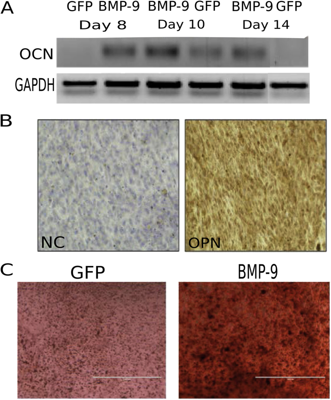
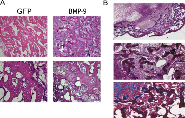
Critical-sized craniofacial defect repair represents a significant challenge to reconstructive surgeons. Many strategies have been employed in an effort to achieve both a functionally and cosmetically acceptable outcome. Bone morphogenetic proteins (BMPs) provide a robust osteoinductive cue to stimulate bony growth and remodeling. Previous studies have suggested that the BMP-9 isoform is particularly effective in promoting osteogenic differentiation of mesenchymal progenitor cells. The aim of this study is to characterize the osteogenic capacity of BMP-9 on calvarial mesenchymal progenitor cell differentiation. Reversibly immortalized murine calvarial progenitor cells (iCALs) were infected with adenoviral vectors encoding BMP-9 or GFP and assessed for early and late stages of osteogenic differentiation in vitro and for osteogenic differentiation via in vivo stem cell implantation studies. Significant elevations in alkaline phosphatase (ALP) activity, osteocalcin (OCN) mRNA transcription, osteopontin (OPN) protein expression, and matrix mineralization were detected in BMP-treated cells compared to control. Specifically, ALP activity was elevated on days 3, 7, 9, 11, and 13 post-infection and OCN mRNA expression was elevated on days 8, 10, and 14 in treated cells. Additionally, treatment groups demonstrated increased OPN protein expression on day 10 and matrix mineralization on day 14 post-infection relative to control groups. BMP-9 also facilitated the formation of new bone in vivo as detailed by gross, microcomputed tomography, and histological analyses. Therefore, we concluded that BMP-9 significantly stimulates osteogenic differentiation in iCALs, and should be considered an effective agent for calvarial tissue regeneration.
Keywords: Bone morphogenetic protein (BMP); Bone morphogenetic protein-9 (BMP-9); Immortalized calvarial cell (iCAL); Mesenchymal progenitor cell; Osteogenic capacity; Osteogenic differentiation.
Figures


Similar articles
-
Characterization of Reversibly Immortalized Calvarial Mesenchymal Progenitor Cells.J Craniofac Surg. 2015 Jun;26(4):1207-13. doi: 10.1097/SCS.0000000000001717. J Craniofac Surg. 2015. PMID: 26080159 Free PMC article.
-
Differentiation of osteoprogenitor cells is induced by high-frequency pulsed electromagnetic fields.J Craniofac Surg. 2012 Mar;23(2):586-93. doi: 10.1097/SCS.0b013e31824cd6de. J Craniofac Surg. 2012. PMID: 22446422
-
Osteogenic activity of the fourteen types of human bone morphogenetic proteins (BMPs).J Bone Joint Surg Am. 2003 Aug;85(8):1544-52. doi: 10.2106/00004623-200308000-00017. J Bone Joint Surg Am. 2003. PMID: 12925636
-
BMP Signaling in the Development and Regeneration of Cranium Bones and Maintenance of Calvarial Stem Cells.Front Cell Dev Biol. 2020 Mar 10;8:135. doi: 10.3389/fcell.2020.00135. eCollection 2020. Front Cell Dev Biol. 2020. PMID: 32211409 Free PMC article. Review.
-
Evaluating Bioassays for the Determination of Simvastatin's Osteogenic Activity: A Systematic Review.J Funct Biomater. 2025 Feb 11;16(2):61. doi: 10.3390/jfb16020061. J Funct Biomater. 2025. PMID: 39997596 Free PMC article. Review.
Cited by
-
Molecular signaling pathways in osteoarthritis and biomaterials for cartilage regeneration: a review.Bioengineered. 2025 Dec;16(1):2501880. doi: 10.1080/21655979.2025.2501880. Epub 2025 May 7. Bioengineered. 2025. PMID: 40336219 Free PMC article. Review.
-
Repair of critical sized cranial defects with BMP9-transduced calvarial cells delivered in a thermoresponsive scaffold.PLoS One. 2017 Mar 1;12(3):e0172327. doi: 10.1371/journal.pone.0172327. eCollection 2017. PLoS One. 2017. PMID: 28249039 Free PMC article.
-
Osteogenic Differentiation Effect of BMP-9 with Phenamil and Simvastatin on Intact Human Amniotic Epithelial Stem Cells.Iran Biomed J. 2022 Nov 1;26(6):463-74. doi: 10.52547/ibj.3748. Iran Biomed J. 2022. PMID: 36437797 Free PMC article.
-
Acceleration of Bone Regeneration in Critical-Size Defect Using BMP-9-Loaded nHA/ColI/MWCNTs Scaffolds Seeded with Bone Marrow Mesenchymal Stem Cells.Biomed Res Int. 2019 Apr 11;2019:7343957. doi: 10.1155/2019/7343957. eCollection 2019. Biomed Res Int. 2019. PMID: 31111065 Free PMC article.
-
Bone Morphogenetic Protein-9-Stimulated Adipocyte-Derived Mesenchymal Progenitors Entrapped in a Thermoresponsive Nanocomposite Scaffold Facilitate Cranial Defect Repair.J Craniofac Surg. 2019 Sep;30(6):1915-1919. doi: 10.1097/SCS.0000000000005465. J Craniofac Surg. 2019. PMID: 30896511 Free PMC article.
References
-
- Turan A., Kostakoglu N., Tuncel U., Gokce E., Markoc F. Scapular bone grafts: good options for craniofacial defects? Ann Plast Surg. 2014 [In press] - PubMed
Grants and funding
LinkOut - more resources
Full Text Sources
Other Literature Sources
Research Materials
