. 2018 Apr 27;5(2):172-184.
doi: 10.1016/j.gendis.2018.04.006. eCollection 2018 Jun.

Characterization of the essential role of bone morphogenetic protein 9 (BMP9) in osteogenic differentiation of mesenchymal stem cells (MSCs) through RNA interference

Affiliations

Characterization of the essential role of bone morphogenetic protein 9 (BMP9) in osteogenic differentiation of mesenchymal stem cells (MSCs) through RNA interference

Shujuan Yan et al. Genes Dis. .

Erratum in

Abstract

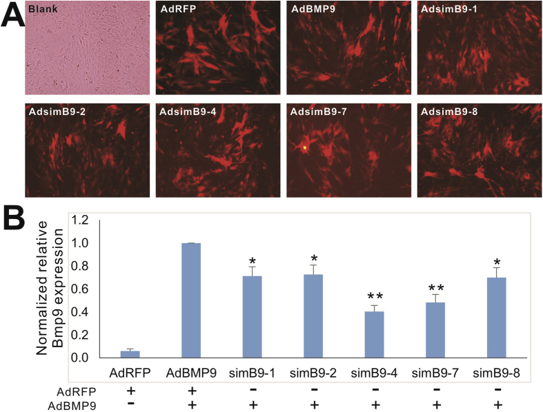
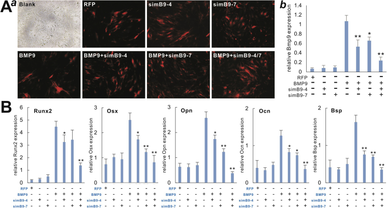
Mesenchymal stem cells (MSCs) are multipotent stem cells and capable of differentiating into multiple cell types including osteoblastic, chondrogenic and adipogenic lineages. We previously identified BMP9 as one of the most potent BMPs that induce osteoblastic differentiation of MSCs although exact molecular mechanism through which BMP9 regulates osteogenic differentiation remains to be fully understood. Here, we seek to develop a recombinant adenovirus system to optimally silence mouse BMP9 and then characterize the important role of BMP9 in osteogenic differentiation of MSCs. Using two different siRNA bioinformatic prediction programs, we design five siRNAs targeting mouse BMP9 (or simB9), which are expressed under the control of the converging H1 and U6 promoters in recombinant adenovirus vectors. We demonstrate that two of the five siRNAs, simB9-4 and simB9-7, exhibit the highest efficiency on silencing exogenous mouse BMP9 in MSCs. Furthermore, simB9-4 and simB9-7 act synergistically in inhibiting BMP9-induced expression of osteogenic markers, matrix mineralization and ectopic bone formation from MSCs. Thus, our findings demonstrate the important role of BMP9 in osteogenic differentiation of MSCs. The characterized simB9 siRNAs may be used as an important tool to investigate the molecular mechanism behind BMP9 osteogenic signaling. Our results also indicate that recombinant adenovirus-mediated expression of siRNAs is efficient and sustained, and thus may be used as an effective delivery vehicle of siRNA therapeutics.

Keywords: BMP9; Bone formation; Mesenchymal stem cells; Osteogenic differentiation; RNA interference; Recombinant adenovirus; siRNA.


Figures

Figure 1
Figure 1
Selection and construction of recombinant adenoviruses expressing optimal siRNAs targeting the mouse BMP9 coding region (simB9s). (A) Schematic representation of mouse Bmp9 coding region and the location and sequences of the five siRNA targeting sites designed by the Dharmacon's siDESIGN and IDT's DsiRNA design tools. (B) Schematic depiction of the expression of siRNAs driven by the convergent H1 and U6 promoters in recombinant adenoviral vectors through the homemade shuttle vector pSES1 or pAdTrace-61, which co-expresses mRFP as a tracking marker. The resultant siRNA-expressing simB9 siRNA adenoviruses are designated as AdsimB9-1, 2, 4, 7 and 8, respectively. AdBMP9 expresses mouse BMP9 and mRFP, while AdRFP was used as a mock control virus.
Figure 2
Figure 2
Characterization of the silencing efficiency of the selected five simB9 siRNAs targeting exogenously expressed mouse BMP9. (A) Efficient transduction of subconfluent iMAD MSCs by recombinant adenoviruses expressing siRNAs targeting mouse Bmp9. RFP signal was recorded at 48 h after infection. Representative results are shown. (B) siRNA-mediated knockdown of exogenously expressed mouse BMP9 in iMADs cells. Subconfluent iMADs were infected with the equal titer of the indicated adenoviruses. At 48 h after infection, total RNA was isolated and subjected to reverse transcription and qPCR analysis of the indicated mouse genes. All samples were normalized with Gapdh. Each assay condition was done in triplicate. “*” p < 0.05, “**” p < 0.01 compared with the AdBMP9-infected group.
Figure 3
Figure 3
Adenovirus-mediated expression of simB9 siRNAs significantly diminishes exogenous BMP9-induced ALP activity in MSCs. (A) Qualitative ALP assay. Subconfluent iMADs were infected with the indicated adenoviruses. At day 5 (a) and day 7 (b), the infected cells were subjected to histochemical staining of ALP activity. Each assay condition was done in duplicate. Representative results are shown. (B) Quantitative ALP assay. Subconfluent iMADs were infected with the indicated adenoviruses. At days 3, 5, and 7, the infected cells were subjected to bioluminescence assay of ALP activity. Each assay was done in triplicate. “*” p < 0.05, “**” p < 0.01 compared with the AdBMP9-infected iMADs group.
Figure 4
Figure 4
BMP9-induced expression of osteogenic regulators and biomarkers is effectively inhibited by the co-expression of simB9-4 and simB9-7 siRNAs in MSCs. (A) Efficient knockdown of exogenous BMP9 by co-expression of simB9-4 and simB9-7. Subconfluent iMADs were infected with the indicated adenoviruses for 48 h (a) and total RNA was isolated from the cells and subjected to qPCR analysis of mouse Bmp9 expression (b). All samples were normalized with Gapdh. Each assay condition was done in triplicate. “*” p < 0.05, “**” p < 0.01 compared with the AdBMP9-infected group. (B) BMP9-induced expression of osteogenic regulators and biomarkers is effectively inhibited by simB9-4 and simB9-7 siRNAs. Subconfluent iMADs were infected with the indicated adenoviruses. Total RNA was isolated from the cells at day 3 (for Runx2 and Osx) and day 5 (for Opn, Ocn and Bsp), and subjected to qPCR analysis of mouse gene expression. All samples were normalized with Gapdh. Each assay condition was done in triplicate. “*” p < 0.05, “**” p < 0.01 compared with the AdBMP9-infected group.
Figure 5
Figure 5
BMP9-induced ALP activity is effectively inhibited by the co-expression of simB9-4 and simB9-7 siRNAs in MSCs. Subconfluent iMADs were infected with the indicated adenoviruses. At days 3, 5, and 7, the infected cells were subjected to ALP activity assays by either histochemical staining (A), or quantitative bioluminescence assay (B). Each assay was done in triplicate. Representative staining results are shown. “*” p < 0.05, “**” p < 0.01 compared with the AdBMP9-infected iMADs group.
Figure 6
Figure 6
BMP9-induced matrix mineralization is effectively inhibited by the co-expression of simB9-4 and simB9-7 siRNAs in MSCs. Subconfluent iMADs were infected with the indicated adenoviruses. At days 7 and 11, the infected cells were subjected to Alizarin Red S staining. Staining results were recorded macrographically (A) or under a low power microscope (B). Each assay condition was done in duplicate. Representative staining results are shown.
Figure 7
Figure 7
BMP9-induced ectopic bone formation is significantly inhibited by the co-expression of simB9-4 and simB9-7 siRNAs in MSCs. Subconfluent iMADs were infected with the indicated adenoviruses for 36 h, and then collected for subcutaneous injection into the flanks of athymic nude mice (n = 5/group). Ectopic bony masses were harvested at 4 weeks after injection and subjected to H & E staining. No masses were retrieved from the groups injected with AdRFP, AdsimB9-4 and AdsimB9-7 infected iMADs. Representative histologic images are shown.

Similar articles

Cited by

References

    1. Prockop D.J. Marrow stromal cells as stem cells for nonhematopoietic tissues. Science. 1997;276(5309):71–74. - PubMed
    1. Caplan A.I., Bruder S.P. Mesenchymal stem cells: building blocks for molecular medicine in the 21st century. Trends Mol Med. 2001;7(6):259–264. - PubMed
    1. Deng Z.L., Sharff K.A., Tang N. Regulation of osteogenic differentiation during skeletal development. Front Biosci. 2008;13:2001–2021. - PubMed
    1. Rastegar F., Shenaq D., Huang J. Mesenchymal stem cells: molecular characteristics and clinical applications. World J Stem Cell. 2010;2(4):67–80. - PMC - PubMed
    1. Shenaq D.S., Rastegar F., Petkovic D. Mesenchymal progenitor cells and their orthopedic applications: forging a path towards clinical trials. Stem Cell Int. 2010;2010:519028. - PMC - PubMed

LinkOut - more resources