. 2018 Oct 10;18(10):6195-6206.
doi: 10.1021/acs.nanolett.8b02179. Epub 2018 Sep 27.

Personalized Tumor RNA Loaded Lipid-Nanoparticles Prime the Systemic and Intratumoral Milieu for Response to Cancer Immunotherapy

Affiliations

Personalized Tumor RNA Loaded Lipid-Nanoparticles Prime the Systemic and Intratumoral Milieu for Response to Cancer Immunotherapy

Elias J Sayour et al. Nano Lett. .

Abstract

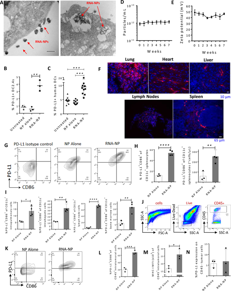
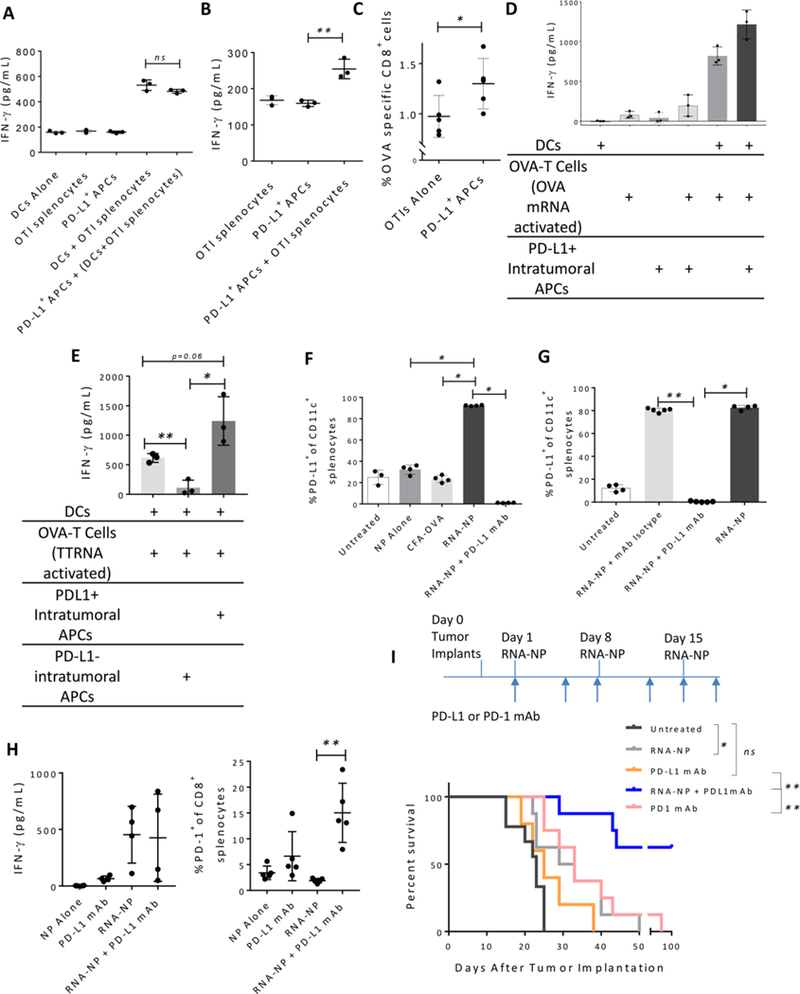
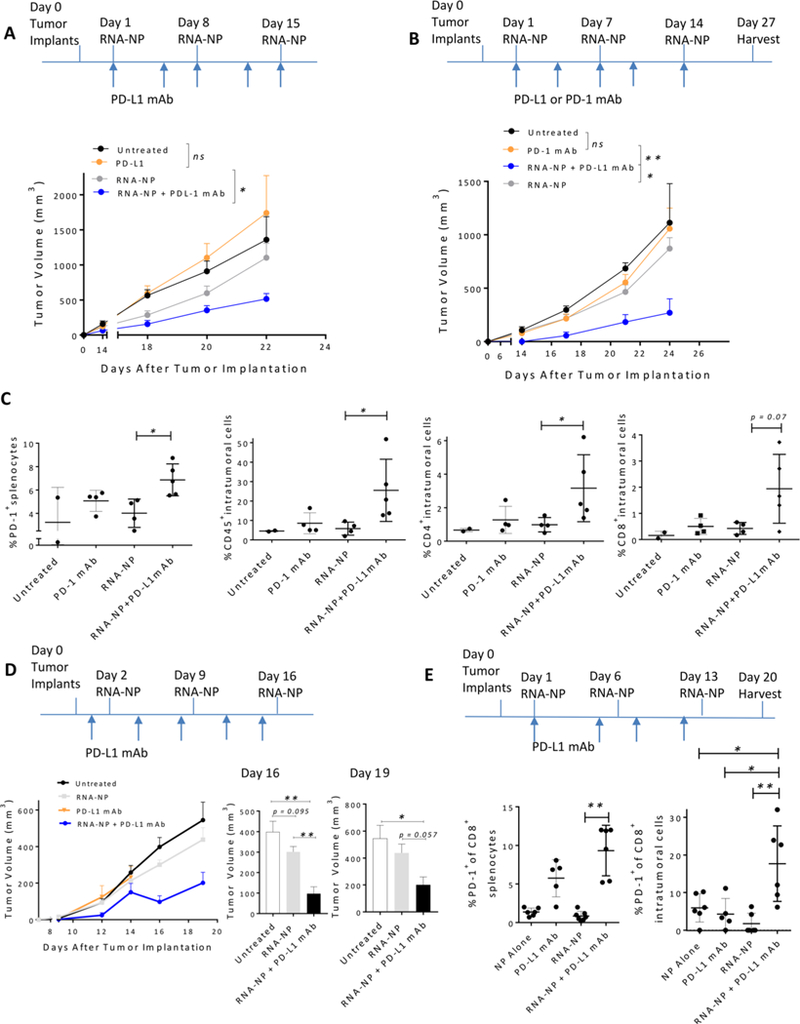
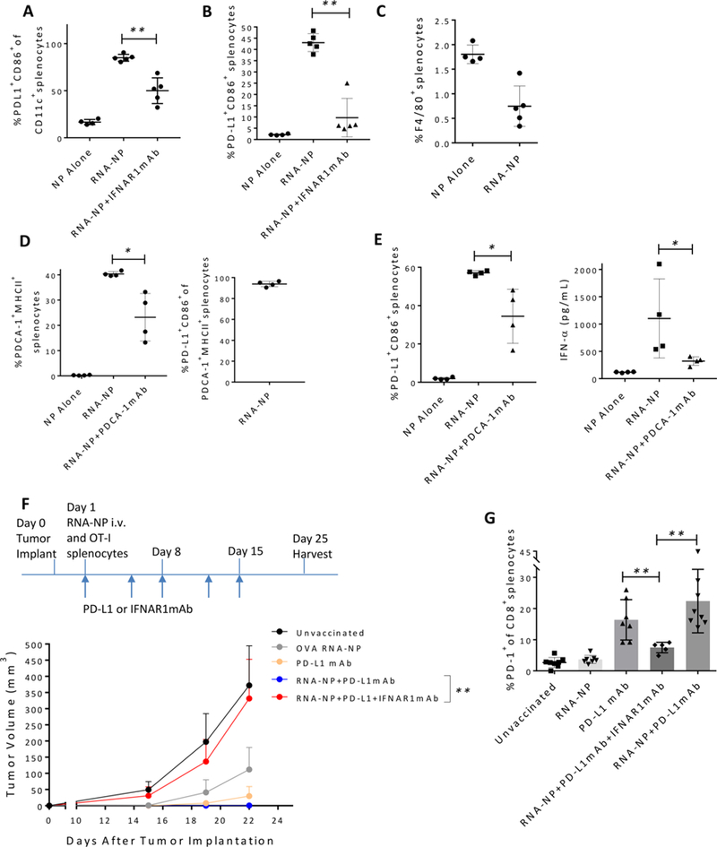
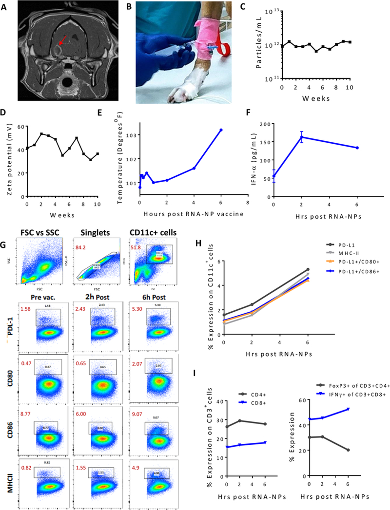
Translation of nanoparticles (NPs) into human clinical trials for patients with refractory cancers has lagged due to unknown biologic reactivities of novel NP designs. To overcome these limitations, simple well-characterized mRNA lipid-NPs have been developed as cancer immunotherapeutic vaccines. While the preponderance of RNA lipid-NPs encoding for tumor-associated antigens or neoepitopes have been designed to target lymphoid organs, they remain encumbered by the profound intratumoral and systemic immunosuppression that may stymie an activated T cell response. Herein, we show that systemic localization of untargeted tumor RNA (derived from whole transcriptome) encapsulated in lipid-NPs, with excess positive charge, primes the peripheral and intratumoral milieu for response to immunotherapy. In immunologically resistant tumor models, these RNA-NPs activate the preponderance of systemic and intratumoral myeloid cells (characterized by coexpression of PD-L1 and CD86). Addition of immune checkpoint inhibitors (ICIs) (to animals primed with RNA-NPs) augments peripheral/intratumoral PD-1+CD8+ cells and mediates synergistic antitumor efficacy in settings where ICIs alone do not confer therapeutic benefit. These synergistic effects are mediated by type I interferon released from plasmacytoid dendritic cells (pDCs). In translational studies, personalized mRNA-NPs were safe and active in a client-owned canine with a spontaneous malignant glioma. In summary, we demonstrate widespread immune activation from tumor loaded RNA-NPs concomitant with inducible PD-L1 expression that can be therapeutically exploited. While immunotherapy remains effective for only a subset of cancer patients, combination therapy with systemic immunomodulating RNA-NPs may broaden its therapeutic potency.

Keywords: RNA nanoparticles; cancer immunotherapy; cancer vaccines; immune checkpoint inhibitors; liposomes; personalized therapy.


Figures

Figure 1.
Figure 1.
RNA-NPs mediate increased PD-L1 upon activation of myeloidAPCs. (A) A cell line of dendritic cells (DC2.4s) was transfected with RNA-NPs. Cells were harvested within 24 hand imaged by TEM (scale bars = 500 nm). (B) DC2.4s were left untransfected or transfected with NPs alone or GFP RNA-NPs and harvested within 24h for assessment of PD-L1 expression (**p < 0.01, unpaired t test). (C) Human DCs were matured from source donor white blood cells, and were left untransfected or transfected with NPs alone or GFP RNA-NPs. Cells were harvested within 24 h for assessment of surface PD-L1 expression by flow cytometry (***p < 0.001, unpaired t test). (D, E) Three different batches of nanoliposomes were generated and assessed for concentration and ζ potential over time with serial measurements (mean ± SEM). (F) Cre encoding mRNA-NPs were administered to Ai14 transgenic mice (which carry a loxP-flanked STOP cassette impeding tdTomato transcription until Cre-recombinase expression), and 1 week later, lungs, hearts, livers, spleen, and lymph nodes were harvested, sectioned, and stained with DAPI before analysis of tdTomato expression by fluorescent microscopy. (G) FACS plots are displayed for PD-L1 (PE, phycoerythrin) and CD86 (APC, allophycocyanins) expression on CD11c+ splenocytes. (H, I) NPs were complexed with OVA-mRNA and injected iv into C57Bl/6 mice (n = 3–4/group) bearing subcutaneous B16–F10 tumors. Spleens, lungs, livers, lymph nodes, and bone marrow were harvested the next day for assessment of CD11c cells expressing CD86/PD-L1 (*p < 0.05, **p < 0.01, ****p < 0.0001, unpaired t test). (J) Gating strategyfor CD45+ intratumoral and CD45- tumor cells: cells are shown by forward and side scatter (left panel); among these, live cells are selected based on live/dead exclusion (middle panel), and CD45+ intratumoral or CD45- tumor cells are selected for analysis (right panel). (K) FACS plots for PD-L1+CD86+ intratumoral cells are shown from mice receiving either NPs alone (left) or RNA-NPs (right). (L–N) Within 3 weeks of subcutaneous B16–F10 tumor cell (500 000 cells) implantation, NPs were complexed with OVA-mRNA and injected iv into C57Bl/6 mice (n = 3/group); tumors were harvested the next day for flow cytometric analysis of CD11c, MHC-I, CD45, CD86, and PD-L1 expression (**p < 0.01, ***p < 0.001, unpaired t test).
Figure 2.
Figure 2.
PD-L1+ APCs do not suppress, but mediate enhancement of antigen-specific T cell immunity. (A, B) OVA RNA-NPs were injected iv into C57Bl/6 mice, and spleens were harvested the next day; spleens were FACS sorted for CD11c+MHCII+CD86+PD-L1+ cells (PD-L1+ APCs) (40 000 cells) which were added to a standard coculture assay (3 replicates/group) of OVA-mRNA electroporated primary murine bone marrow derived DCs (40 000 cells) and/or OTI derived splenocytes (400 000 cells) followed by serum assessment of IFN-γ by ELISA (**p < 0.01, unpaired t test). (C) OVA RNA-NPs were injected iv into C57Bl/6 mice, and spleens were harvested the next day; spleens were FACS sorted for CD11c+MHCII+CD86+PD-L1+ (PD-L1+ APCs) cells which were adoptively transferred to naïve C57Bl/6 mice (n = 5/group) “spiked” with a responder T cell population from OTI transgenic mice (1 × 10⁷ OTI splenocytes per mouse) (*p < 0.05, Mann–Whitney test). (D, E) OVA RNA-NPs were injected iv into tumor-bearing C57Bl/6 mice. Tumors were harvested within 3 weeks, FACS sorted for CD11c+MHCII+CD86+PD-L1+ cells (PD-L1+ intratumoral APCs) or CD 11 c+MHCII+CD86+PDL1- cells (PD-L1- intratumoral APCs) (40 000 cells) which were added to a standard coculture assay (n = 3 replicates/group) of OVA-mRNA electroporated primary murine bone marrow derived DCs (40 000 cells) and OVA mRNA activated T cells (400 000 cells). T cell activation was assessed by ELISA for IFN-γ production after 48 h (*p < 0.05, **p <0.01, unpaired t test). (F, G) PD-L1 expression by flow cytometry from splenocytes of C57Bl/6 mice (n = 3–5/group) vaccinated with iv GFP RNA-NPs, CFA-OVA peptide (administered intradermal), NPs alone, or iv GFP RNA-NPs with anti-PD-L1 mAbs or isotype mAbs (administered intraperitoneal (ip) 24 h prior to RNA-NPs) (*p < 0.05, **p < 0.01, Mann–Whitney test). (H) C57Bl/6 mice (n = 4–5/group) were vaccinated with pp65 RNA-NPs iv once weekly (×3) before spleens were harvested a week later for analysis of IFN-γ by ELISA (from soups of restimulated splenocytes with overlapping pp65 peptide pool) and by flow cytometric analysis ofpercent PD-1+ CD8+cells (**p < 0.01, Mann–Whitney test). (I) B16F10-OVA(1 000 000 cells) was implanted subcutaneously in the flanks of C57Bl/6 mice (n = 5–9/ group), and PD-1 mAbs, PD-L1 mAbs, OVA RNA-NPs, or OVA RNA-NP+ PD-L1 mAbs were administered the following day. OVA RNA-NPs were injected iv once weekly (×3), and PD-1/PD-L1 mAbs were injected ip twice weekly (×3 weeks). Survival is plotted on a Kaplan–Meier curve (*p < 0.05, **p < 0.01, Gehan–Breslow–Wilcoxon test).
Figure 3.
Figure 3.
RNA-NPs sensitize poorly immunogenic murine tumor models to ICIs. (A) B16F0 melanomas (50 000 cells) were implanted in the flanks of C57B1/6 mice (n = 5–7/group), and the following day, PD-L1 mAbs, TTRNA-NPs, or TTRNA-NP+PD-L1 mAbs were administered. TTRNA-NPs were injected iv once weekly (×3); PD-L1 mAbs were injected ip twice weekly (until last RNA-NP vaccine). Early time-point measurements of tumor volumes were plotted (day 22 tumor volume: RNA-NP versus RNA-NP + PD L1 mAb, *p < 0.05, Mann–Whitney test). (B) B16F0 melanomas were implanted in the flanks of C57Bl/6 mice (n = 5–7/group), and the following day, PD-1 mAbs, TTRNA-NPs, or TTRNA-NP with PD-L1 mAbs were administered. TTRNA-NPs were injected ivonce weekly (×3); PD-1 and PD-L1 mAbs were injected ip twice weekly (until last RNA-NP vaccine). Early time-point measurements of tumor volumes were plotted (day 21 and day 24 tumor volumes: PD-1 mAb versus RNA-NP + PD-L1 mAb, *p < 0.05, **p < 0.01, Mann–Whitney test), and (C) on day 27, spleens were harvested for assessment of peripheral PD-1 expression, and intratumoral CD45, CD4, and CD8 expression (*p < 0.05, Mann–Whitney test). (D) B16–F10 tumors (150 000 cells) were implanted in the flanks of C57Bl/6 mice (n = 5), and in the following days, PD-L1 mAbs, TTRNA-NPs, or TTRNA-NP+PD-L1 mAbs were administered. TTRNA-NPs were injected iv once weekly (×3); PD-L1 mAbs were injected ip twice weekly (until last RNA-NP vaccine). Tumor volume measurements were plotted over time (*p < 0.05, **p < 0.01, Mann–Whitney test, mean ± SEM). Animals receiving PD-L1 mAbs alone were removed from analysis on days 16–19 after >50% of animals were euthanized. (E) LLC (300 000 cells) was implanted in the flanks of C57Bl/6 mice (n = 7–8/group), and the next day, PD-L1 mAbs, TTRNA-NPs, or TTRNA-NP +PD-L1 mAbs were administered. TTRNA-NPs were injected iv once weekly (×3); PD-L1 mAbs were injected ip twice weekly (until last RNA-NP vaccine). Spleens and tumors were harvested on day 20 for assessment of PD-1 and CD8 expression (*p < 0.05, **p < 0.01, Mann–Whitney test).
Figure 4.
Figure 4.
Type I IFN drives synergistic activity from RNA-NP and ICIs. (A, B) NPs alone, GFP RNA-NPs, or GFP RNA-NPs with IFNAR1 blocking antibodies (500 μg administered ip) were administered systemically into naïve C57Bl/6 mice (n = 4–5/group), and spleens were harvested the next day for flow cytometric analysis of CD86, PD-L1, and CD11c expression (**p < 0.01, Mann–Whitney test). (C) RNA-NPs were administered iv to naïve C57Bl/6 mice (n = 4–5/group), and spleens were harvested the next day for assessment of F4/80 expression. (D, E) NPs alone, luciferase RNA-NPs, or luciferase RNA-NPs with PDCA-1 blocking antibodies were administered systemically into naïve C57Bl/6 mice (n = 4/group). Within 24 h, serum was collected for IFN-α detection by ELISA (overflow values assigned concentration of 2,000 pg/mL), and spleens were harvested for flow cytometric analysis of PDCA-1, MHCII, CD86, and PD-L1 expression (*p < 0.05, Mann–Whitney test). (F) B16F10-OVA melanomas (1 000 000 cells) were implanted in the flanks of C57Bl/6 mice (n = 7–8), and the following day, all mice received 1 × 10⁷ splenocytes iv obtained from OTI mice. Animals were left unvaccinated or received treatment with PD-L1 mAbs, OVA RNA-NPs, or OVA RNA-NP+PD-L1 mAbs with or without concomitant IFNAR1 mAbs. PD-L1 and IFNAR1 mAbs were administered ip twice weekly, and RNA-NPs were administered iv on day 1. Early time-point measurements of tumor volumes were plotted (day 22 tumor volumes: RNA-NP+PD-L1 mAb versus RNA-NP+ PD-L1 mAb + IFNAR1 mAb, **p< 0.01, Mann–Whitney test). (G) Spleens were harvested from all groups on day 25 for assessment of PD-1 expression on CD8+ splenocytes (**p < 0.01, Mann–Whitney test).
Figure 5.
Figure 5.
Personalized mRNA-NPs are safe and active in a translational disease model for spontaneous canine glioma. (A) A 9-year old spayed female Boxer was diagnosed with a malignant glioma on MRI (confirmed by histopathology) and enrolled to receive personalized mRNA-NPs per UFIACUC#201609430. (B) mRNA was amplified from a cDNA library prepared from extracted total RNA material from the patient’s biopsy. mRNA was sterilely complexed with DOTAP nanoliposomes, and drawn up from a vacutainer with a needle syringe. The RNA-NPs (>~12 mL) were then administered via iv push over 5 min through an intravenous catheter placed in the patient’s right front leg. (C, D) Nanoliposomes were generated for the canine patient and assessed for concentration and ζ potential over time with serial measurements. (E) Temperatures were plotted over time during the patient’s initial 6 h observation period postvaccination. (F) IFN-α ELISA was performed in duplicate on serum from the canine at baseline, 2 and 6 h postvaccination. (G) Blood was drawn at baseline, 2 and 6 h postvaccination for assessment of PD-L1, MHCII, CD80, and CD86 on CD11c+ cells. Gating strategy is shown for canine CD11c+ cells and expression of PD-L1, CD80, CD86, and MHCII. (H) CD11c expression of PD-L1, MHC-II, PD-L1/CD80, and PD-L1/CD86 is plotted over time during the canine’s initial observation period. (I) CD3+ cells were analyzed over time during the canine’s initial observation period for percent CD4 and CD8, and these subsets were assessed for expression of FoxP3 and IFN-γ, respectively.

Similar articles

Cited by

References

    1. Filley AC; Henriquez M; Dey M Oncotarget 2017, 8 (53), 91779–91794. - PMC - PubMed
    1. Brahmer JR; Tykodi SS; Chow LQ; Hwu WJ; Topalian SL; Hwu P; Drake CG; Camacho LH; Kauh J; Odunsi K; Pitot HC; Hamid O; Bhatia S; Martins R; Eaton K; Chen S; Salay TM; Alaparthy S; Grosso JF; Korman AJ; Parker SM; Agrawal S; Goldberg SM; Pardoll DM; Gupta A; Wigginton JM N. Engl. J. Med. 2012, 366 (26), 2455–65. - PMC - PubMed
    1. Borghaei H; Paz-Ares L; Horn L; Spigel DR; Steins M; Ready NE; Chow LQ; Vokes EE; Felip E; Holgado E; Barlesi F; Kohlhaufl M; Arrieta O; Burgio MA; Fayette J; Lena H; Poddubskaya E; Gerber DE; Gettinger SN; Rudin CM; Rizvi N; Crino L; Blumenschein GR Jr.; Antonia SJ; Dorange C; Harbison CT; Graf Finckenstein F; Brahmer JR N. Engl. J. Med. 2015, 373 (17), 1627–39. - PMC - PubMed
    1. Topalian SL; Hodi FS; Brahmer JR; Gettinger SN; Smith DC; McDermott DF; Powderly JD; Carvajal RD; Sosman JA; Atkins MB; Leming PD; Spigel DR; Antonia SJ; Horn L; Drake CG; Pardoll DM; Chen L; Sharfman WH; Anders RA; Taube JM; McMiller TL; Xu H; Korman AJ; Jure-Kunkel M; Agrawal S; McDonald D; Kollia GD; Gupta A; Wigginton JM; Sznol M N. Engl. J. Med. 2012, 366 (26), 2443–54. - PMC - PubMed
    1. Gros A; Robbins PF; Yao X; Li YF; Turcotte S; Tran E; Wunderlich JR; Mixon A; Farid S; Dudley ME; Hanada K; Almeida JR; Darko S; Douek DC; Yang JC; Rosenberg SA J. Clin. Invest. 2014, 124 (5), 2246–59. - PMC - PubMed

Publication types

MeSH terms