

Longitudinal In Vivo Diffusion Magnetic Resonance Imaging Remote from the Lesion Site in Rat Spinal Cord Injury
- PMID: 30259800
- PMCID: PMC6482909
- DOI: 10.1089/neu.2018.5964
Longitudinal In Vivo Diffusion Magnetic Resonance Imaging Remote from the Lesion Site in Rat Spinal Cord Injury
Abstract
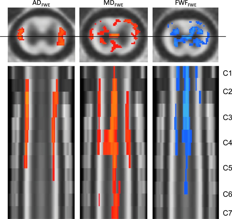
Diffusion tensor imaging (DTI) has demonstrated success as a biomarker of spinal cord injury (SCI) severity as shown from numerous pre-clinical studies. However, artifacts from stabilization hardware at the lesion have precluded its use for longitudinal assessments. Previous research has documented ex vivo diffusion changes in the spinal cord both caudal and cranial to the injury epicenter. The aim of this study was to use a rat contusion model of SCI to evaluate the utility of in vivo cervical DTI after a thoracic injury. Forty Sprague-Dawley rats underwent a thoracic contusion (T8) of mild, moderate, severe, or sham severity. Magnetic resonance imaging (MRI) of the cervical cord was performed at 2, 30, and 90 days post-injury, and locomotor performance was assessed weekly using the Basso, Bresnahan, and Beattie (BBB) scoring scale. The relationships between BBB scores and MRI were assessed using region of interest analysis and voxel-wise linear regression of DTI, and free water elimination (FWE) modeling to reduce partial volume effects. At 90 days, axial diffusivity (ADFWE), mean diffusivity (MDFWE), and free water fraction (FWFFWE) using the FWE model were found to be significantly correlated with BBB score. FWE was found to be more predictive of injury severity than conventional DTI, specifically at later time-points. This study validated the use of FWE technique in spinal cord and demonstrated its sensitivity to injury remotely.
Keywords: diffusion tensor imaging; free water elimination; spinal cord injury.
Conflict of interest statement
No competing financial interest exist.
Figures
References
-
- Burns A.S., Lee B.S., Ditunno J.F., and Tessler A. (2003). Patient selection for clinical trials: the reliability of the early spinal cord injury examination. J. Neurotrauma 20, 477–82 - PubMed
-
- Fawcett J.W., Curt A., Steeves J.D., Coleman W.P., Tuszynski M.H., Lammertse D., Bartlett P.F., Blight A.R., Dietz V., Ditunno J., Dobkin B.H., Havton L.A., Ellaway P.H., Fehlings M.G., Privat A., Grossman R., Guest J.D., Kleitman N., Nakamura M., Gaviria M., and Short D. (2007). Guidelines for the conduct of clinical trials for spinal cord injury as developed by the ICCP panel: spontaneous recovery after spinal cord injury and statistical power needed for therapeutic clinical trials. Spinal Cord 45, 190–205 - PubMed
-
- Bottomley P.A., Hardy C.J., Argensinger R.E., and Allen-Moore G. (1987). A review of 1H nuclear magnetic resonance relaxation in pathology: are T1 and T2 diagnostic? Med. Phys. 14, 1–37 - PubMed
-
- Tu T.W. and Frank J.A. (2013). Assessing white matter integrity in experimental spinal cord injury using diffusion tensor imaging. J. Neurosci. Neuroengineering 2, 415–430
-
- Ellingson B.M., Kurpad S.N., and Schmit B.D. (2008). Ex vivo diffusion tensor imaging and quantitative tractography of the rat spinal cord during long-term recovery from moderate spinal contusion. J. Magn. Reson. Imaging 28, 1068–1079 - PubMed
Publication types
MeSH terms
Grants and funding
LinkOut - more resources
Full Text Sources
Other Literature Sources
Medical
