. 2019 Jan;111(1):82-95.
doi: 10.1111/mmi.14140. Epub 2018 Oct 26.

Ail provides multiple mechanisms of serum resistance to Yersinia pestis

Affiliations

Ail provides multiple mechanisms of serum resistance to Yersinia pestis

Joshua J Thomson et al. Mol Microbiol. 2019 Jan.

Abstract

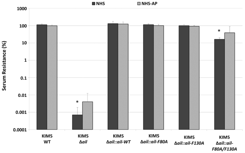
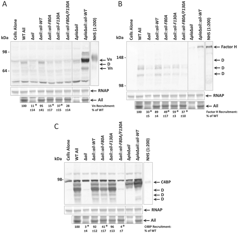
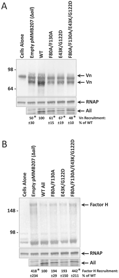
Ail, a multifunctional outer membrane protein of Yersinia pestis, confers cell binding, Yop delivery and serum resistance activities. Resistance to complement proteins in serum is critical for the survival of Y. pestis during the septicemic stage of plague infections. Bacteria employ a variety of tactics to evade the complement system, including recruitment of complement regulatory factors, such as factor H, C4b-binding protein (C4BP) and vitronectin (Vn). Y. pestis Ail interacts with the regulatory factors Vn and C4BP, and Ail homologs from Y. enterocolitica and Y. pseudotuberculosis recruit factor H. Using co-sedimentation assays, we demonstrate that two surface-exposed amino acids, F80 and F130, are required for the interaction of Y. pestis Ail with Vn, factor H and C4BP. However, although Ail-F80A/F130A fails to interact with these complement regulatory proteins, it still confers 10,000-fold more serum resistance than a Δail strain and prevents C9 polymerization, potentially by directly interfering with MAC assembly. Using site-directed mutagenesis, we further defined this additional mechanism of complement evasion conferred by Ail. Finally, we find that at Y. pestis concentrations reflective of early-stage septicemic plague, Ail weakly recruits Vn and fails to recruit factor H, suggesting that this alternative mechanism of serum resistance may be essential during plague infection.


Figures

Figure 1.
Figure 1.. Killing of Y. pestis Δail by human serum is mediated by the alternative pathway of complement.
~7.5 × 10⁵ CFU of mid-log cultures of Y. pestis strains containing wild-type Ail, a chromosomal deletion of ailail), or chromosomal ail recombinants were treated with 80% NHS, 80% HIS (Heat-inactivated serum), or 80% NHS-AP (NHS treated with 5mM EGTA and 10mM MgCl2 to inactivate CP/LP) for one hour at 37°C. Surviving bacteria were plated and enumerated by colony counting. Percent serum resistance was calculated as the number of surviving colonies in NHS/HIS or NHS-AP/HIS x 100 and is displayed on a logarithmic scale. Strains were tested a minimum of 3 times for each condition in separate experiments. Significance was determined using the two-way ANOVA with Tukey’s post hoc test. *, p-value < 0.05 when compared to the parental KIM5 wild-type (WT) strain in the same serum condition.
Figure 2.
Figure 2.. Co-sedimentation of complement regulatory factors with Y. pestis is mediated by Ail extracellular loop residues F80 and F130.
Overnight cultures of Y. pestis KIM5 strains containing wild-type Ail, a chromosomal deletion of ail, or specific chromosomal alleles of ail were mixed with 50% NHS to a final OD620 = 50. Mixtures were shaken vigorously at 37°C for 30 minutes. Cells were centrifuged, washed, and analyzed by Western blot for the presence of membrane-associated complement regulators: A) vitronectin (Vn) B) factor H C) C4b-binding protein (C4BP). Levels of expressed Ail were determined by Coomassie staining. ΔailΔpla strains are included to show full-length, un-degraded complement regulatory proteins. The cells alone lane indicates Y. pestis KIM5 cross-reactive bands recognized in the absence of NHS. Blots are one representative of at least three independent experiments and are shown with the Coomassie-stained gel showing Ail expression from the same experiment, as well as the loading control anti-E. coli RNA polymerase alpha. Molecular weight markers are indicated on the left of the blot. Quantification of band intensity was performed using at least 3 independent experiments with ImageJ software (NIH). Intensity of bands corresponding to complement regulator recruitment is shown as a percentage of WT recruitment (normalized to 100%) in each individual blot. Significance was determined using one-way ANOVA with Tukey’s post hoc test. *, p-value < 0.05 when compared to the wild-type strain of Y. pestis KIM5. Abbreviations: Vn=vitronectin, C4BP=C4b-binding protein, D=degraded form of protein (degraded by Pla).
Figure 3.
Figure 3.. Multiple Ail substitutions required to reveal a serum sensitivity phenotype comparable to Δail deletion.
A) Amino acid substitutions of Y. pestis Ail residues corresponding to homologous residues that no longer confer full serum resistance in Yersinia enterocolitica Ail (Miller et al., 2001) and Salmonella enterica Rck (Cirillo et al., 1996). B) Resistance of Y. pestis KIM5 Δail expressing plasmid-borne Ail or Ail derivatives, to killing by normal human serum (NHS). ~7.5 × 10⁵ CFU of mid-log culture grown with 500μM IPTG (to induce Ail expression) was added to 80% heat-inactivated serum (HIS) or 80% NHS for one hour at 37oC. Surviving bacteria were plated and enumerated by colony counting. Percent serum resistance was calculated by (number of surviving colonies in NHS or NHS-AP/HIS) x 100 and is displayed on a logarithmic scale. Strains were tested a minimum of 3 times in separate experiments. Ail expression and stability was determined by Coomassie staining shown beneath the graph. Significance was assessed using the one-way ANOVA with Tukey’s post hoc test. *, p-value < 0.05 when compared to serum resistance of a strain expressing wild-type Ail.
Figure 4.
Figure 4.. Co-sedimentation of alternative pathway regulatory factors is mediated by various extracellular loop residues of Y. pestis.
Overnight cultures grown in the presence of 100μM IPTG (to induce Ail expression) were mixed with 50% NHS to a final OD620 = 50. Mixtures were shaken vigorously at 37°C for 30 minutes. Mixtures were centrifuged and cell pellets were washed, then subjected to Western blotting for complement regulatory factors: A) vitronectin (Vn) and B) factor H. All Western blots are accompanied by Coomassie-stained gel showing Ail expression in the same samples. Molecular weight markers are indicated on the left. The cells alone lane represents Y. pestis in the absence of NHS. Quantification of band intensity was performed using at least 3 independent experiments with ImageJ software (NIH). Intensity of bands corresponding to complement regulator recruitment is shown as a percentage of WT recruitment (normalized to 100%) in each individual blot. Significance was determined using one-way ANOVA with Tukey’s post hoc test. *, p-value < 0.05 when compared to a strain expressing wild-type Ail.
Figure 5.
Figure 5.. Loss of Ail-mediated recruitment of complement regulatory factors at lower bacterial concentration.
Overnight cultures grown in the presence of 100μM IPTG (to induce Ail expression) were mixed with 50% NHS to a final OD620 = 0.25. Mixtures were shaken vigorously at 37°C for 30 minutes. Samples were centrifuged and cell pellets were washed, then subjected to Western blotting for complement regulatory factors: A) vitronectin (Vn) and B) factor H. Western blots are accompanied by Coomassie-stained gel showing Ail expression in the same samples, as well as the loading control anti-E. coli RNA polymerase alpha. Molecular weight markers are indicated on the left. The cells alone lane represents Y. pestis in the absence of NHS. Quantification of band intensity was performed using at least 3 independent experiments with ImageJ software (NIH). Intensity of bands corresponding to complement regulator recruitment is shown as a percentage of wild-type Ail-mediated recruitment (normalized to 100%) in each individual blot. Significance was determined using one-way ANOVA with Tukey’s post hoc test. *, p-value < 0.05 when compared to a strain expressing wild-type Ail.
Figure 6.
Figure 6.. Membrane-association of polymerized C9 is increased in strains lacking ail or containing multiple mutations to extracellular loops.
Overnight cultures grown in the presence of 100μM IPTG (to induce Ail expression) were mixed with 50% NHS to a final OD620 = 0.25. Mixtures were shaken vigorously at 37°C for 30 minutes. Samples were centrifuged, cell pellets were washed, and subjected to Western blotting under non-reducing conditions using an anti-C9 polyclonal antibody. Western blot is one representative of at least three independent experiments. Ail expression was determined by Coomassie staining and is shown beneath the blot. Molecular weight markers are indicated on the left of the blot. Cells alone lane represents Y. pestis in the absence of NHS. Zymosan activated NHS is a positive control for the formation of polymerized C9 compared to NHS alone (monomeric C9). Quantification of band intensity from at least 3 independent experiments was performed using ImageJ software (NIH). Intensity of bands corresponding to C9 polymerization is shown as a percentage of the Δail mutant (a strain with no ability to inhibit C9 polymerization), which was normalized to 100%. Significance was determined using one-way ANOVA with Tukey’s post hoc test. *, p-value < 0.05 when compared to a strain expressing wild-type Ail.

Similar articles

Cited by

References

    1. Bartra SS, Ding Y, Fujimoto LM, Ring JG, Jain V, Ram S, Marassi FM & Plano GV, (2015) Yersinia pestis uses the Ail outer membrane protein to recruit vitronectin. Microbiology 161: 2174–2183. - PMC - PubMed
    1. Bartra SS, Styer KL, O’Bryant DM, Nilles ML, Hinnebusch BJ, Aballay A & Plano GV, (2008) Resistance of Yersinia pestis to Complement-Dependent Killing Is Mediated by the Ail Outer Membrane Protein. Infect. Immun 76: 612–622. - PMC - PubMed
    1. Biedzka-Sarek M, Jarva H, Hyytiainen H, Meri S & Skurnik M, (2008a) Characterization of complement factor H binding to Yersinia enterocolitica serotype O:3. Infect Immun 76: 4100–4109. - PMC - PubMed
    1. Biedzka-Sarek M, Salmenlinna S, Gruber M, Lupas AN, Meri S & Skurnik M, (2008b) Functional mapping of YadA- and Ail-mediated binding of human factor H to Yersinia enterocolitica serotype O:3. Infect Immun 76: 5016–5027. - PMC - PubMed
    1. Bliska J & Falkow S, (1992) Bacterial Resistance to Complement Killing Mediated by the Ail Protein of Yersinia enterocolitica. Proc Natl Acad Sci USA 89: 3561–3565. - PMC - PubMed

Publication types

MeSH terms

Substances