

doi: 10.1093/ons/opy270.
A Connectomic Atlas of the Human Cerebrum-Chapter 16: Tractographic Description of the Vertical Occipital Fasciculus
Affiliations
- PMID: 30260427
- PMCID: PMC6890522
- DOI: 10.1093/ons/opy270
Item in Clipboard
A Connectomic Atlas of the Human Cerebrum-Chapter 16: Tractographic Description of the Vertical Occipital Fasciculus
Oper Neurosurg.
.
Abstract
In this supplement, we show a comprehensive anatomic atlas of the human cerebrum demonstrating all 180 distinct regions comprising the cerebral cortex. The location, functional connectivity, and structural connectivity of these regions are outlined, and where possible a discussion is included of the functional significance of these areas. In this chapter, we specifically address regions integrating to form the vertical occipital fasciculus.
Figures
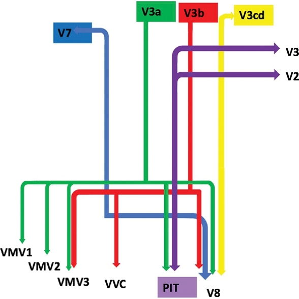
Simplified tract map showing the structural connections that integrate within the VOF. Connections between cortical areas are color-coded based on the parcellation of origin (eg, red arrows indicate structural connections from origin V3b to areas VMV3, VVC, and V8). Note that arrows are not meant to imply the direction of information transmit.
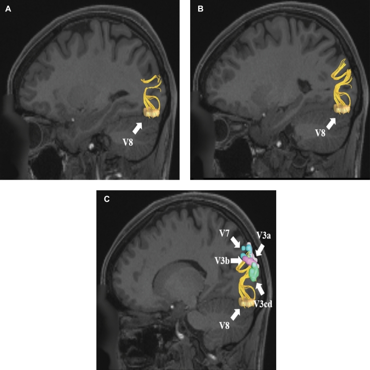
VOF connections from region V8. Area V8 is located in the lateral occipital cortex and has structural connections to areas V3a, V3b, V3cd, and V7. These connections are shown in the left cerebral hemisphere on T1-weighted MR images in the sagittal plane: A, medial view, B, lateral view without regions of interest, C, lateral view with regions of interest. All parcellations are identified with white arrows and corresponding labels.
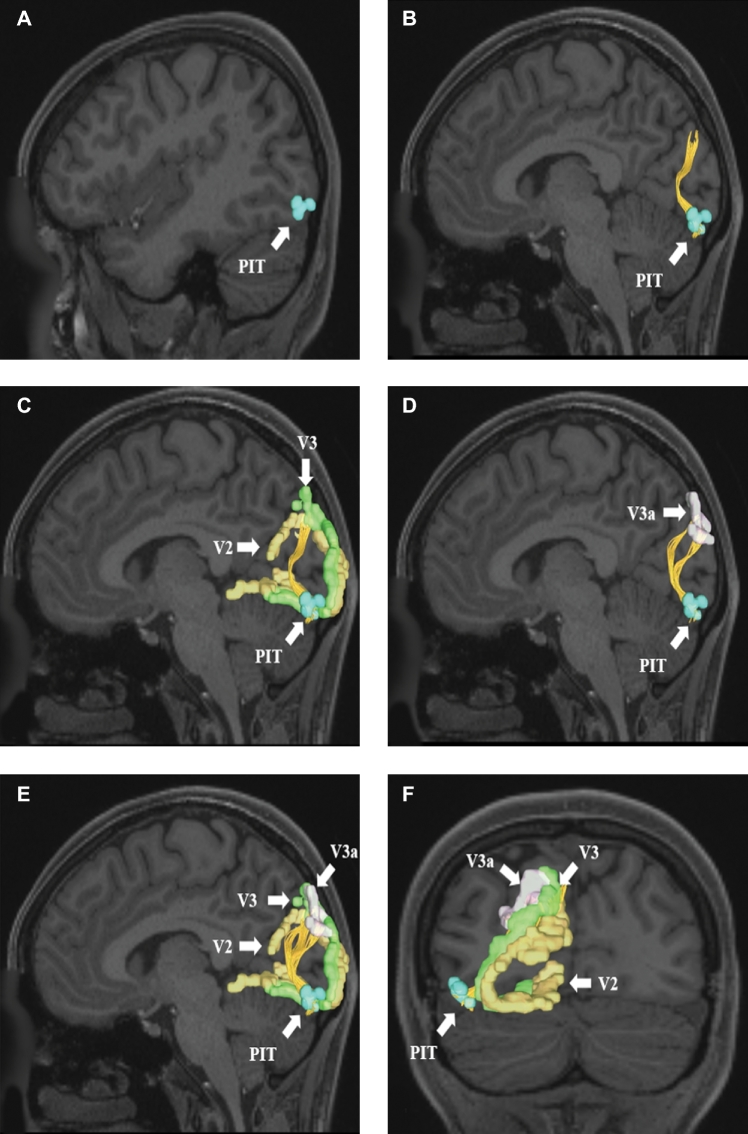
VOF connections from region PIT. Area PIT is located in the lateral occipital cortex and has structural connections to areas V2, V3, and V3a. These connections are shown in the left cerebral hemisphere on T1-weighted MR images in the A–E, sagittal and F, coronal planes: A, medial view of PIT, B, lateral view of PIT with the VOF readily identified, C, VOF connections to V2 and V3, D, VOF connections to V3a, E, entire set of VOF connections from area PIT, and F, posterior coronal view of the early visual processing regions connected to PIT. All parcellations are identified with white arrows and corresponding labels.
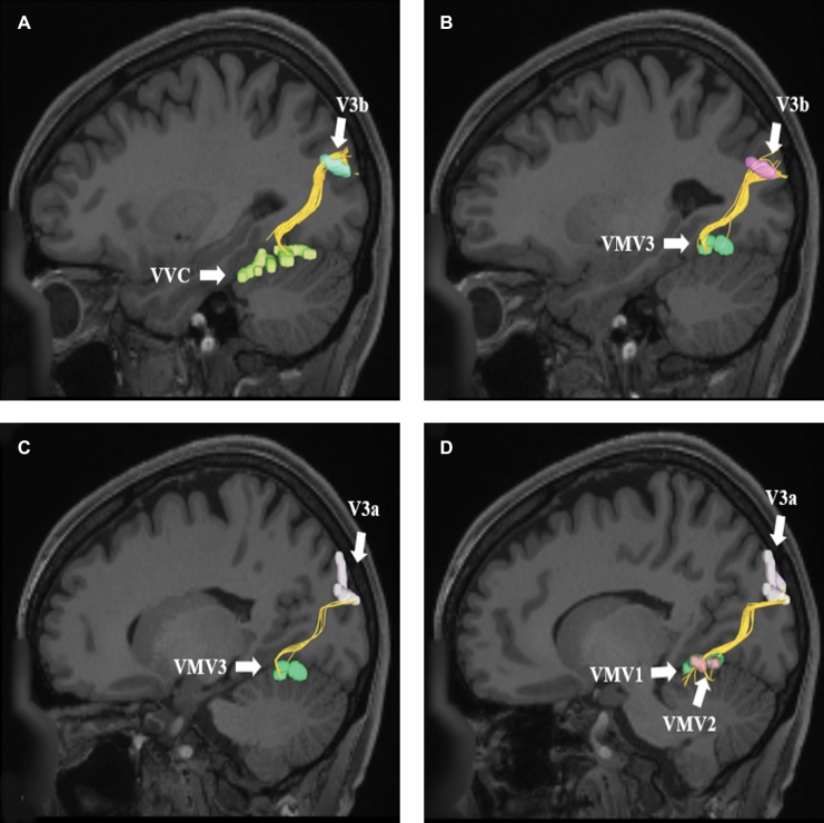
VOF connections from regions A and B, V3b and C and D, V3a. Area V3b exhibits structural connections to areas A, VVC and B, VMV3. These connections are shown in the left cerebral hemisphere on T1-weighted MR images in the sagittal plane. Area V3a exhibits structural connections to areas C, VMV3 and D, VMV1 and VMV2. These connections are shown in the left cerebral hemisphere on T1-weighted MR images in the sagittal plane. All parcellations are identified with white arrows and corresponding labels.
Similar articles
-
A Connectomic Atlas of the Human Cerebrum-Chapter 11: Tractographic Description of the Inferior Longitudinal Fasciculus.Oper Neurosurg. 2018 Dec 1;15(suppl_1):S423-S428. doi: 10.1093/ons/opy265. Oper Neurosurg. 2018. PMID: 30260434 Free PMC article.
-
A Connectomic Atlas of the Human Cerebrum-Chapter 15: Tractographic Description of the Uncinate Fasciculus.Oper Neurosurg. 2018 Dec 1;15(suppl_1):S450-S455. doi: 10.1093/ons/opy269. Oper Neurosurg. 2018. PMID: 30260439 Free PMC article.
-
A Connectomic Atlas of the Human Cerebrum-Chapter 14: Tractographic Description of the Frontal Aslant Tract.Oper Neurosurg. 2018 Dec 1;15(suppl_1):S444-S449. doi: 10.1093/ons/opy268. Oper Neurosurg. 2018. PMID: 30260440 Free PMC article.
-
The white matter architecture underlying semantic processing: A systematic review.Neuropsychologia. 2020 Jan;136:107182. doi: 10.1016/j.neuropsychologia.2019.107182. Epub 2019 Sep 27. Neuropsychologia. 2020. PMID: 31568774
-
Imaging connectivity: MRI and the structural networks of the brain.Funct Neurol. 2013 Jul-Sep;28(3):197-203. Funct Neurol. 2013. PMID: 24139656 Free PMC article. Review.
Cited by
-
Connectivity-based parcellation of normal and anatomically distorted human cerebral cortex.Hum Brain Mapp. 2022 Mar;43(4):1358-1369. doi: 10.1002/hbm.25728. Epub 2021 Nov 26. Hum Brain Mapp. 2022. PMID: 34826179 Free PMC article.
-
Anatomy and white matter connections of the lateral occipital cortex.Surg Radiol Anat. 2020 Mar;42(3):315-328. doi: 10.1007/s00276-019-02371-z. Epub 2019 Nov 16. Surg Radiol Anat. 2020. PMID: 31734739
-
Stroke disconnectome decodes reading networks.Brain Struct Funct. 2022 Dec;227(9):2897-2908. doi: 10.1007/s00429-022-02575-x. Epub 2022 Oct 3. Brain Struct Funct. 2022. PMID: 36192557 Free PMC article.
-
White matter variability, cognition, and disorders: a systematic review.Brain Struct Funct. 2022 Mar;227(2):529-544. doi: 10.1007/s00429-021-02382-w. Epub 2021 Nov 3. Brain Struct Funct. 2022. PMID: 34731328 Free PMC article.
-
Occipital Intralobar fasciculi: a description, through tractography, of three forgotten tracts.Commun Biol. 2021 Mar 30;4(1):433. doi: 10.1038/s42003-021-01935-3. Commun Biol. 2021. PMID: 33785859 Free PMC article.
References
-
- Gungor A, Baydin S, Middlebrooks EH, Tanriover N, Isler C, Rhoton AL Jr. The white matter tracts of the cerebrum in ventricular surgery and hydrocephalus. J Neurosurg. 2017;126(3):945-971. - PubMed
-
- Wu Y, Sun D, Wang Y, Wang Y, Wang Y. Tracing short connections of the temporo-parieto-occipital region in the human brain using diffusion spectrum imaging and fiber dissection. Brain Res. 2016;1646:152-159. - PubMed
Publication types
MeSH terms
Grants and funding
LinkOut - more resources
Full Text Sources
Other Literature Sources
