

A Connectomic Atlas of the Human Cerebrum-Chapter 13: Tractographic Description of the Inferior Fronto-Occipital Fasciculus
- PMID: 30260438
- PMCID: PMC6890527
- DOI: 10.1093/ons/opy267
A Connectomic Atlas of the Human Cerebrum-Chapter 13: Tractographic Description of the Inferior Fronto-Occipital Fasciculus
Abstract
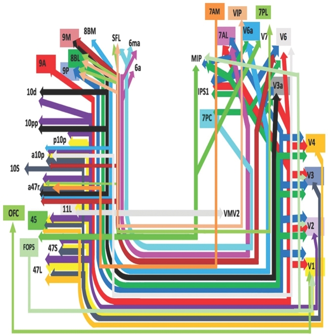
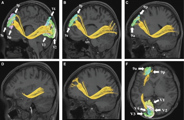
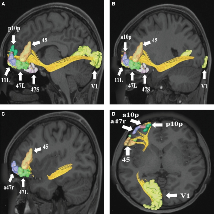
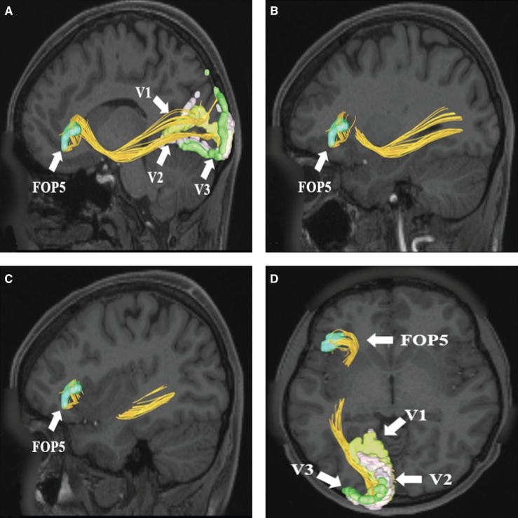
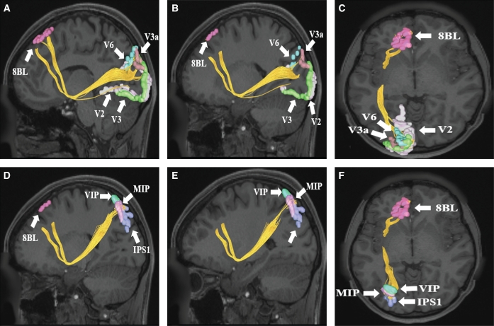
The inferior fronto-occipital fasciculus (IFOF) is a large white matter tract of the human cerebrum with functional connectivity associated with semantic language processing and goal-oriented behavior. However, little is known regarding the overall connectivity of this tract. Recently, the Human Connectome Project parcellated the human cortex into 180 distinct regions. In our other work, we have shown these various regions in relation to clinically applicable anatomy and function. Utilizing Diffusion Spectrum Magnetic Resonance Imaging tractography coupled with the human cortex parcellation data presented earlier in this supplement, we aim to describe the macro-connectome of the IFOF in relation to the linked parcellations present within the human cortex. The purpose of this study is to present this information in an indexed, illustrated, and tractographically aided series of figures and tables for anatomic and clinical reference.
Figures
Similar articles
-
A Connectomic Atlas of the Human Cerebrum-Chapter 10: Tractographic Description of the Superior Longitudinal Fasciculus.Oper Neurosurg. 2018 Dec 1;15(suppl_1):S407-S422. doi: 10.1093/ons/opy264. Oper Neurosurg. 2018. PMID: 30260421 Free PMC article.
-
A Connectomic Atlas of the Human Cerebrum-Chapter 12: Tractographic Description of the Middle Longitudinal Fasciculus.Oper Neurosurg. 2018 Dec 1;15(suppl_1):S429-S435. doi: 10.1093/ons/opy266. Oper Neurosurg. 2018. PMID: 30260450 Free PMC article.
-
A Connectomic Atlas of the Human Cerebrum-Chapter 16: Tractographic Description of the Vertical Occipital Fasciculus.Oper Neurosurg. 2018 Dec 1;15(suppl_1):S456-S461. doi: 10.1093/ons/opy270. Oper Neurosurg. 2018. PMID: 30260427 Free PMC article.
-
The inferior fronto-occipital fasciculus: bridging phylogeny, ontogeny and functional anatomy.Brain. 2025 May 13;148(5):1507-1525. doi: 10.1093/brain/awaf055. Brain. 2025. PMID: 39932875 Free PMC article. Review.
-
Anatomical variability of the arcuate fasciculus: a systematical review.Surg Radiol Anat. 2019 Aug;41(8):889-900. doi: 10.1007/s00276-019-02244-5. Epub 2019 Apr 26. Surg Radiol Anat. 2019. PMID: 31028450
Cited by
-
Pinpointing Neural Correlates of Attachment in Poly-Drug Use: A Diffusion Tensor Imaging Study.Front Neurosci. 2020 Jun 11;14:596. doi: 10.3389/fnins.2020.00596. eCollection 2020. Front Neurosci. 2020. PMID: 32595448 Free PMC article.
-
Surgical Technique and Outcome of Extensive Frontal Lobectomy for Treatment of Intracable Non-lesional Frontal Lobe Epilepsy.Neurol Med Chir (Tokyo). 2020 Jan 15;60(1):17-25. doi: 10.2176/nmc.oa.2018-0286. Epub 2019 Dec 5. Neurol Med Chir (Tokyo). 2020. PMID: 31801933 Free PMC article.
-
Inferior fronto-occipital fascicle displacement in temporoinsular gliomas using diffusion tensor imaging.J Neuroimaging. 2022 Jul;32(4):638-646. doi: 10.1111/jon.12992. Epub 2022 Mar 30. J Neuroimaging. 2022. PMID: 35352437 Free PMC article.
-
White matter biomarker for predicting de novo Parkinson's disease using tract-based spatial statistics: a machine learning-based model.Quant Imaging Med Surg. 2024 Apr 3;14(4):3086-3106. doi: 10.21037/qims-23-1478. Epub 2024 Mar 28. Quant Imaging Med Surg. 2024. PMID: 38617147 Free PMC article.
-
Tumor location and neurocognitive function-Unravelling the association and identifying relevant anatomical substrates in intra-axial brain tumors.Neurooncol Adv. 2024 Feb 9;6(1):vdae020. doi: 10.1093/noajnl/vdae020. eCollection 2024 Jan-Dec. Neurooncol Adv. 2024. PMID: 38464948 Free PMC article.
References
-
- de Schotten MT, Dell’Acqua F, Valabregue R, Catani M. Monkey to human comparative anatomy of the frontal lobe association tracts. Cortex. 2012;48(1):82-96. - PubMed
-
- Motomura K, Fujii M, Maesawa S, Kuramitsu S, Natsume A, Wakabayashi T. Association of dorsal inferior frontooccipital fasciculus fibers in the deep parietal lobe with both reading and writing processes: a brain mapping study. J Neurosurg. 2014;121(1):142-148. - PubMed
-
- Almairac F, Herbet G, Moritz-Gasser S, de Champfleur NM, Duffau H. The left inferior fronto-occipital fasciculus subserves language semantics: a multilevel lesion study. Brain Struct Funct. 2015;220(4):1983-1995. - PubMed
Publication types
MeSH terms
Grants and funding
LinkOut - more resources
Full Text Sources
Other Literature Sources
