

doi: 10.1093/ons/opy269.
A Connectomic Atlas of the Human Cerebrum-Chapter 15: Tractographic Description of the Uncinate Fasciculus
Affiliations
- PMID: 30260439
- PMCID: PMC6890529
- DOI: 10.1093/ons/opy269
Item in Clipboard
A Connectomic Atlas of the Human Cerebrum-Chapter 15: Tractographic Description of the Uncinate Fasciculus
Oper Neurosurg.
.
Abstract
In this supplement, we show a comprehensive anatomic atlas of the human cerebrum demonstrating all 180 distinct regions comprising the cerebral cortex. The location, functional connectivity, and structural connectivity of these regions are outlined, and where possible a discussion is included of the functional significance of these areas. In this chapter, we specifically address the regions integrating to form the uncinate fasciculus.
Figures
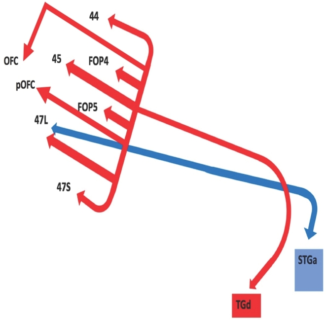
Simplified tract map showing the structural connections that integrate within the UF. Connections between cortical areas are color-coded based on the parcellation of origin (eg, red arrows indicate structural connections from origin TGd to areas 44, FOP4, 45, FOP5, OFC, pOFC, 47L, and 47S). Note that arrows are not meant to imply the direction of information transmit.
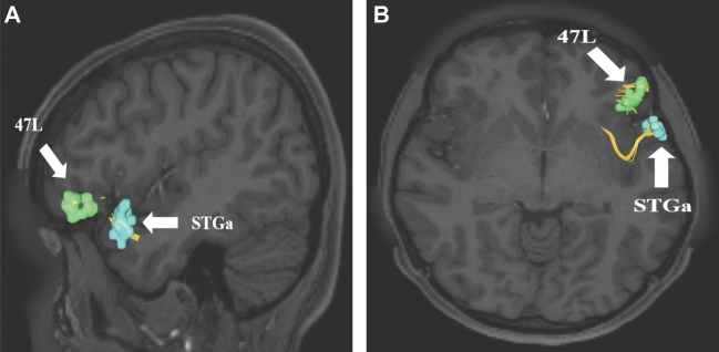
Uncinate connections from temporal polar region STGa. Area STGa only has 1 structural connection integrated within the uncinate to area 47L. This connection is shown in the left cerebral hemisphere on T1-weighted MR images in the A, sagittal and B, axial planes. All parcellations are identified with white arrows and corresponding labels.
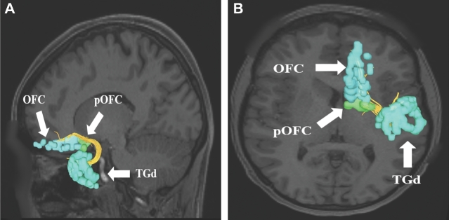
Uncinate connections from temporal polar region TGd. Area TGd has multiple structural connections integrated within the uncinate to areas 44, FOP4, 45, FOP5, OFC, pOFC, 47L, and 47S. A subset of these connections to regions OFC and pOFC are shown in the left cerebral hemisphere on T1-weighted MR images in the A, sagittal and B, axial planes. All parcellations are identified with white arrows and corresponding labels.
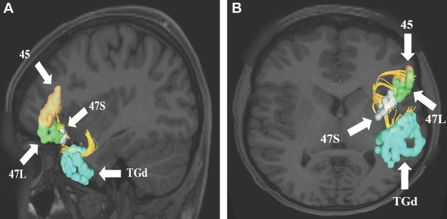
Uncinate connections from temporal polar region TGd. Area TGd has multiple structural connections integrated within the uncinate to areas 44, FOP4, 45, FOP5, OFC, pOFC, 47L, and 47S. A subset of these connections to regions 45, 47S, and 47L are shown in the left cerebral hemisphere on T1-weighted MR images in the A, sagittal and B, axial planes. All parcellations are identified with white arrows and corresponding labels.
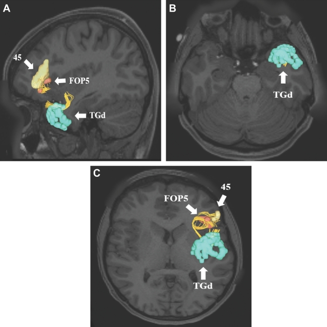
Uncinate connections from temporal polar region TGd. Area TGd has multiple structural connections integrated within the uncinate to areas 44, FOP4, 45, FOP5, OFC, pOFC, 47L, and 47S. A subset of these connections to regions 45 and FOP5 are shown in the left cerebral hemisphere on T1-weighted MR images in the A, sagittal and B and C, axial planes. All parcellations are identified with white arrows and corresponding labels.
Similar articles
-
A Connectomic Atlas of the Human Cerebrum-Chapter 11: Tractographic Description of the Inferior Longitudinal Fasciculus.Oper Neurosurg. 2018 Dec 1;15(suppl_1):S423-S428. doi: 10.1093/ons/opy265. Oper Neurosurg. 2018. PMID: 30260434 Free PMC article.
-
A Connectomic Atlas of the Human Cerebrum-Chapter 16: Tractographic Description of the Vertical Occipital Fasciculus.Oper Neurosurg. 2018 Dec 1;15(suppl_1):S456-S461. doi: 10.1093/ons/opy270. Oper Neurosurg. 2018. PMID: 30260427 Free PMC article.
-
A Connectomic Atlas of the Human Cerebrum-Chapter 17: Tractographic Description of the Cingulum.Oper Neurosurg. 2018 Dec 1;15(suppl_1):S462-S469. doi: 10.1093/ons/opy271. Oper Neurosurg. 2018. PMID: 30260430 Free PMC article.
-
Asymmetries in the human brain.Handb Clin Neurol. 2025;208:15-36. doi: 10.1016/B978-0-443-15646-5.00030-0. Handb Clin Neurol. 2025. PMID: 40074393 Review.
-
The white matter architecture underlying semantic processing: A systematic review.Neuropsychologia. 2020 Jan;136:107182. doi: 10.1016/j.neuropsychologia.2019.107182. Epub 2019 Sep 27. Neuropsychologia. 2020. PMID: 31568774
Cited by
-
Connectivity-based parcellation of normal and anatomically distorted human cerebral cortex.Hum Brain Mapp. 2022 Mar;43(4):1358-1369. doi: 10.1002/hbm.25728. Epub 2021 Nov 26. Hum Brain Mapp. 2022. PMID: 34826179 Free PMC article.
-
Tumor location and neurocognitive function-Unravelling the association and identifying relevant anatomical substrates in intra-axial brain tumors.Neurooncol Adv. 2024 Feb 9;6(1):vdae020. doi: 10.1093/noajnl/vdae020. eCollection 2024 Jan-Dec. Neurooncol Adv. 2024. PMID: 38464948 Free PMC article.
-
Evolution of white matter damage in amyotrophic lateral sclerosis.Ann Clin Transl Neurol. 2020 May;7(5):722-732. doi: 10.1002/acn3.51035. Epub 2020 May 4. Ann Clin Transl Neurol. 2020. PMID: 32367696 Free PMC article.
-
Clinical and anatomical analysis of the epileptogenic spread patterns in focal cortical dysplasia patients.Surg Neurol Int. 2023 Aug 18;14:291. doi: 10.25259/SNI_210_2023. eCollection 2023. Surg Neurol Int. 2023. PMID: 37680931 Free PMC article.
-
Open label pilot of personalized, neuroimaging-guided theta burst stimulation in early-stage Alzheimer's disease.Front Neurosci. 2024 Dec 9;18:1492428. doi: 10.3389/fnins.2024.1492428. eCollection 2024. Front Neurosci. 2024. PMID: 39717698 Free PMC article.
References
Publication types
MeSH terms
Grants and funding
LinkOut - more resources
Full Text Sources
Other Literature Sources
