. 2019 Feb;20(2):254-269.
doi: 10.1111/mpp.12752. Epub 2018 Oct 31.

Plant responses underlying nonhost resistance of Citrus limon against Xanthomonas campestris pv. campestris

Affiliations

Plant responses underlying nonhost resistance of Citrus limon against Xanthomonas campestris pv. campestris

María A Chiesa et al. Mol Plant Pathol. 2019 Feb.

Abstract

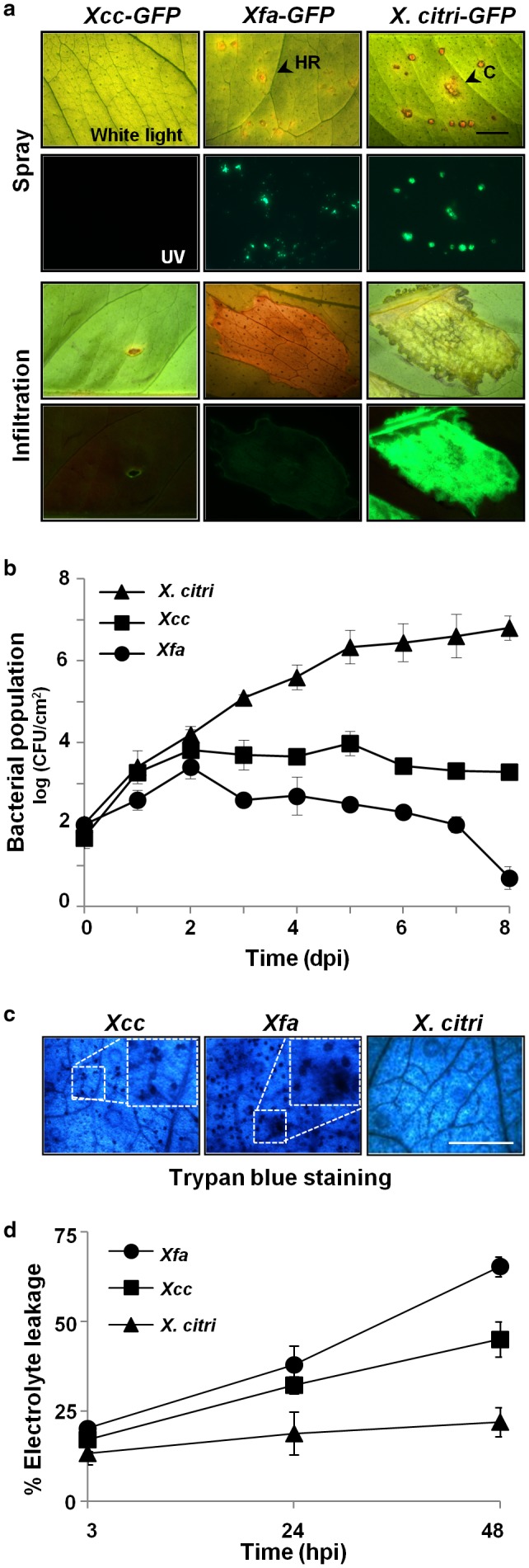
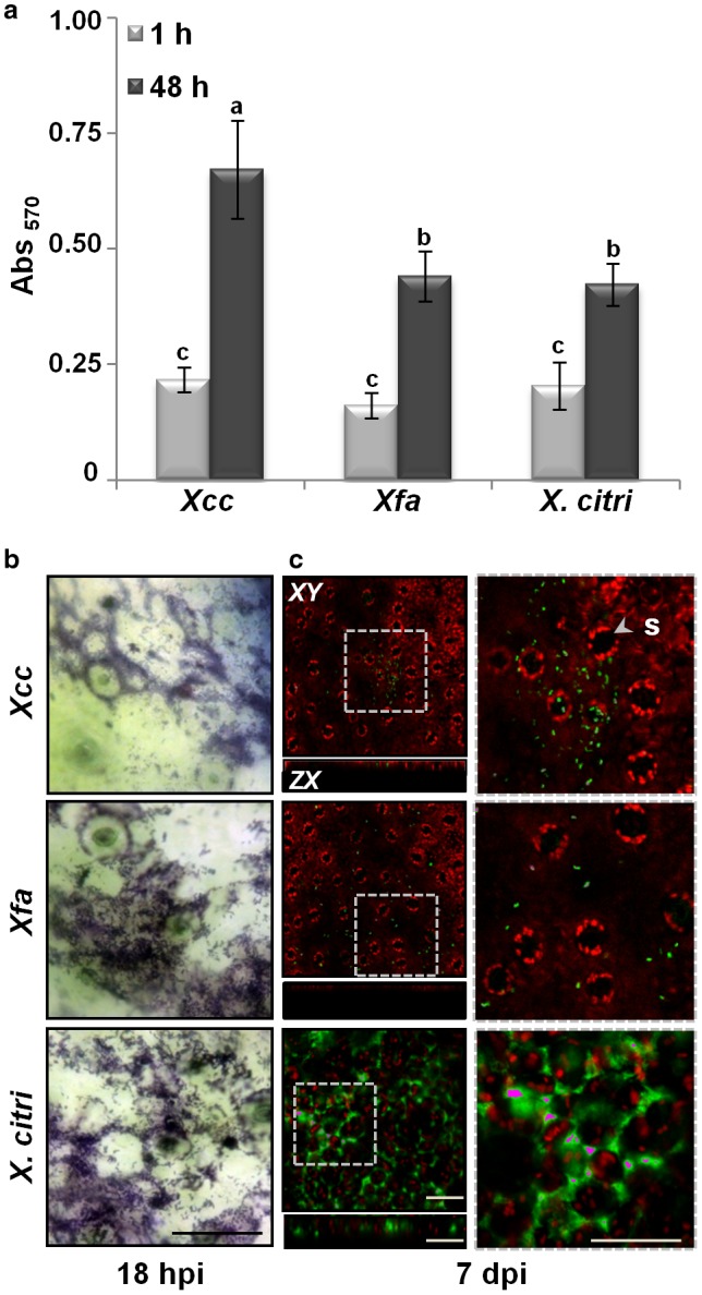
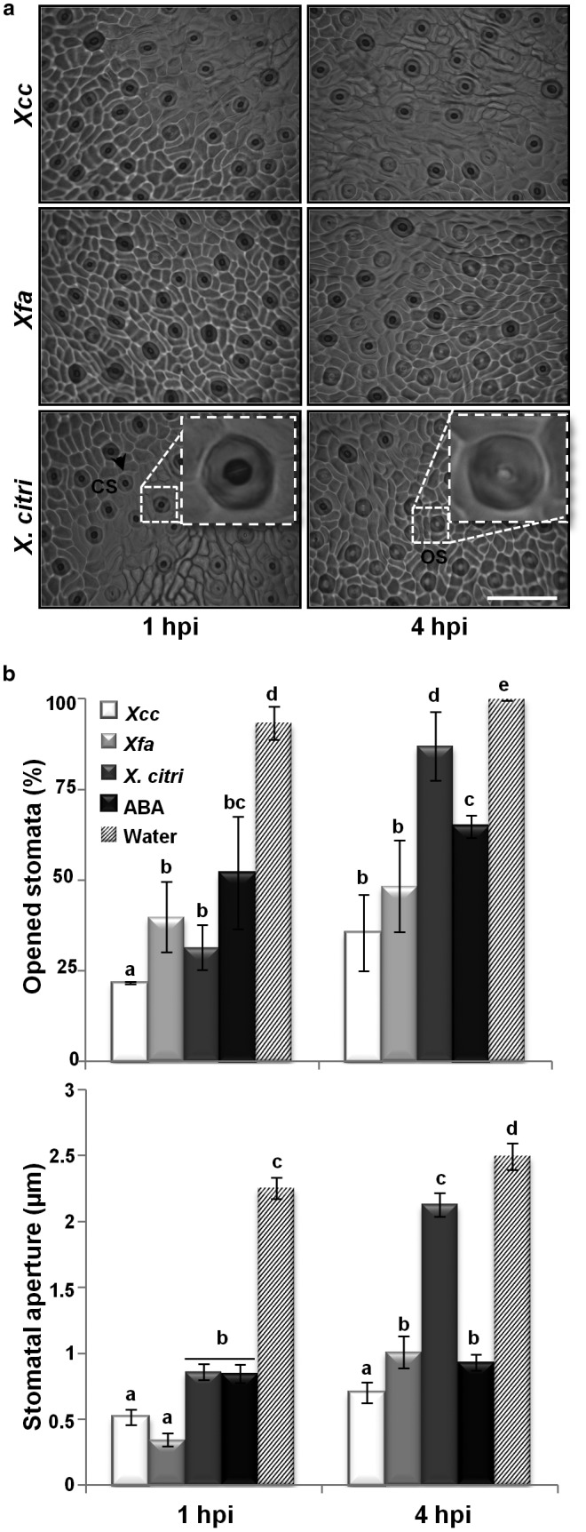
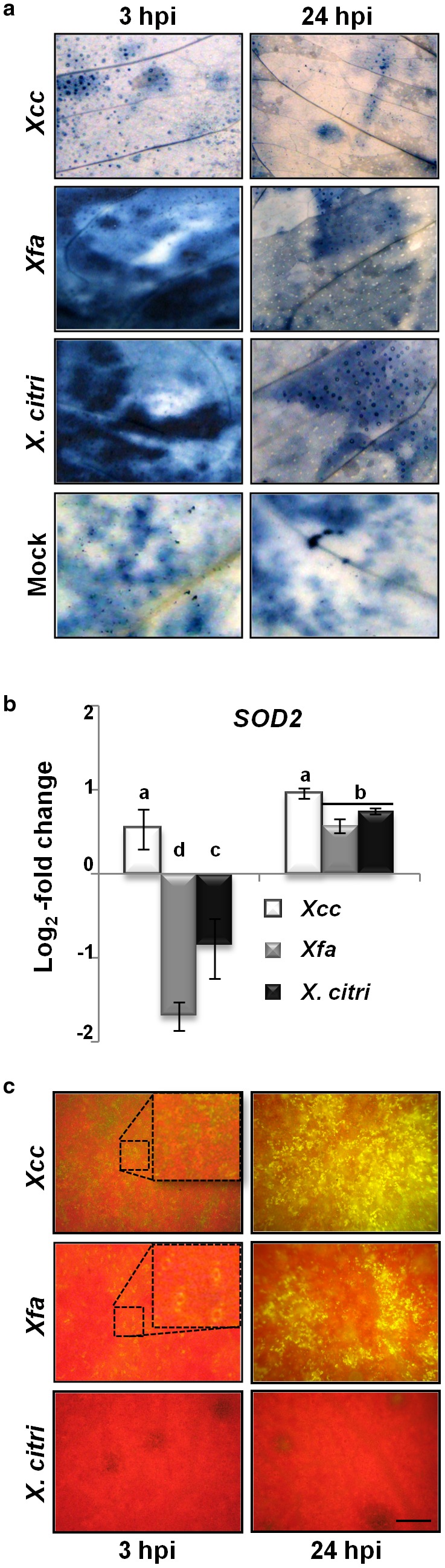
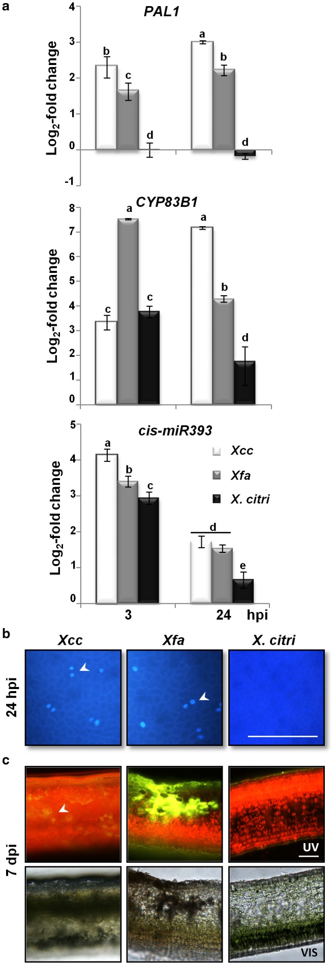
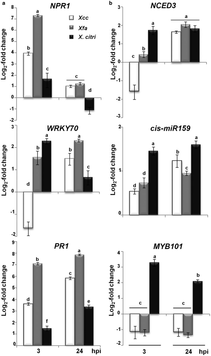
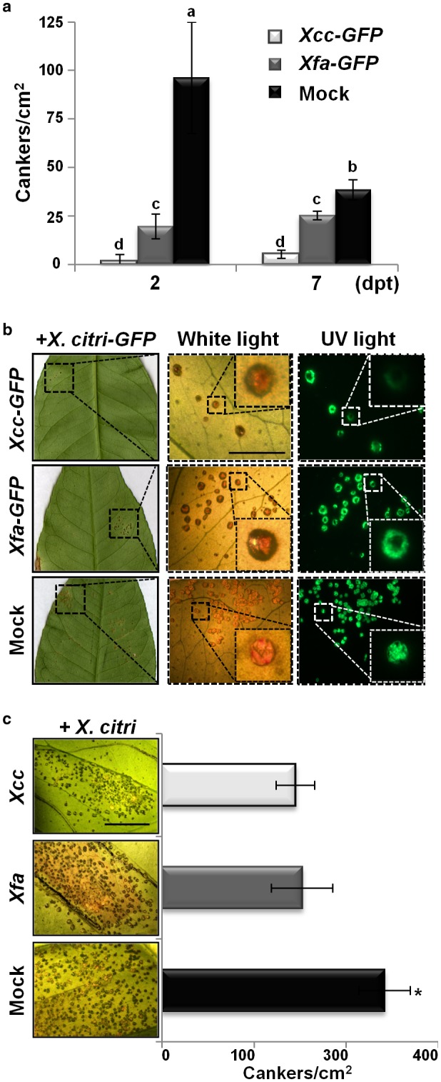
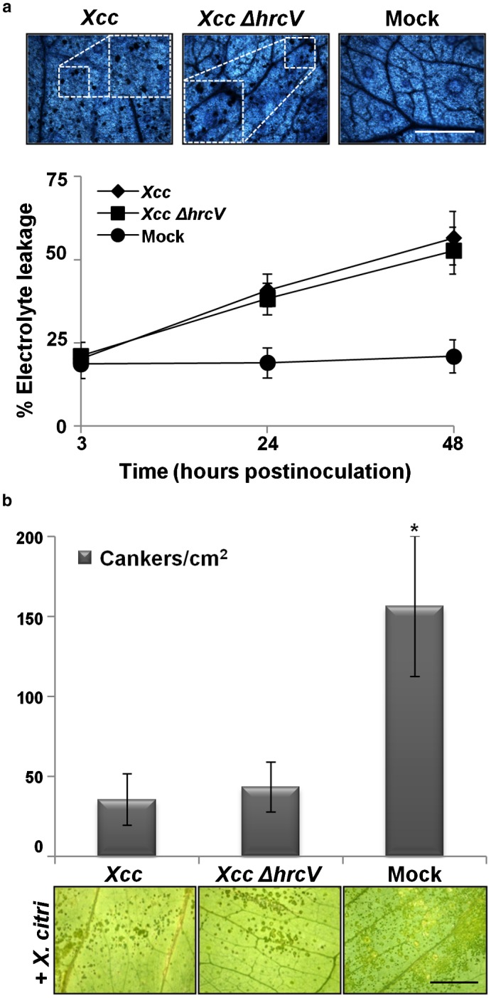
Citrus is an economically important fruit crop that is severely afflicted by citrus canker, a disease caused by Xanthomonas citri ssp. citri (X. citri); thus, new sustainable strategies to manage this disease are needed. Although all Citrus spp. are susceptible to this pathogen, they are resistant to other Xanthomonas species, exhibiting non-host resistance (NHR), for example, to the brassica pathogen X. campestris pv. campestris (Xcc) and a gene-for-gene host defence response (HDR) to the canker-causing X. fuscans ssp. aurantifolii (Xfa) strain C. Here, we examine the plant factors associated with the NHR of C. limon to Xcc. We show that Xcc induced asymptomatic type I NHR, allowing the bacterium to survive in a stationary phase in the non-host tissue. In C. limon, this NHR shared some similarities with HDR; both defence responses interfered with biofilm formation, and were associated with callose deposition, induction of the salicylic acid (SA) signalling pathway and the repression of abscisic acid (ABA) signalling. However, greater stomatal closure was seen during NHR than during HDR, together with different patterns of accumulation of reactive oxygen species and phenolic compounds and the expression of secondary metabolites. Overall, these differences, independent of Xcc type III effector proteins, could contribute to the higher protection elicited against canker development. We propose that Xcc may have the potential to steadily activate inducible defence responses. An understanding of these plant responses (and their triggers) may allow the development of a sustained and sustainable resistance to citrus canker.

Keywords: PAMP-triggered immunity; biofilm formation; canker disease; glucosinolates; protection; salicylic acid; stomatal immunity.


Figures

Figure 1
Figure 1
Phenotypic responses induced by Xanthomonas spp. in Citrus limon. (a) Macroscopic phenotype on abaxial surface of ‘Eureka´ lemon leaves at 16 days post‐inoculation (dpi) with Xanthomonas campestris pv. campestris (Xcc), X. fuscans ssp. aurantifolii (Xfa) strain C and X. citri ssp. citri (X. citri) by spraying [109 colony‐forming units (CFU)/mL] and pressure infiltration (107 CFU/mL). Leaves were photographed under white and UV light. Arrows: hypersensitive response (HR) and canker (C). Scale bar, 10 mm. (b) Bacterial population growth on C. limon leaves inoculated by spraying. Values are expressed as means ± standard deviation (SD) of three independent biological replicates. (c) Microscopic cell death phenotype observed at 48 h post‐inoculation (hpi). Insets show the amplification of microscopic cell death. Scale bar, 150 mm. (d) Quantification of cell death in leaves treated as described in (c) by measuring the percentage of electrolyte leakage at 3, 24 and 48 hpi. Values are expressed as means ± SD of three independent biological replicates. [Colour figure can be viewed at wileyonlinelibrary.com]
Figure 2
Figure 2
Non‐host resistance (NHR) and the host defence response (HDR) disrupt biofilm formation in Citrus limon. (a) Bacterial adhesion and biofilm formation of Xanthomonas campestris pv. campestris (Xcc), X. fuscans ssp. aurantifolii (Xfa) strain C and X. citri ssp. citri (X. citri) on an inert plastic surface after 1 and 48 h of incubation. Values are expressed as means ± standard deviation (SD), n = 24. Different letters indicate significant differences at P < 0.05 [two‐way analysis of variance (ANOVA), Tukey’s test]. (b) Bacterial adhesion of Xcc, Xfa and X. citri on abaxial surfaces of C. limon leaves assessed by crystal violet staining at 18 h post‐inoculation (hpi). Stained cells attached to the leaf surface were analysed microscopically. (c) Biofilm formation of Xanthomonas spp. on leaves inoculated by spraying [109 colony‐forming units (CFU)/mL], observed using confocal laser scanning microscopy at 7 days post‐inoculation (dpi). Red indicates chlorophyll fluorescence and green indicates green fluorescent protein (GFP)‐tagged bacteria. XY and ZX are the XY and ZX axis projected images, respectively. Sections from the left panels are shown magnified in the right panels. Arrow: stomata (S). Scale bar, 50 μm. [Colour figure can be viewed at wileyonlinelibrary.com]
Figure 3
Figure 3
Non‐host resistance (NHR) and the host defence response (HDR) involve the maintenance of stomatal closure in Citrus limon. (a) Dried‐gel imprint of intact C. limon epidermis showing opened (OS) and closed (CS) stomata, shown enlarged in the insets, in leaves inoculated by spraying with bacterial suspension [109 colony‐forming units (CFU)/mL] of Xanthomonas campestris pv. campestris (Xcc), X. fuscans ssp. aurantifolii (Xfa) strain C and X. citri ssp. citri (X. citri). Scale bar, 100 μm. (b) Quantification of percentage of open stomata and stomatal aperture at 1 and 4 h post‐inoculation (hpi) in leaves exposed to Xcc, Xfa, X. citri infection and abscisic acid (ABA) or water treatments by spraying. Values are expressed as the means ± standard deviation (SD) from three independent biological replicates (n = 60 stomata). Different letters above the bars indicate significant differences at P < 0.05 [two‐way analysis of variance (ANOVA), Tukey’s test].
Figure 4
Figure 4
Different patterns of reactive oxygen species (ROS) accumulation are triggered in non‐host resistance (NHR) and the host defence response (HDR). (a) In situ accumulation of superoxide radicals (O2 •−) detected by dark blue formazan precipitate in Citrus limon leaves inoculated by pressure infiltration with bacterial suspensions of Xanthomonas campestris pv. campestris (Xcc), X. fuscans ssp. aurantifolii (Xfa) strain C and X. citri ssp. citri (X. citri), plus mock. (b) Quantitative reverse transcription‐polymerase chain reaction analysis of copper/zinc superoxide dismutase (SOD2). mRNAs measured at 3 and 24 h post‐inoculation (hpi). Relative gene expression (ΔΔCt) fold change of mRNA levels was performed considering mock‐treated plants as reference sample and histone H4 transcript as an endogenous control. Values are expressed as means ± standard deviation (SD) from three independent biological replicates. Different letters indicate significant differences at P < 0.05 [two‐way analysis of variance (ANOVA), Tukey’s test]. (c) H2O2 accumulation at 3 and 24 hpi in C. limon leaves infiltrated with bacterial suspensions of the different Xanthomonas strains [107 colony‐forming units (CFU)/mL]. Leaves were stained with 2′,7′‐dichlorofluorescein diacetate (DCFH‐DA) and observed by fluorescence microscopy. H2O2 accumulation in the guard cells is shown enlarged in the top insets. Scale bar, 100 μm. [Colour figure can be viewed at wileyonlinelibrary.com]
Figure 5
Figure 5
Cell wall reinforcement through phenolic compounds and callose deposition is involved in Citrus limon non‐host resistance (NHR) and the host defence response (HDR). (a) Quantitative reverse transcription‐polymerase chain reaction analysis of phenylalanine ammonia lyase (PAL1), CYP83B1 and cis‐miR393 in Citrus limon leaves inoculated by pressure infiltration with bacterial suspensions [107 colony‐forming units (CFU)/mL] of Xanthomonas campestris pv. campestris (Xcc), X. fuscans ssp. aurantifolii (Xfa) strain C and X. citri ssp. citri (X. citri). mRNAs were measured at 3 and 24 h post‐inoculation (hpi). Relative gene expression (ΔΔCt) fold change of mRNA levels was performed considering mock‐treated plants as reference sample and histone H4 transcript as an endogenous control. Values are expressed as means ± standard deviation (SD) from three independent biological replicates. Different letters indicate significant differences at P < 0.05 [two‐way analysis of variance (ANOVA), Tukey’s test]. (b) Callose deposition in C. limon inoculated leaves, stained with aniline blue (arrows). Scale bar, 100 mm. (c) Light microscopic images of C. limon inoculated leaves, photographed at 7 days post‐inoculation (dpi) under white and UV light. Green fluorescent polyphenolic compounds and red chlorophyll fluorescence are observed. Scale bar, 10 μm. [Colour figure can be viewed at wileyonlinelibrary.com]
Figure 6
Figure 6
Salicylic acid (SA) signalling pathway is induced, whereas abscisic acid (ABA) synthesis and signalling are repressed, in Citrus limon non‐host resistance (NHR) and the host defence response (HDR). Quantitative reverse transcription‐polymerase chain reaction in leaves inoculated by pressure infiltration with bacterial suspensions [107 colony‐forming units (CFU)/mL] of Xanthomonas campestris pv. campestris (Xcc), X. fuscans ssp. aurantifolii (Xfa) strain C and X. citri ssp. citri (X. citri). mRNAs were measured at 3 and 24 h post‐inoculation (hpi). Relative gene expression (ΔΔCt) fold change of mRNA levels was performed considering mock‐treated plants as reference sample and histone H4 transcript as an endogenous control. Values are expressed as means ± standard deviation (SD) from three independent biological replicates. Different letters indicate significant differences at P < 0.05 [two‐way analysis of variance (ANOVA), Tukey’s test]. (a) Expression profiles of SA signalling pathway genes encoding non‐expressor of pathogenesis‐related genes 1 (NPR1), WRKY70 transcription factor and pathogenesis‐related 1 (PR1). (b) Expression profiles of ABA biosynthesis (9‐cis‐epoxycarotenoid dioxygenase, NCED3) and signalling (MYB101 and csi‐miR159) encoding genes.
Figure 7
Figure 7
Non‐host resistance (NHR) and the host defence response (HDR) protect Citrus limon from canker development. (a) Number of canker lesions per square centimetre in leaves pretreated by cotton swab with bacterial suspensions [109 colony‐forming units (CFU)/mL] of Xanthomonas campestris pv. campestris (Xcc) and X. fuscans ssp. aurantifolii (Xfa) strain C tagged with green fluorescent protein (GFP), or 10 mm MgCl2 (mock). At 2 and 7 days post‐treatment (dpt), the leaves were challenged via spraying with the pathogenic X. citri ssp. citri (X. citri)‐GFP (109 CFU/mL), and canker lesions were quantified at 18 days post‐inoculation (dpi). Values are expressed as means ± standard deviation (SD) from three independent biological replicates. Different letters indicate significant differences at P < 0.05 [two‐way analysis of variance (ANOVA), Tukey’s test]. (b) Phenotypic response of C. limon leaves pretreated with XccGFP, Xfa‐GFP or mock, and subsequently challenged with X. citriGFP strain at 2 dpt, as described in (a). Sections from the left panels are shown magnified in the right panels under white and UV light. (c) Phenotypic response and canker quantification (18 dpi) of C. limon leaves pretreated by pressure infiltration with XccGFP and Xfa‐GFP bacterial suspensions (106 CFU/mL) or mock, and subsequently challenged (2 dpt) via infiltration with X. citriGFP (106 CFU/mL). Scale bar, 10 mm. [Colour figure can be viewed at wileyonlinelibrary.com]
Figure 8
Figure 8
Citrus limon non‐host resistance (NHR) to Xanthomonas campestris pv. campestris (Xcc) is independent of type III effector proteins. (a) Microscopic cell death phenotype observed at 48 h post‐inoculation (hpi) in leaves inoculated by cotton swab with bacterial suspensions [109 colony‐forming units (CFU)/mL] of Xcc or the hrcV‐deficient mutant strain of Xcc 8004 (Xcc ΔhrcV), or 10 mm MgCl2 (mock). Insets show the amplification of microscopic cell death. Scale bar, 150 mm. Quantification of cell death in leaves treated as described previously by measurement of percentage electrolyte leakage at 3, 24 and 48 hpi. Values are expressed as means ± standard deviation (SD) of three independent biological replicates. (b) Number of canker lesions per square centimetre in leaves pretreated by cotton swab with Xcc and Xcc ΔhrcV bacterial suspensions (109 CFU/mL) or mock. At 2 days post‐treatment (dpt), the leaves were challenged via spraying with the pathogenic X. citri ssp. citri (X. citri) (109 CFU/mL) and canker lesions were quantified at 18 days post‐inoculation. Values are expressed as means ± SD from three independent biological replicates. The dataset marked with an asterisk is significantly different as assessed by Tukey’s test (P < 0.05). Scale bar, 10 mm. [Colour figure can be viewed at wileyonlinelibrary.com]

Similar articles

Cited by

References

    1. Adlung, N. , Prochaska, H. , Thieme, S. , Banik, A. , Bluher, D. , John, P. , Nagel, O. , Schulze, S. , Gantner, J. , Delker, C. , Stuttmann, J. and Bonas, U. (2016) Non‐host resistance induced by the Xanthomonas effector XopQ is widespread within the genus Nicotiana and functionally depends on EDS1. Front Plant Sci. 7, 1796. - PMC - PubMed
    1. An, C. and Mou, Z. (2012) Non‐host defense response in a novel Arabidopsis–Xanthomonas citri subsp. citri pathosystem. PLoS One, 7, e31130. - PMC - PubMed
    1. An, C. , Wang, C. and Mou, Z. (2017) The Arabidopsis Elongator complex is required for nonhost resistance against the bacterial pathogens Xanthomonas citri subsp. citri and Pseudomonas syringae pv. phaseolicola NPS3121. New Phytol. 214, 1245–1259. - PubMed
    1. Araujo, W.L. , Marcon, J. , Maccheroni, W. Jr , Van Elsas, J.D. , Van Vuurde, J.W. and Azevedo, J.L. (2002) Diversity of endophytic bacterial populations and their interaction with Xylella fastidiosa in citrus plants. Appl. Environ. Microbiol. 68, 4906–4914. - PMC - PubMed
    1. Arnaud, D. and Hwang, I. (2015) A sophisticated network of signaling pathways regulates stomatal defenses to bacterial pathogens. Mol. Plant, 8, 566–581. - PubMed

Publication types

MeSH terms