

Stabilized collagen matrix dressing improves wound macrophage function and epithelialization
- PMID: 30260708
- PMCID: PMC6338656
- DOI: 10.1096/fj.201800352R
Stabilized collagen matrix dressing improves wound macrophage function and epithelialization
Abstract
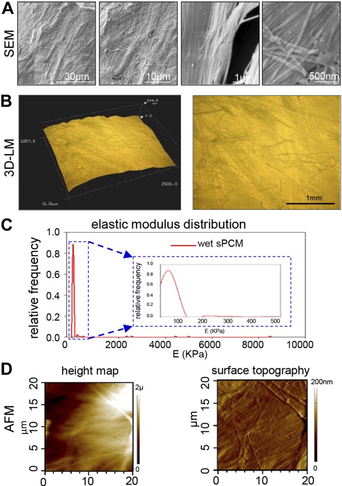
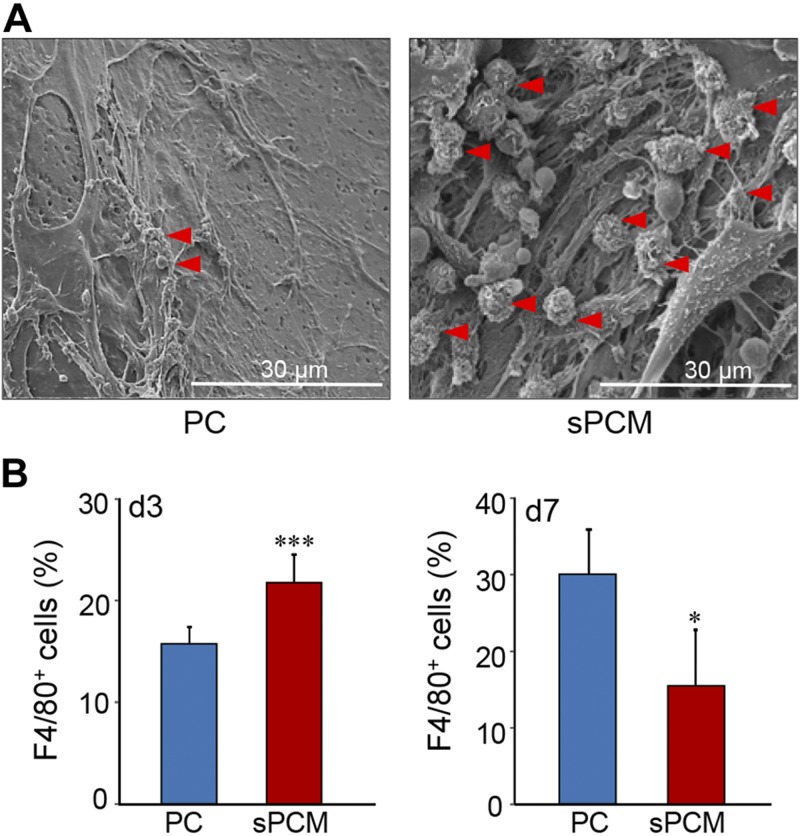
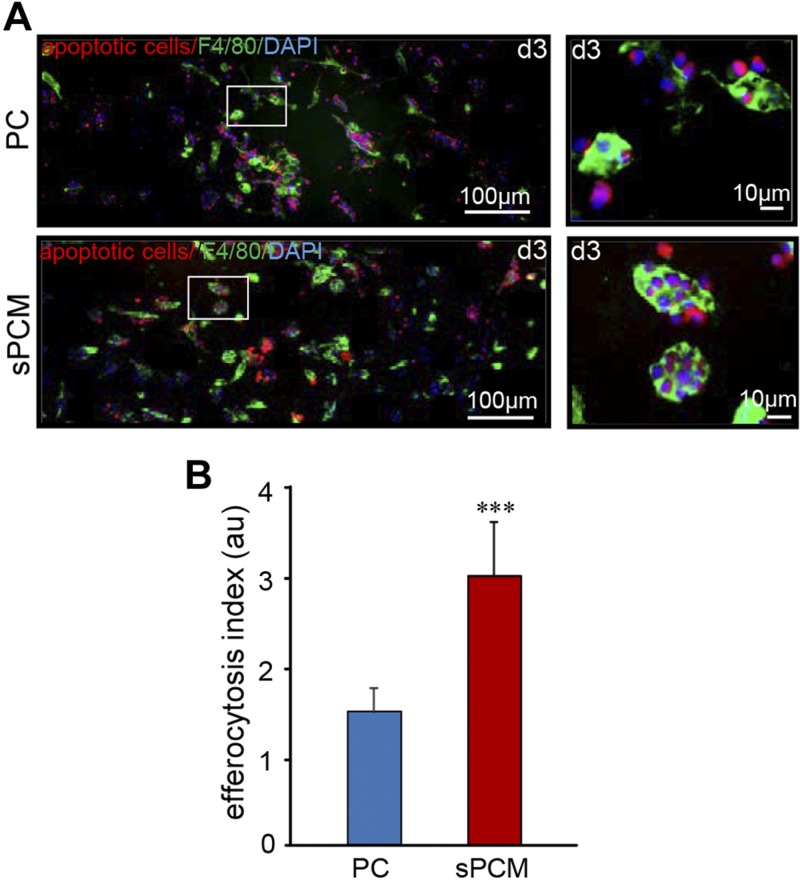
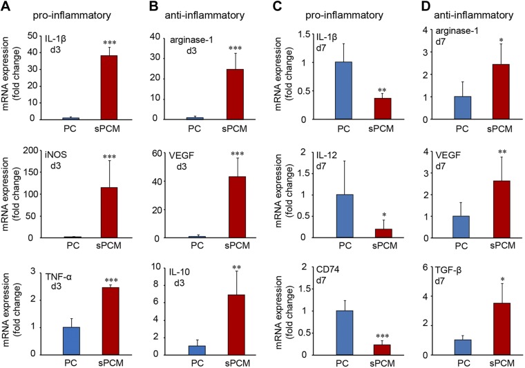
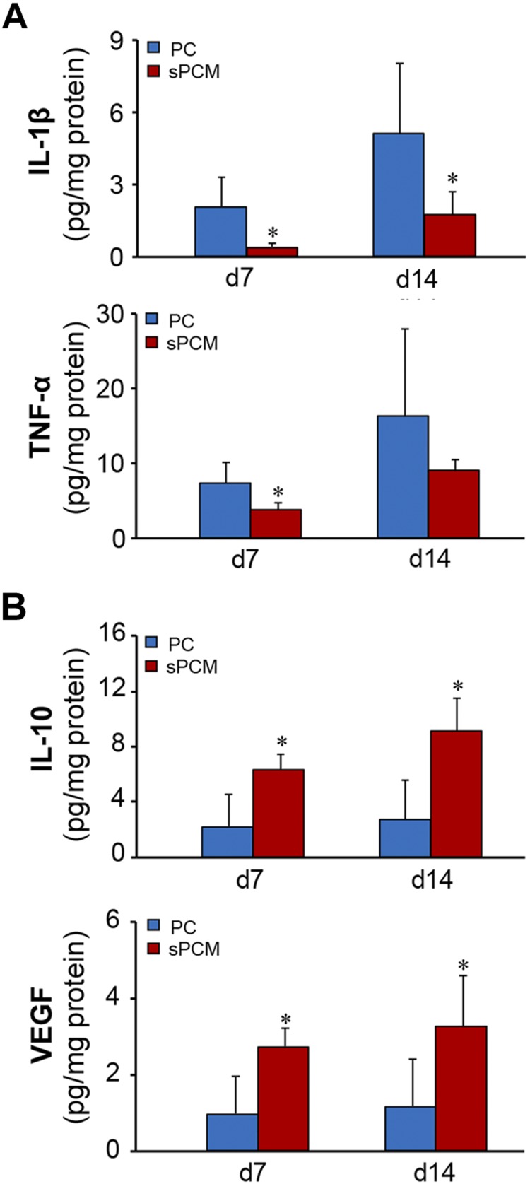
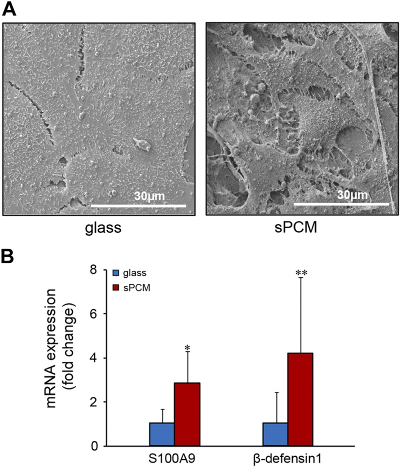
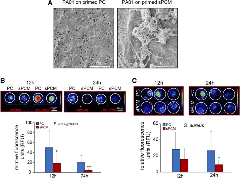
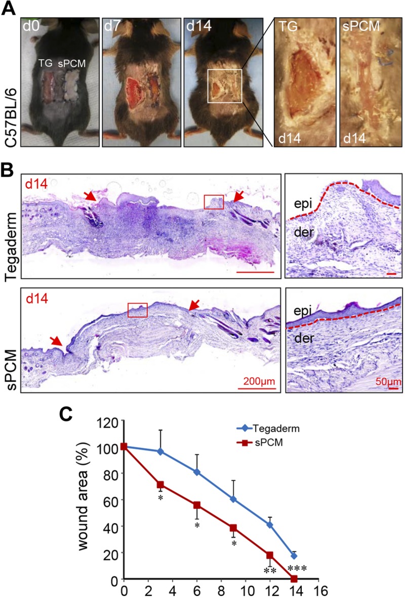
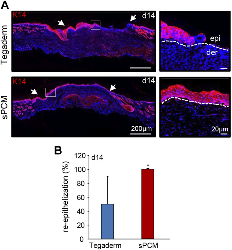
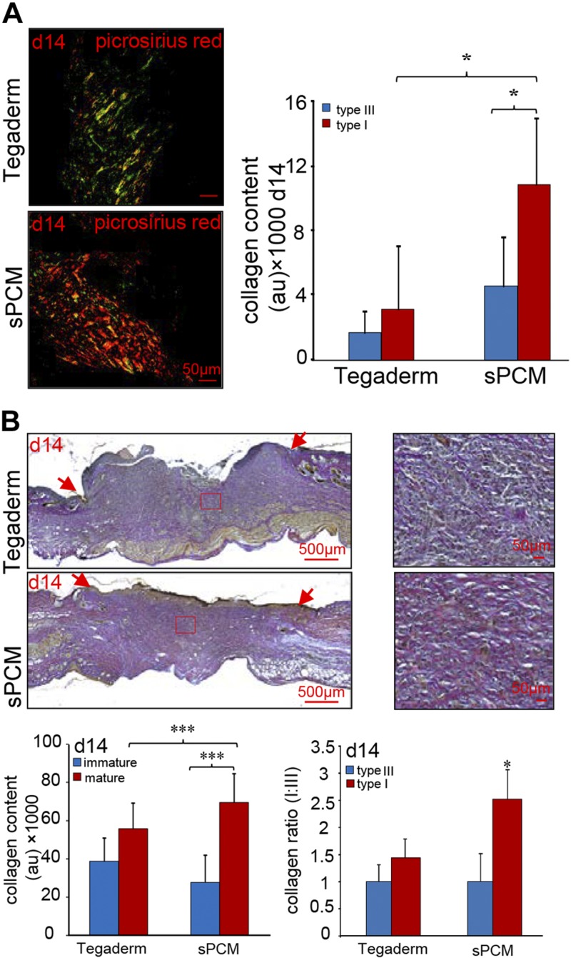
Decellularized matrices of biologic tissue have performed well as wound care dressings. Extracellular matrix-based dressings are subject to rapid degradation by excessive protease activity at the wound environment. Stabilized, acellular, equine pericardial collagen matrix (sPCM) wound care dressing is flexible cross-linked proteolytic enzyme degradation resistant. sPCM was structurally characterized utilizing scanning electron and atomic force microscopy. In murine excisional wounds, sPCM was effective in mounting an acute inflammatory response. Postwound inflammation resolved rapidly, as indicated by elevated levels of IL-10, arginase-1, and VEGF, and lowering of IL-1β and TNF-α. sPCM induced antimicrobial proteins S100A9 and β-defensin-1 in keratinocytes. Adherence of Pseudomonas aeruginosa and Staphylococcus aureus on sPCM pre-exposed to host immune cells in vivo was inhibited. Excisional wounds dressed with sPCM showed complete closure at d 14, while control wounds remained open. sPCM accelerated wound re-epithelialization. sPCM not only accelerated wound closure but also improved the quality of healing by increased collagen deposition and maturation. Thus, sPCM is capable of presenting scaffold functionality during the course of wound healing. In addition to inducing endogenous antimicrobial defense systems, the dressing itself has properties that minimize biofilm formation. It mounts robust inflammation, a process that rapidly resolves, making way for wound healing to advance.-El Masry, M. S., Chaffee, S., Das Ghatak, P., Mathew-Steiner, S. S., Das, A., Higuita-Castro, N., Roy, S., Anani, R. A., Sen, C. K. Stabilized collagen matrix dressing improves wound macrophage function and epithelialization.
Keywords: ECM; antimicrobial peptides; biofilm; cytokines; scaffold.
Conflict of interest statement
The authors thank Dr. B. Deng (Center for Electron Microscopy and Analysis College of Engineering, The Ohio State University) for her assistance with light microscopy studies on sPCM. This work was supported by U.S. National Institutes of Health Grants GM077185 and GM069589 (National Institute of General Medical Sciences), NR013898 and NR015676 (National Institute of Nursing Research), and DK076566 (National Institute of Diabetes and Digestive and Kidney Diseases). This work was supported, in part, by an unrestricted gift from Harbor MedTech (Irvine, CA, USA). The authors declare no conflicts of interest.
Figures
References
Publication types
MeSH terms
Substances
Grants and funding
LinkOut - more resources
Full Text Sources
Other Literature Sources
Research Materials
Miscellaneous
