. 2019 Feb 1;126(2):341-353.
doi: 10.1152/japplphysiol.00719.2018. Epub 2018 Sep 27.

Adult skeletal muscle deletion of Mitofusin 1 and 2 impedes exercise performance and training capacity

Affiliations

Adult skeletal muscle deletion of Mitofusin 1 and 2 impedes exercise performance and training capacity

Margaret B Bell et al. J Appl Physiol (1985). .

Abstract

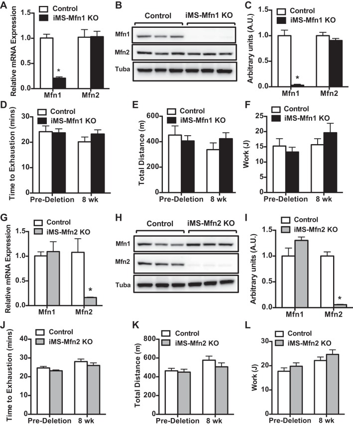
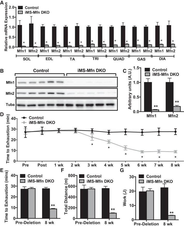
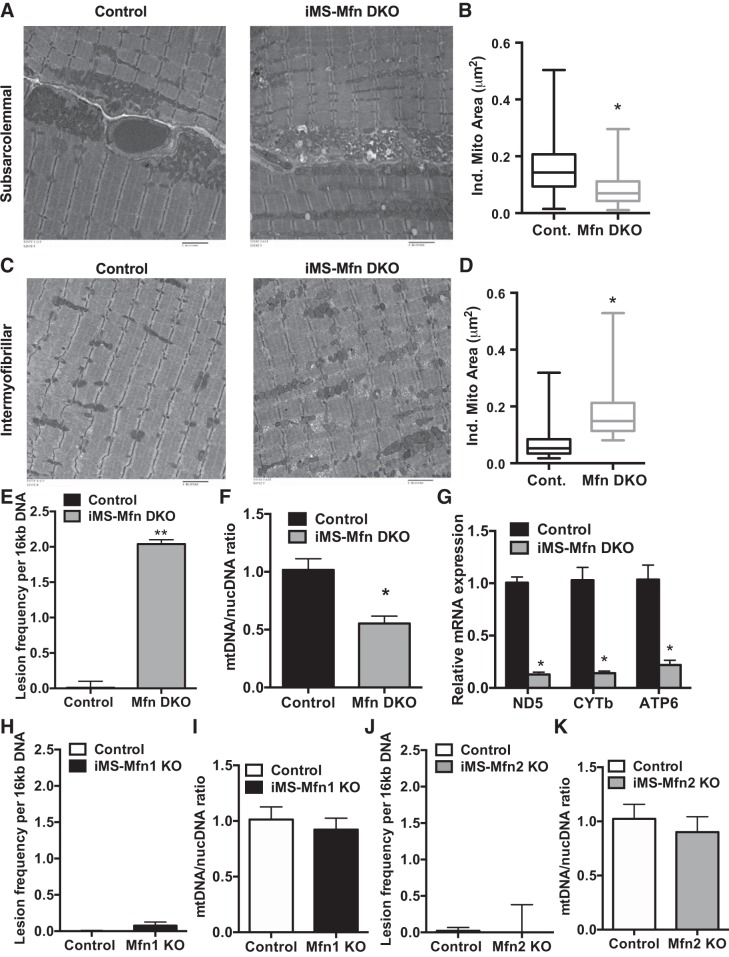
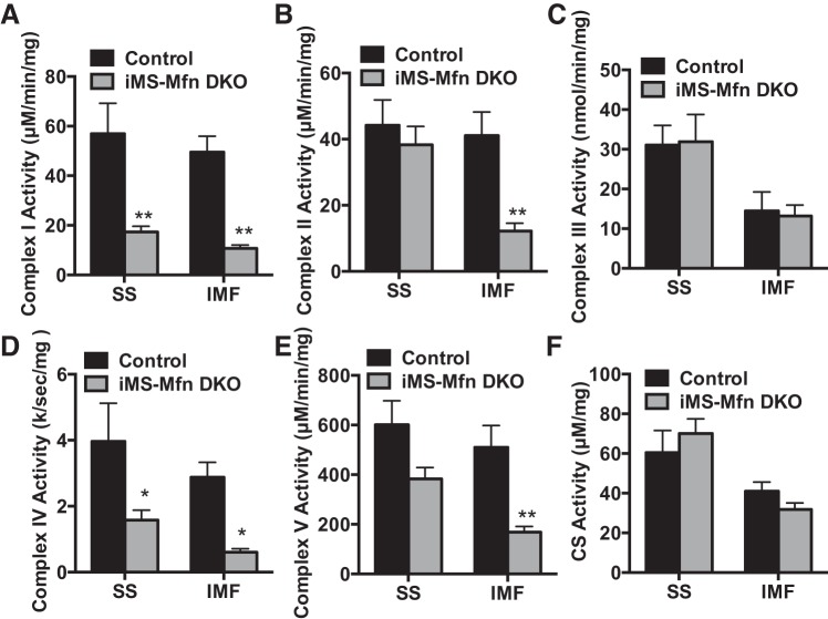
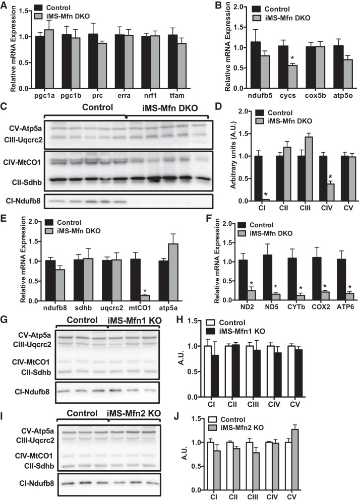
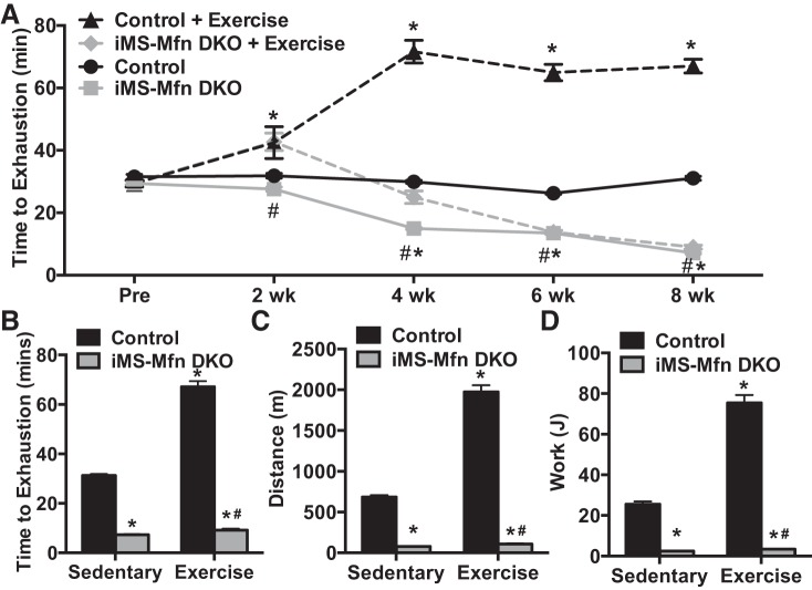
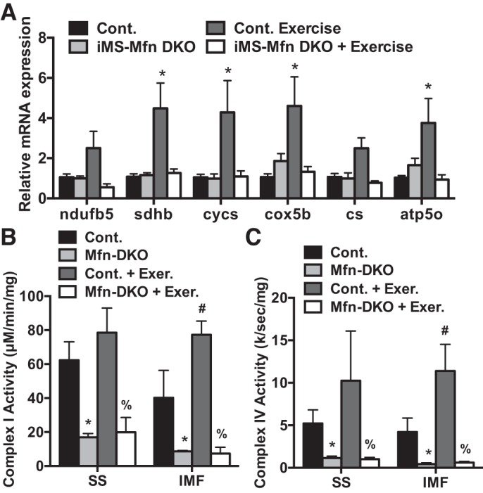
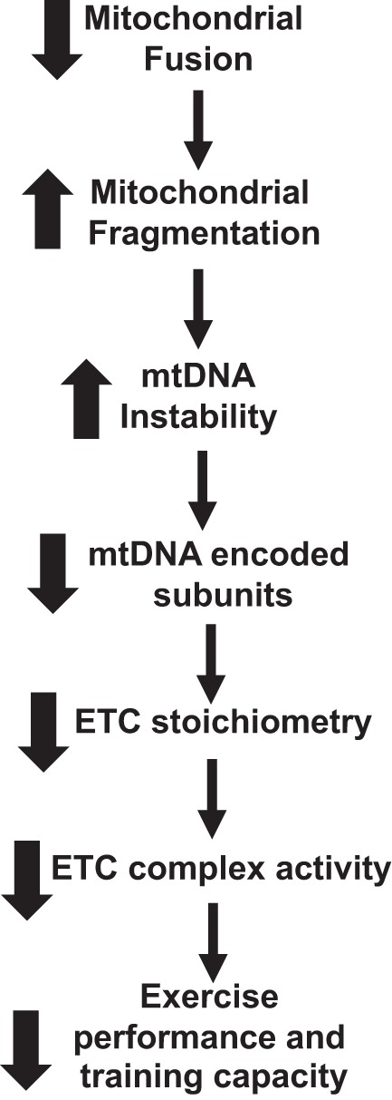
Endurance exercise has been shown to be a positive regulator of skeletal muscle metabolic function. Changes in mitochondrial dynamics (fusion and fission) have been shown to influence mitochondrial oxidative capacity. We therefore tested whether genetic disruption of mitofusins (Mfns) affected exercise performance in adult skeletal muscle. We generated adult-inducible skeletal muscle-specific Mfn1 (iMS-Mfn1KO), Mfn2 (iMS-Mfn2KO), and Mfn1/2 (iMS-MfnDKO) knockout mice. We assessed exercise capacity by performing a treadmill time to exhaustion stress test before deletion and up to 8 wk after deletion. Analysis of either the iMS-Mfn1KO or the iMS-Mfn2KO did not reveal an effect on exercise capacity. However, analysis of iMS-MfnDKO animals revealed a progressive reduction in exercise performance. We measured individual electron transport chain (ETC) complex activity and observed a reduction in ETC activity in both the subsarcolemmal and intermyofibrillar mitochondrial fractions specifically for NADH dehydrogenase (complex I) and cytochrome- c oxidase (complex IV), which was associated with a decrease in ETC subunit expression for these complexes. We also tested whether voluntary exercise training would prevent the decrease in exercise capacity observed in iMS-MfnDKO animals ( n = 10/group). However, after 8 wk of training we did not observe any improvement in exercise capacity or ETC subunit parameters in iMS-MfnDKO animals. These data suggest that the decrease in exercise capacity observed in the iMS-MfnDKO animals is in part the result of impaired ETC subunit expression and ETC complex activity. Taken together, these results provide strong evidence that mitochondrial fusion in adult skeletal muscle is important for exercise performance. NEW & NOTEWORTHY This study is the first to utilize an adult-inducible skeletal muscle-specific knockout model for Mitofusin (Mfn)1 and Mfn2 to assess exercise capacity. Our findings reveal a progressive decrease in exercise performance with Mfn1 and Mfn2 deletion. The decrease in exercise capacity was accompanied by impaired oxidative phosphorylation specifically for complex I and complex IV. Furthermore, voluntary exercise training was unable to rescue the impairment, suggesting that normal fusion is essential for exercise-induced mitochondrial adaptations.

Keywords: electron transport chain; exercise; mitochondria; mitofusin; skeletal muscle.


Conflict of interest statement

No conflicts of interest, financial or otherwise, are declared by the authors.

Figures

Fig. 1.
Fig. 1.
Effect of adult deletion of either Mitofusin (Mfn)1 or Mfn2 on exercise performance. A–F: Mfn1 and Mfn2 mRNA expression in different skeletal muscle beds (A); Mfn1 and Mfn2 protein expression (B); densitometry quantification of Mfn1 and Mfn2 protein expression normalized to tubulin and relative to control in gastrocnemius (GAS) muscle (C; n = 3 per group), baseline time to exhaustion (D); total distance run (E); and calculated total work (F; n = 7 per group) in adult-inducible skeletal muscle-specific Mfn1 knockout mice (iMS-Mfn1KO) and control littermates. G–L: Mfn1 and Mfn2 mRNA expression (G); Mfn1 and Mfn2 protein expression (H); densitometry quantification of Mfn1 and Mfn2 protein expression normalized to tubulin (Tuba) and relative to control in GAS muscle (I); baseline time to exhaustion (J); total distance run (K); and calculated total work (K) in adult-inducible skeletal muscle-specific Mfn2 knockout mice (iMS-Mfn2KO) and control littermates. Data are means ± SE. *P < 0.05 compared with control as determined by unpaired t-test.
Fig. 2.
Fig. 2.
Effect of adult deletion of both Mitofusin (Mfn)1 and Mfn2 on exercise performance: Mfn1 and Mfn2 mRNA expression in various skeletal muscle beds: soleus (SOL), extensor digitorum longus (EDL), tibialis anterior (TA), triceps (TRI), quadriceps (QUAD), gastrocnemius (GAS), and diaphragm (DIA) (A); Mfn1 and Mfn2 protein expression (B); densitometry quantification of Mfn1 and Mfn2 protein expression normalized to tubulin and relative to control in GAS muscle (C; n = 3 per group); longitudinal exercise stress test (D); time to exhaustion (E); total distance run (F); and calculated total work in adult-inducible skeletal muscle-specific Mfn1 and Mfn2 double knockout mice (iMS-MfnDKO) and control littermates (G; n = 7 per group) before deletion and 8 wk after deletion. Data are means ± SE. *P < 0.05; **P < 0.01 compared with control as determined by unpaired t-test.
Fig. 3.
Fig. 3.
Mild impairment to gait kinematics with muscle deletion of Mitofusin (Mfn)1 and Mfn2: paw print intensity (A); stand time (B); swing time (C); step cycle (D); swing speed (E); and stride length (F) in adult-inducible skeletal muscle-specific Mfn1 and Mfn2 double knockout mice (iMS-MfnDKO; n = 5) and control littermates (n = 5) 8 wk after deletion. Data are means ± SE. **P < 0.01 compared with control as determined by unpaired t-test.
Fig. 4.
Fig. 4.
Mitochondrial morphology with deletion of mitofusins (Mfns) in skeletal muscle. A–F: representative transmission electron micrograph (TEM) of subsarcolemmal (SS) mitochondria (A); quantification of individual mitochondrial area in the SS fraction (B); representative TEM of intermyofibrillar (IMF) mitochondria (C); quantification of individual mitochondrial area in the IMF fraction (D); mitochondrial DNA (mtDNA) damage represented as lesion frequency (E); mtDNA-to-nuclear DNA (nucDNA) ratio (F); and mRNA expression of mitochondrial-encoded electron transport chain subunits NADH dehydrogenase 5 (ND5), cytochrome b (CYTb), and ATP synthase subunit a (ATP6) in soleus (G) of adult-inducible skeletal muscle-specific Mfn1 and Mfn2 double knockout mice (iMS-MfnDKO) and control littermates. H and I: mitochondrial DNA damage represented as lesion frequency (H) and mtDNA-to-nucDNA ratio (I) of adult-inducible skeletal muscle-specific Mfn1 knockout mice (iMS-Mfn1KO) and control littermates. J and K: mtDNA damage represented as lesion frequency (J) and mtDNA-to-nucDNA ratio (I) of adult-inducible skeletal muscle-specific Mfn2 knockout mice (iMS-Mfn2KO) and control littermates. Data in B and D are presented as a whisker plot with the minimum and maximum value, median, lower and upper quartile based on n = 4; N = 500–1,000 mitochondria. Data in E and F are means ± SE; n = 3–7 per group; *P < 0.05; **P < 0.01 compared with control as determined by unpaired t-test.
Fig. 5.
Fig. 5.
Electron transport chain complex activity with skeletal muscle deletion of Mitofusin (Mfn): NADH dehydrogenase (complex I) activity (A); succinate dehydrogenase (complex II) activity (B); cytochrome c reductase (complex III) activity (C); cytochrome-c oxidase (complex IV) activity (D); ATP synthase (complex V) activity (E); and citrate synthase (CS) activity (F) in the subsarcolemmal (SS) and intermyofibrillar (IMF) mitochondrial fractions from gastrocnemius muscle of adult-inducible skeletal muscle-specific Mfn1 and Mfn2 double knockout mice (iMS-MfnDKO; n = 5) and control littermates (n = 5). Data are means ± SE. *P < 0.05; **P < 0.01 compared with control as determined by unpaired t-test.
Fig. 6.
Fig. 6.
Loss of complex I and complex IV subunit expression with Mitofusin (Mfn) deletion in skeletal muscle. A–F: gastrocnemius (GAS) muscle of adult-inducible skeletal muscle-specific Mfn1 and Mfn2 double knockout mice (iMS-MfnDKO; n = 5) and control (n = 5) littermates. A: mRNA expression of transcriptional regulators peroxisome proliferator-activated receptor γ coactivator 1 [PGC-1α, PGC-1β, and PGC-1-related coactivator (PRC)], estrogen-related receptor alpha (Errα), nuclear respiratory factor 1 (Nrf1), and mitochondrial transcription factor A (Tfam). B: mRNA expression of nuclear-encoded electron transport chain (ETC) subunits NADH dehydrogenase 1 beta subunit 5 (ndufb5), cytochrome c, somatic (cycs), cytochrome c oxidase subunit 5B (cox5b), and ATP synthase subunit O (atp5o). C: protein expression of ETC subunits ATP synthase F1 subunit alpha (Atp5a), Ubiquinol-cytochrome c reductase protein 2 (Uqcrc2), mitochondrial-encoded cytochrome-c oxidase 1 (MtCO1), Succinate dehydrogenase complex iron sulfur subunit B (Sdhb), and NADH:Ubiquinone oxidoreductase subunit B8 (Ndufb8). D: densitometry quantification of ETC subunit protein expression normalized to tubulin (Fig. 2B) and relative to control. E: mRNA expression of ETC subunits represented in C. F: mRNA expression of mitochondrial-encoded ETC subunits NADH dehydrogenase 2 and 5 (ND2 and ND5), cytochrome b (CYTb), cytochrome oxidase 2 (COX2), and ATP synthase subunit membrane subunit 6 (ATP6). G–J: GAS muscle of adult-inducible skeletal muscle-specific Mfn2 knockout mice (iMS-Mfn2KO; n = 3) and control (n = 3) littermates. G: protein expression of ETC subunits. H: densitometry quantification of ETC subunit protein expression normalized to tubulin (Fig. 1B) and relative to control. I: protein expression of ETC subunits. J: densitometry quantification of ETC subunit protein expression normalized to tubulin (Fig. 1H) and relative to control. Data are presented as means ± SE. *P < 0.05 compared with control as determined by unpaired t-test.
Fig. 7.
Fig. 7.
Decreased exercise performance with training in Mitofusin (Mfn) skeletal muscle-deficient animals. A–D: 8-wk longitudinal exercise stress test (A), time to exhaustion (B), total distance run (C), and calculated total work performed (D) in adult-inducible skeletal muscle-specific Mfn1 and Mfn2 double knockout mice (iMS-MfnDKO) and control littermates. Data are presented as means ± SE; n = 10 per group; *P < 0.05 compared with sedentary control, #P < 0.05 compared with exercised control as determined by 2-way ANOVA with pairwise comparisons (Tukey adjustment).
Fig. 8.
Fig. 8.
Impaired mitochondrial oxidative phosphorylation response to exercise training with Mitofusin (Mfn) deletion. A–C: mRNA expression of nuclear-encoded electron transport chain (ETC) subunits NADH dehydrogenase 1 beta subunit 5 (ndufb5), succinate dehydrogenase complex iron sulfur subunit B (sdhb), cytochrome c, somatic (cycs), cytochrome-c oxidase subunit 5B (cox5b), citrate synthase (cs), and ATP synthase subunit O (atp5o) (A) and NADH dehydrogenase (complex I) (B) and cytochrome-c oxidase (complex IV) (C) activity in the subsarcolemmal (SS) and intermyofibrillar (IMF) mitochondrial fractions from gastrocnemius muscle of adult-inducible skeletal muscle-specific Mfn1 and Mfn2 double knockout mice (iMS-MfnDKO; n = 5) and control littermates (n = 4). Data are presented as means ± SE. *P < 0.05 compared with sedentary control; #P < 0.05 compared with sedentary iMS-MfnDKO; %P < 0.05 compared with exercised control. P values were determined by 2-way ANOVA with pairwise comparisons (Tukey adjustment).
Fig. 9.
Fig. 9.
Flow chart of the effect of disrupting mitochondrial fusion on exercise capacity. Deleting both Mitofusin (Mfn)1 and Mfn2 in skeletal muscle impairs exercise performance and training capacity. ETC, electron transport chain; mtDNA, mitochondrial DNA.

References

    1. Abel ED. Mitochondrial dynamics and metabolic regulation in cardiac and skeletal muscle. Trans Am Clin Climatol Assoc 129: 266–278, 2018. - PMC - PubMed
    1. Baar K, Wende AR, Jones TE, Marison M, Nolte LA, Chen M, Kelly DP, Holloszy JO. Adaptations of skeletal muscle to exercise: rapid increase in the transcriptional coactivator PGC-1. FASEB J 16: 1879–1886, 2002. doi:10.1096/fj.02-0367com. - DOI - PubMed
    1. Ballinger SW, Patterson C, Knight-Lozano CA, Burow DL, Conklin CA, Hu Z, Reuf J, Horaist C, Lebovitz R, Hunter GC, McIntyre K, Runge MS. Mitochondrial integrity and function in atherogenesis. Circulation 106: 544–549, 2002. doi:10.1161/01.CIR.0000023921.93743.89. - DOI - PubMed
    1. Ballmann C, Tang Y, Bush Z, Rowe GC. Adult expression of PGC-1α and -1β in skeletal muscle is not required for endurance exercise-induced enhancement of exercise capacity. Am J Physiol Endocrinol Metab 311: E928–E938, 2016. doi:10.1152/ajpendo.00209.2016. - DOI - PMC - PubMed
    1. Bannerman P, Burns T, Xu J, Miers L, Pleasure D. Mice hemizygous for a pathogenic mitofusin-2 allele exhibit hind limb/foot gait deficits and phenotypic perturbations in nerve and muscle. PLoS One 11: e0167573, 2016. [Erratum in PLoS One 13: e0204536, 2018.] doi:10.1371/journal.pone.0167573. - DOI - PMC - PubMed

Publication types